Download presentation
Presentation is loading. Please wait.

1
UNSW Embryology Outflow Tract Heart (cut) Week 5, right oblique view
 Week 5, right oblique view")
2
atrioventricular canals right and left atria right and left conotruncus lumen Heart (cut) Week 5, right oblique view
 Week 5, right oblique view")
3
ventricular septum (muscular) endocardial cushions ventricles right and left Heart (cut) Week 5, right oblique view conotruncus lumen
 endocardial cushions ventricles right and left Heart (cut) Week 5, right oblique view conotruncus lumen")
5
Endocardial cushion development (truncal and bulbar) Endocardial cushion development (truncal and bulbar)
 Endocardial cushion development (truncal and bulbar)")
6
bulbar ridges truncal ridges Endocardial cushion development (truncal and bulbar) Endocardial cushion development (truncal and bulbar)
 Endocardial cushion development (truncal and bulbar)")
7
bulbar ridges truncal ridges Truncal mesenchyme expands due to migration from: ventral pharyngeal area, pharyngeal arches and endoderm Truncal mesenchyme expands due to migration from: ventral pharyngeal area, pharyngeal arches and endoderm

9
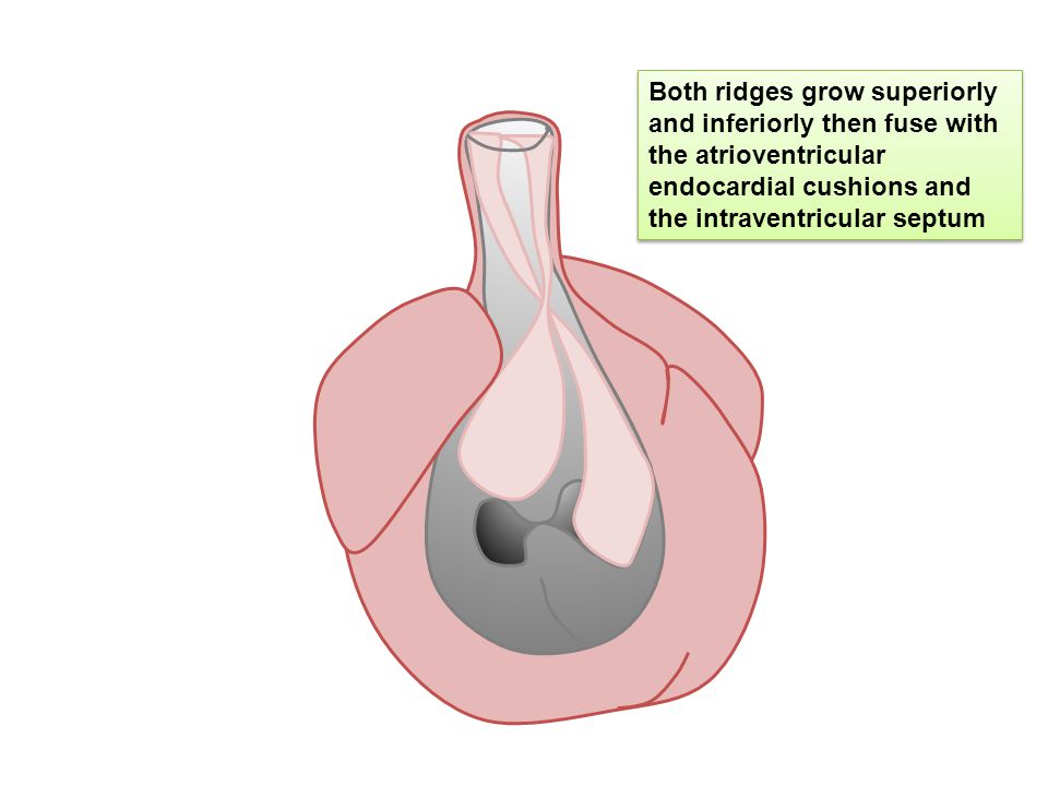
Both ridges grow superiorly and inferiorly then fuse with the atrioventricular endocardial cushions and the intraventricular septum

10
Cardiac neural crest migration from pharyngeal arches contributes to division of the outflow tract

11
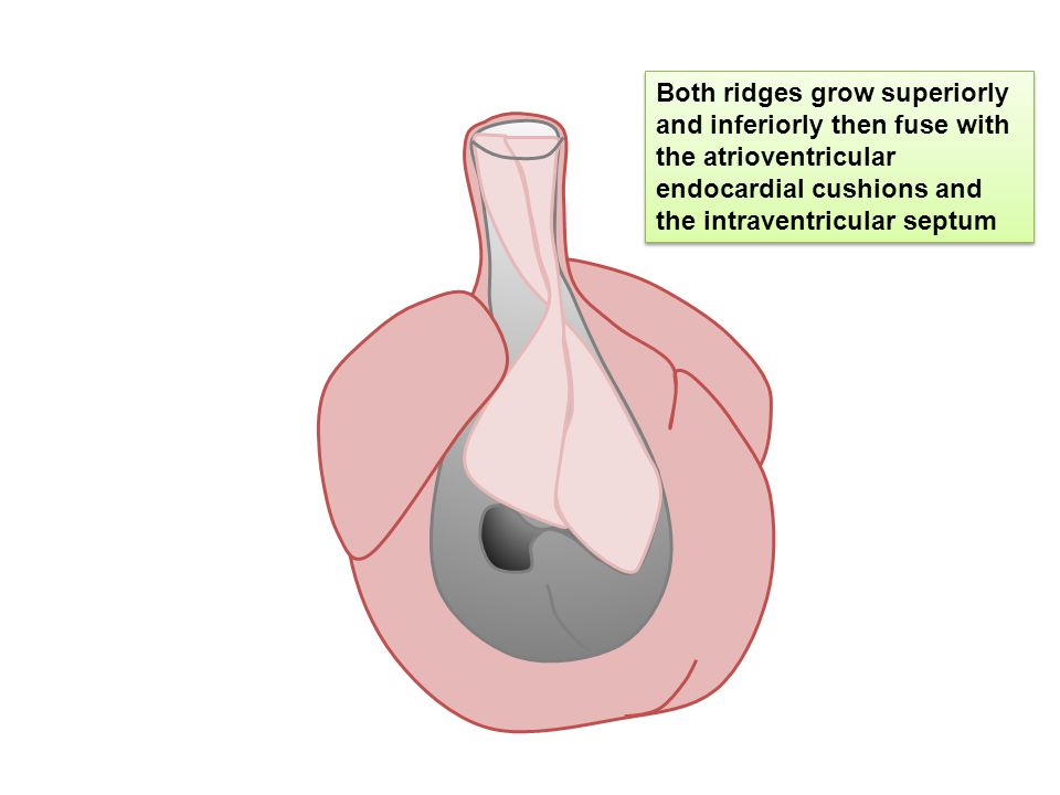
Both ridges grow superiorly and inferiorly then fuse with the atrioventricular endocardial cushions and the intraventricular septum

16
Aorta and pulmonary trunk are then initially established from this septation

17
primodial pulmonary trunk primodial aorta Aorta and pulmonary trunk are then initially established from this septation

18
Aorta and pulmonary trunk development

21
pulmonary trunkaorta

Similar presentations


, UNSEPARATED VENTRICLE (18), LIVER (12), UMBILICAL VEIN (17), TRANSVERSE.>")


