Download presentation
Presentation is loading. Please wait.

1
Radiology Packet 6 Acquired cardiac diseases

2
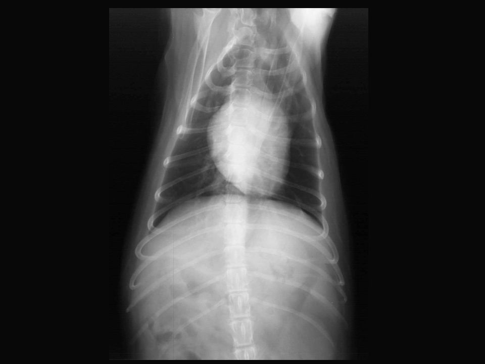
13 year old M Miniature Poodle “Carlos” Hx: Presented for evaluation of coughing that has been getting progressively worse and is worse at night. On PE a grade 2-3 of 6 murmur is ausculted. The murmur is systolic and is heard best on the left side of the chest.

4
13 year old M Miniature Poodle “Carlos” RF –Caudal mainstem bronchi are elevated and there is straightening of the the caudal cardiac waist. –The trachea is elevated and the caudal cardiac margin is elongated. –In the VD view the cardiac silhouette is rounded. –There is a bulge at the area of the left auricular appendage and rounding of the cardiac apex. –There is increased interstitial infiltrates in the lungs in the perihilar region and caudal lung fields. –Hepatomegaly. RD –Moderate generalized cardiac enlargement is present –Left-sided cardiac enlargement predominates –Pulmonary vascular congestion and cardiogenic pulmonary edema are evidence of left-sided congestive heart failure R/O –Mitral valve endocardiosis with mitral regurgitation –Right sided cardiac enlargement is likely due to concurrent tricuspid endocardiosis

5
16-month old Jack Russell Terrier “Bandit” Hx: Presented with a history of exercise intolerance and polycythemia (PCV 65-70%)
")
6
16-month old Jack Russell Terrier “Bandit” RF –In the lateral view the heart is ~4 ICS wide and the VHS is 10.7 on the Buchanan scale. –The heart is widened and there is increased sternal contact. –Loss of the cranial cardiac waist. –In the VD view the heart has a “reverse D” appearance. –There is a prominent bulge at 1-2 o’clock indicating enlargement of the main pulmonary artery. –There is a bulge in the aortic arch at the same level as the pulmonary artery bulge. RD –Right ventricular enlargement R/O –Pulmonic stenosis –Right-to-left PDA –Heartworm disease

7
14-year old M Miniature Poodle “Tuffy” Hx: presented for evaluation of intervertebral disc disease. On PE a grade 1-2 of 6 murmur is ausculted. The murmur is systolic and is heard best on the left side of the chest.

9
14-year old M Miniature Poodle “Tuffy” RF –Caudal mainstem bronchi are elevated and there is subtle straightening of the caudal cardiac waist. –There is a diffuse, mild broncho-interstitial lung pattern present that is consistent with normal aging change. –The liver extends beyond the costal arch indicating that it is enlarged. RD –Left atrium is mildly enlarged R/O –Mitral valve endocardiosis and secondary mitral regurgitation

10
12-year old FS Shih Tzu “Pepper” Hx: Presented for cataract surgery. During PE a grade 3 of 6 holosystolic murmur with the point of maximal intensity on the left was auscultated.

12
12-year old FS Shih Tzu “Pepper” RF –There is elevation of the caudal mainstem bronchi and straightening of the caudal cardiac margin. –Trachea is elevated and the caudal margin of the heart is elongated. –In the VD view the cardiac apex is slightly rounded. –The cardiac silhouette is slightly wide in the lateral view. –There is a diffuse mild broncho-interstitial lung pattern present which is consistent with normal aging change. RD –Mild left-sided cardiac enlargement +/- mild right-sided cardiac enlargement R/O –Mitral valve endocardiosis –Tricuspid valve endocardiosis

13
12-year old cat “Garlock” Hx: Presented with coughing and exercise intolerance.

15
12-year old cat “Garlock” RF –Diffuse increased opacity of the entire lung field. –Increase in size of the pulmonary vasculature. –In the DV view the caudal pulmonary arteries are much larger than the veins. R/O –Feline heartworm disease Note – on these films it is hard to see the changes. But note, for heartworm disease is cats – it is the vasculature and lungs that show the changes primarily. The heart changes we see in dogs are often not seen in cats.

16
13-year old MN DSH “Snowball” Hx: History of weight loss, tachypnea and tachycardia. A mass ~ 1cm x 1cm is palpable in the cervical region.

18
13-year old MN DSH “Snowball” RF –The cardiac silhouette is slightly enlarged. The VHS is ~ 8.0. –In the lateral view the cardiac silhouette is somewhat elongated and there is bulging of the heart base. –In the VD view the atrial region of the heart (base) is wide. –There is a mild diffuse interstitial lung pattern present that is consistent with normal age change. –Several sites of spondylosis are present in the thoracic spine. –Large osteochondral fragments are visisble in both of the elbows. RD –Mild ventricular hypertrophy R/O –Hyperthyroidism –Hypertrophic cardiomyopathy
 is wide. –There is a mild diffuse interstitial lung pattern present that is consistent with normal age change. –Several sites of spondylosis are present in the thoracic spine. –Large osteochondral fragments are visisble in both of the elbows. RD –Mild ventricular hypertrophy R/O –Hyperthyroidism –Hypertrophic cardiomyopathy.")
19
4-year old MN boxer Hx: Presented for evaluation of exercise intolerance and coughing.

21
4-year old MN boxer RF –In the lateral view the base of the heart appears wide and there is bulging of the cardiac silhouette at the cranial dorsal margin. –In the VD view there is a prominent bulge in the area of the main pulmonary artery with rounding of the right heart margin. –The left ventricular area is normal creating a flat appearance to the left side of the heart and a classic “reverse-D” shape. –In the lateral view a very large and somewhat tortuous cranial pulmonary artery is visible. Some of the caudal pulmonary vessels are large and tortuous. –Enlarged pulmonary arteries are visible in the VD view. –Increased opacity of the perihilar region and displacement of the cranial mainstem bronchi from the trachea are noted. –Focal area of interstitial pulmonary opacity is visible in the caudal lung tip in the lateral view.

22
4-year old MN boxer RD –Right-sided cardiac enlargement –Enlargement of the main pulmonary artery segment –Enlarged, tortuous and truncated pulmonary arteries –Focal area of interstitial pulmonary opacity R/O –Severe canine heartworm disease –Allergic pneumonitits –Thromboembolic disease

23
3-year old MN DSH “Socrates” Hx: Presented for re-evaluation of previously diagnosed hypetrophic cardiomyopathy.

25
3-year old MN DSH “Socrates” RF –The cardiac silhouette is enlarged. –In the lateral view of the heart base is enlarged making the heart appear wide with bulging of the cranial cardiac margin. –In the VD view the atrial region of the heart is very wide but the ventricular region remains relatively normal. –The pulmonary vessels are greater than normal diameter and are larger than the pulmonary arteries. RD –Cardiomegaly (r/o HCM) –Vascular congestion
 –Vascular congestion.")
Similar presentations


 MRCVS European and RCVS Specialist in Veterinary.>")


