Download presentation
Presentation is loading. Please wait.

1
ABDOMEN Lu Xiaoli Regional Anatomy & Operative Surgery
China Medical University

2
BOUNDARIES

3
Abdominopelvic Cavity
Abdominal Cavity Pelvic Cavity P242-fig.4.21

4
DIVISIONS P242-fig.4.22

5
P243-fig.4.23

6
P243-fig.4.23

7
Which one of the following is not one of the 9 regions of the abdomen?
Right hypochondriac Left inguinal or iliac Epigastric Right upper Left lumbar

8
Which of the following is NOT true concerning the peritoneal cavity?
The peritoneal cavity is a potential space. The peritoneal cavity contains organs inside of it. The peritoneal cavity is filled with fluid that lubricates its contents. The parietal and visceral peritoneum are linings of the peritoneal cavity.

9
The usual location for an appendectomy incision is the:
left lower quadrant left upper quadrant right lower quadrant right upper quadrant

10
You were asked to assist in a surgical operation on a young patient to treat an ulcer in the first part of the duodenum. You would expect that the surgeon will approach the ulcer by doing an anterior abdominal wall incision in the following region:

11
Epigastric Left inguinal Left lumbar Right hypochondrial Hypogastric

12
ABDOMINAL WALL

13
Abdominal wall Anterolateral abdominal wall Posterior abdominal wall

14
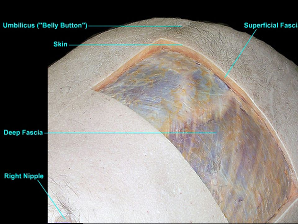
LAYERS Skin Superficial fascia Transversalis fascia Deep fascia
Muscles Transversalis fascia Extraperitoneal fascia Peritoneum

16
Superficial fascia Camper’s fascia Scarpa's fascia P245-fig.4.25~4.26

17
SUPERFICIAL ARTERIES Lateral Median Inferior Posterior intercostal a.
Subcostal a. Lumbar a. Median Epigastric a. hypogastric a. Inferior Superficial epigastric a. Superficial iliac a. P255-fig.4.39

18
Superficial veins lateral thoracic subclavian thoracoepigastric
paraumbilical portal S epigastric femoral S circumflex iliac

20
Caput Medusae (Medusa Head )
")
21
INNERVATIONS Intercostal Nerve T7-T12 10th Intercostal Nerve

22
MUSCLES Anterior Group Lateral Group External Oblique Internal Oblique
Transversus Rectus Abdominis Pyramidalis

23
RECTUS ABDOMINIS Tendinous Intersection (3) Linea Semilunaris
 Linea Semilunaris")
24
Rectus Sheath

25
Arcuate line

26
PYRAMIDALIS

27
LINEA ALBA

28
External Oblique Abdominis

29
Oblique Internal Abdominis

30
Transversus Abdominis

31
Arteries 5 intercostal arteries subcostal arteries 4 lumbar arteries
Superior epigastric artery—internal thoracic artery Inferior epigastric artery -external iliac artery Deep iliac circumflex artery- external iliac artery

32
Inferior epigastric artery

34
Lymphatic Drainage Anterior →
Intercostal Lymphatic Nodes Parasternal Lymphatic Nodes Middle Lumbar Lymphatic Nodes Lower External Iliac Lymphatic Nodes

35
Innervations Intercostal n. Anterior cutaneous branch
Lateral cutaneous branch

36
T7-12 thoracic n. Iliohypogastric n. Ilioinguinal n. Genitofemoral n.

37
Transversalis Fascia

38
Extraperitoneal Fascia

39
Parietal Peritoneum

40
Umbilical Folds Median -- median umbilical lig. Medial
-- chorda arteriae umbilicalis Lateral -- inferior epigastric a. & v.

41
INCISIONS Longitudinal Midline Paramedian Transrectal Oblique
Subcostal McBurney’s Transverse Pfannenstiel Combined Thoracal-abdominal

43
The inferior border of the rectus sheath posteriorly is called the:
Falx inguinalis Inguinal ligament Internal inguinal ring Arcuate line Linea alba

44
Following an emergency appendectomy your patient complained of having paresthesia (numbness) of the skin at the pubic region. The most likely nerve that has been injured during the operation is: Genitofemoral Iliohypogastric Subcostal Spinal nerve T10 Spinal nerve T9

45
An obstetrician decides to do a Caesarean section on a 25-year-old pregnant woman. A transverse suprapubic incision is chosen for that purpose. All of the following abdominal wall layers will be encountered during the incision EXCEPT the:

46
Anterior rectus sheath
Posterior rectus sheath Rectus abdominis muscle Skin and subcutaneous tissue Transversalis fascia, extraperitoneal fat, and peritoneum

47
Surgical approaches to the abdomen sometimes necessitate a midline incision between the two rectus sheaths, i.e., through the: Linea aspera Arcuate line Semilunar line Iliopectineal line Linea alba

48
The internal thoracic artery is sometimes surgically cut near the caudal end of the sternum and used to supply blood to a region of the heart. In these cases, maintenance of adequate blood flow to the rectus abdominis may be dependent on increased flow through which artery?

49
Superficial epigastric
Inferior epigastric Umbilical Superficial circumflex iliac Deep circumflex iliac

50
INGUINAL REGION

52
Boundaries

53
LAYERS Skin Superficial layer Camper’s Scarpa’s

54
External Oblique Abdominis
Inguinal Lig. Lacunar Lig. Pectineal Lig. (cooper’s Lig.)
")
55
Superficial Inguinal Ring
Reflected Ligament Intercrural Fibers Lateral Crus Superficial Inguinal Ring Medial Crus

56
Internal oblique abdominis
transverse abdominis

57
Cremaster Conjoint Tendon

58
Cremaster Conjoint Tendon

59
Transverse Abdominal Fascia
abdominal inguinal ring (deep inguinal ring)
")
60
Extraperitoneal fascia Parietal peritoneum
Medial inguinal fossa lateral inguinal fossa

61
Descent Of Testis

62
4 lunar month 11 weeks 8 lunar month

66
Inguinal Canal Roof internal oblique abdominis transversus abdominis
Floor inguinal ligament lacunar ligament anterior wall external abdominal oblique aponeurosis internal abdominal oblique aponeurosis posterior wall transversalis fascia conjoint tendon (falx inguinalis)
")
67
Contents (male) spermatic cord ilioinguinal nerve
arteries: testicular artery, deferential artery, cremasteric artery nerves: genital branch of the genitofemoral nerve, nerve to cremaster, sympathetic nerves vas deferens pampiniform plexus lymphatic vessels ilioinguinal nerve

68
Contents (female) round ligament of the uterus ilioinguinal nerve
 round ligament of the uterus ilioinguinal nerve")
69
Spermatic Fascia Internal spermatic fascia Middle External
Transversalis fascia Middle cremaster External aponeurosis of external oblique abdominis

70
HERNIA Inguinal hernia Indirect Direct

71
Inguinal Triangle (Hesselbach's triangle )
Direct Hernia

72
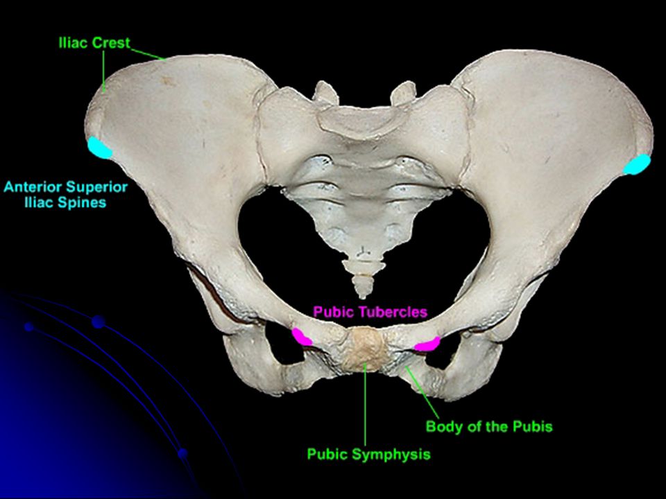
A medical student was asked by her preceptor to palpate the margin of the superficial inguinal ring of a healthy male patient. After passing her finger down the edge of the medial crus of the superficial inguinal ring, she felt a bony protuberance deep to the lateral edge of the spermatic cord, which she correctly identified as the :

73
pecten pubis pubic symphysis pubic tubercle iliopubic eminence iliopectineal line

74
In order to reduce a hernia (return it to the abdominal cavity), a surgeon finds it necessary to ligate an artery in the extraperitoneal connective tissue (preperitoneal fat) running vertically just medial to the bowel as the bowel passes through the abdominal wall. This artery is the:

75
Deep circumflex iliac Inferior epigastric Superficial circumflex iliac Superficial epigastric Superficial external pudendal

76
During a laparoscopic examination of the deep surface of the lower anterior abdominal wall (using a lighted scope on a thin tube inserted through the wall), the attending physician noted something of interest and asked the young resident to look at the medial inguinal fossa. To do so, the young doctor would have to look at the area between the:

77
inferior epigastric artery and urachus
medial umbilical ligament and urachus inferior epigastric artery and lateral umbilical fold medial umbilical ligament and inferior epigastric artery median umbilical ligament and medial umbilical ligament

78
If one were to make an incision parallel to and 2 inches above the inguinal ligament, one would find the inferior epigastric vessels between which layers of the abdominal wall?

79
Camper's and Scarpa's fascias
External abdominal oblique and internal abdominal oblique muscles Internal abdominal oblique and transversus abdominis muscles Skin and deep fascia of the abdominal wall Tranversus abdominis muscle and peritoneum

80
Which structure passes through the deep inguinal ring?
Iliohypogastric nerve Ilioinguinal nerve Inferior epigastric artery Medial umbilical ligament Round ligament of the uterus

81
A loop of bowel protrudes through the abdominal wall to form a direct inguinal hernia; viewed from the abdominal side, the hernial sac would be found in which region?

82
Deep inguinal ring Lateral inguinal fossa Medial inguinal fossa Superficial inguinal ring Supravesical fossa

83
In a female with an indirect inguinal hernia, the herniated mass lies along side of which structure as it traverses the inguinal canal? Iliohypogastric nerve Inferior epigastric artery Ovarian artery and vein Pectineal ligament Round ligament of the uterus

84
The skin of the mons pubis is supplied by which nerve?
Anterior scrotal Anterior labial Femoral branch of the genitofemoral Iliohypogastric nerve Subcostal nerve

85
During your peer presentation of the inguinal region dissection, you would indicate the position of the deep inguinal ring to be: Above the anterior superior iliac spine Above the midpoint of the inguinal ligament Above the pubic tubercle In the supravesical fossa Medial to the inferior epigastric artery

86
A 45-year-old porter develops a direct inguinal hernia
A 45-year-old porter develops a direct inguinal hernia. If the hernia extended through the superficial inguinal ring, it would be surrounded by all of the abdominal wall layers EXCEPT the:

87
External spermatic fascia
Internal spermatic fascia Peritoneum and extraperitoneal connective tissue Weak fascia of the transversus abdominis muscle lateral to the falx

88
The boundaries of the inguinal triangle include all except:
Arcuate line Inferior epigastric vessels Inguinal ligament Lateral border of rectus abdominus muscle

89
The superficial inguinal ring is an opening in which structure?
External abdominal oblique aponeurosis Falx inguinalis Internal abdominal oblique muscle Scarpa's fascia Transversalis fascia

90
Which nerve passes through the superficial inguinal ring and may therefore be endangered during inguinal hernia repair? Femoral branch of the genitofemoral Ilioinguinal Iliohypogastric Obturator Subcostal

91
During exploratory surgery of the abdomen, an incidental finding was a herniation of bowel between the lateral edge of the rectus abdominis muscle, the inguinal ligament and the inferior epigastric vessels. These boundaries defined the hernia as a(n):
:.")
92
Congenital inguinal hernia
Direct inguinal hernia Femoral hernia Indirect inguinal hernia Umbilical hernia

93
Anterolateral abdominal wall Superficial layers Skin Superficial skin Deep layers Muscles Transversalis fascia Subperitoneal fascia Parietal peritoneum Inguinal region Inguinal canal triangle 4 walls 2 openings Contens

Similar presentations