Download presentation
Presentation is loading. Please wait.

1
Orthopaedic Neurology
Diagnostic Guide to Neurological Levels

2
Motor Power Interruption of the nerve root causes denervation and paralysis of its myotome. Pressure on a nerve root can cause a decrease in muscle strength. Muscle testing is utilized to evaluate whether or not a lesion is present and to what degree it is effecting the muscle strength.

3
Muscle Grading Chart Muscle gradations Description.
5 – normal 4 – good 3 – fair 2 – poor 1 – trace 0 - zero Description. Complete range of motion against gravity with full resistance. Complete range of motion against gravity with some resistance. Complete range of motion against gravity. Complete range of motion with gravity eliminated. Evidence of slight contractility. No joint motion. No evidence of contractility.

4
Sensation Pathology to the cord or nerve root results in loss of light touch, followed by loss of sensation of pain. During recovery from nerve root injury, sensation of pain returns before light touch.

5
Sensation The 2 sensations are tested separately, light touch with a cotton swab, pain with pinpricks. Pinwheels can be utilized to evaluate sensation. Results can be recorded on a dermatome chart as normal, hyperesthetic (increased), hyposthetic (decreased), dyesthetic (altered), or anesthetic (absent).
, hyposthetic (decreased), dyesthetic (altered), or anesthetic (absent).")
6
Reflex Interruption in the basic reflex arc results in the loss of reflex, while pressures on the nerve root itself may decrease its intensity (hyporeflexia). Interruption of the upper motor neuron’s regulatory control results in a hyperactive nerve (hyperreflexia). Reflexes should be reported as normal, increased, or decreased utilizing bilateral comparison.
. Interruption of the upper motor neuron’s regulatory control results in a hyperactive nerve (hyperreflexia). Reflexes should be reported as normal, increased, or decreased utilizing bilateral comparison.")
7
Stretch Reflex Arc

8
Nerve Root Lesions by Neurologic Level

9
Evaluation of Nerve Root Lesions
Upper Extremity

10
Cervical Spine C5 is the 1st significant contribution to the brachial plexus. C1-4 are difficult to test; However, C4 is the major innervation to the diaphragm (via the phrenic nerve).
.")
11
The Cervical Spine

14
Deltoid & Supraspinatous

15
Elbow Flexion and Extension

16
Biceps Brachii & Brachialis

17
Functions of the Biceps

18
Muscle Test for the Biceps

19
Biceps Reflex Test

20
Memory Trick

21
Muscle Test Shoulder Abduction

22
Sensory Distribution C5

24
Wrist Extension and Flexion

25
Extensor Carpi Ulnaris (Left), Extensor Carpi Radialis (Right)
, Extensor Carpi Radialis (Right)")
26
Muscle Test Wrist Extension

27
Brachioradialis Reflex Test

28
Memory Trick

30
Triceps Brachii

31
Walking With a Crutch Utilizes the Triceps Muscle

32
Muscle Test Wrist Flexors

33
Flexor Carpi Radialis (Left), Flexor Carpi Ulnaris (Right)
, Flexor Carpi Ulnaris (Right)")
34
Finger Extension and Flexion

35
Extensor Digitorum

36
Muscle Test Finger Extension

37
Triceps Reflex Test

39
Flexor Digitorum Superficialis (Left), and Profundus (Right)
, and Profundus (Right)")
40
Lumbricales

41
Muscle Test Finger Flexors

42
Memory Trick

44
Finger Abduction and Adduction

45
Muscle Test Finger Abduction

46
Muscle Test Finger Adduction

47
Summary of Muscle Testing for the Upper Extremity

48
Summary of Reflex Testing for the Upper Extremity

49
Summary of Sensation for the Upper Extremity

50
Cervical Vertebrae and Nerve Roots

51
Herniated Cervical Disc

52
Occiput & C1 Articulation

53
C1 and C2 Articulation

54
Anatomic Basis for Posterior Cervical Disc Herniation

60
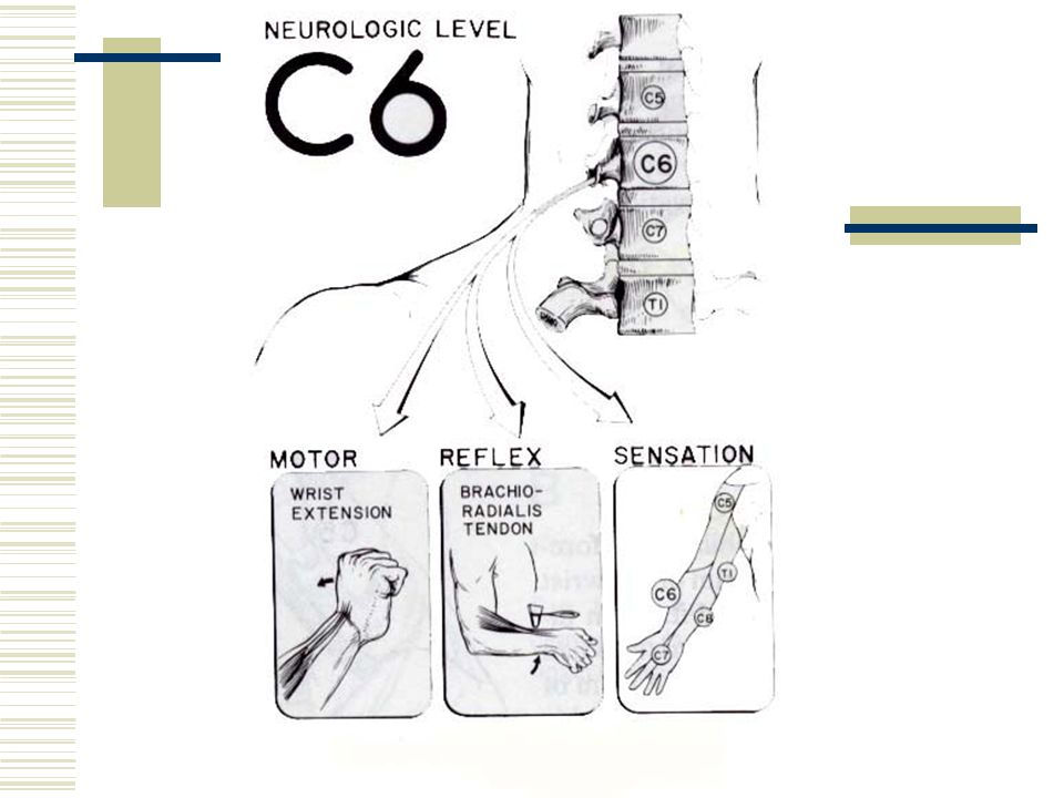
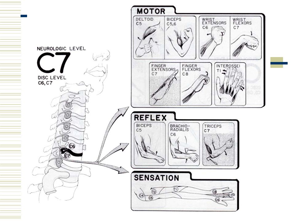
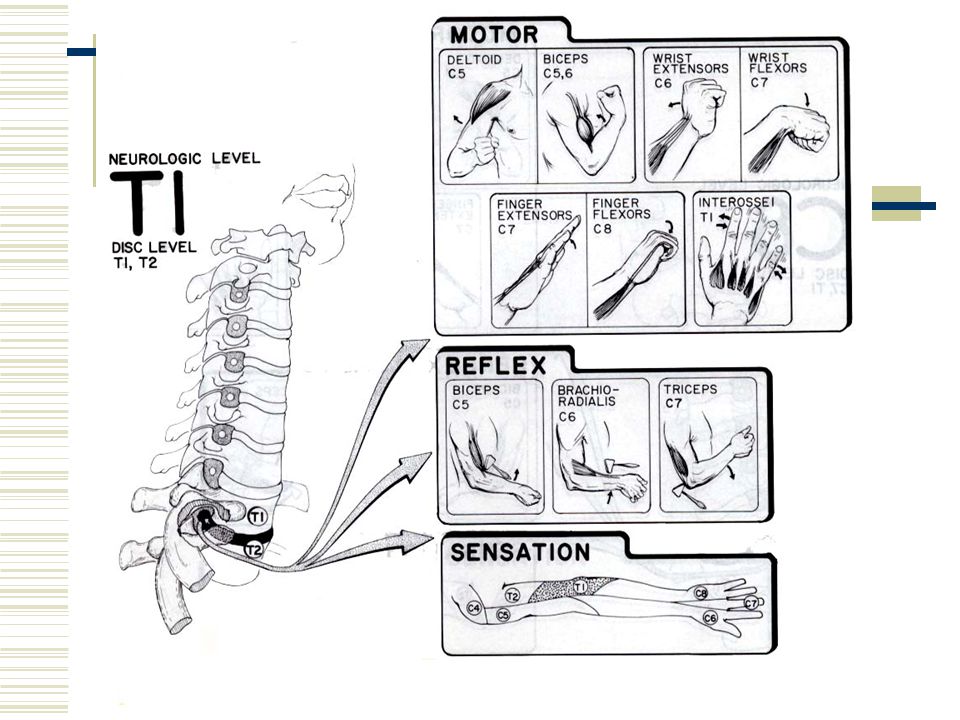
Neurologic Levels in Upper Extremity
Motor C5 – shoulder abduction C6 – wrist extension C7 – wrist flexion and finger extension C8 – finger flexion T1 – finger abduction, adduction

61
Neurologic Levels in Upper Extremity
Sensation C5 – lateral arm C6 – lateral forearm, thumb, and index finger C7 – middle finger (variable) C8 – medial forearm, ring, and small finger T1 – medial arm T2 - Axilla
 C8 – medial forearm, ring, and small finger. T1 – medial arm. T2 - Axilla.")
62
Neurologic Levels in Upper Extremity
Reflex C5 – biceps C6 – Brachioradialis C7 - triceps

64
Whiplash Injury to the Cervical Spine

65
Anatomy of a Cervical Vertebrae

66
Orthopedic Tests Cervical Spine
Valsalva test Distraction test Compression test

67
Valsalva Test

68
Distraction Test

69
Compression Test

70
Thoracic Spine

71
Beevor’s Spine

72
Hip Flexion

73
Iliopsoas

74
Muscle Test Iliopsoas

75
Knee Extension

76
Rectus Femoris (Left), Vastus Intermedius and Lateralis (Right)
, Vastus Intermedius and Lateralis (Right)")
77
Extension Lag

78
Muscle Test Quadriceps

79
Hip Adduction

80
Adductors

81
Muscle Test Hip Adductors

82
Dermatomes of the Lower Extremity

84
Foot Inversion

85
Muscle Test Tibialis Anterior

86
Patellar Tendon Reflex

87
Memory Trick

89
Foot Dorsiflexion (Ankle Extension)
")
90
Extensor Hallucis Longus

91
Muscle Test Extensor Hallucis Longus

92
Muscle Test Toe Extensors

93
Memory Trick

94
Hip Abduction

95
Gluteus Medius

96
Muscle Test Gluteus Medius

97
L5 Sensory Dermatome

99
Foot Eversion

100
Peroneus Longus & Brevis

101
Muscle Test Peronei Muscles

102
Foot Plantarflexion

103
Gastrocnemius & Soleus

104
Muscle Test Gastrocnemius

105
Hip Extension

106
Gluteus Maximus

107
Muscle Test Gluteus Maximus

108
Achilles Reflex Test

109
Memory Trick

110
Sensory Dermatomes S2, S3, S4, S5

111
Sensory Dermatomes L4-S1

112
Testing Sensation

113
Anatomic Basis for Posterior Lumbar Disc Herniation

117
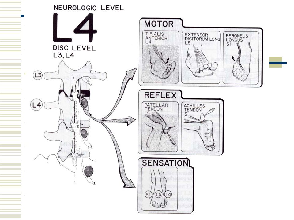
Neurologic Levels in Lower Extremity
Motor L3 – quadriceps (L2, L3, L4) L4 – Tibialis anterior L5 – toe extensors S1 - Peronei
 L4 – Tibialis anterior. L5 – toe extensors. S1 - Peronei.")
118
Neurologic Levels in Lower Extremity
Sensation T12 – lower abdomen just proximal to inguinal ligament L1 – upper thigh just distal to inguinal ligament L2 – mid thigh L3 – lower thigh L4 – medial leg – medial side of foot L5 – lateral leg – dorsum of foot S1 – lateral side of foot S2 – longitudinal strip, posterior thigh

120
Neurologic Levels in Lower Extremity
Reflex L4 – patellar L5 – Tibialis posterior (difficult to obtain) S1 – Achilles tendon
 S1 – Achilles tendon.")
Similar presentations


>")
