Download presentation
Presentation is loading. Please wait.

1
Organization of the Cell

2
Cell theory Cells are the basic living units of organization and function All cells come from other cells Work of Schleiden, Schwann, and Virchow contributed to this theory Each cell is a microcosm of life

3
Cell organization and size permit homeostasis
Homeostasis: The balanced internal environment of the body; the automatic tendency of an organism to maintain such a steady state.

4
Cell surface area-to-volume ratio
Plasma membrane must be large enough relative to cell volume to regulate passage of materials

5
Organization is basically the same in all cells
All cells have a plasma membrane, which is a selective barrier Cells have internal organelles that are specialized for various functions Cell size is limited by the surface to volume ratio, so cells remain small Cell shape and size are related to function.

6
Surface to volume ratio
As the cell size increases, the volume becomes too large for the surface membrane to handle.

7
Cell surface area-to-volume ratio

9
Biological size and cell diversity

10
Small size… (add) Molecules must be transported to the locations where they are converted into other forms With small cell size, distances molecules travel within them are relatively short which speeds up many cellular activities

11
Cells are studied by a combination of methods
Robert Hooke: identified the cell walls and gave the structure the name “cell”. Study of the cell was not possible until microscopes.

12
Microscopes Light microscope, referred to as compound microscope, used by most students Two features determine how clearly an object is viewed Magnification: how much larger Resolution: how clear Light microscope has 500 times more resolution than human eye

13
Allows study of the ultrastructure of cells
Electron microscope Developed in the 1950s Allows study of the ultrastructure of cells 10,000 times more resolution than human eye

14
Types of electron microscope
Transmission electron microscope: used for studying ultra structure. Specimen must be specially prepared and sliced very thin. Scanning electron microscope: bounces electrons off a thin metallic coating on the object. Enables surface area to be viewed.

15
Comparing light and electron microscopy

16
Cell fractionation Used to determine function of organelles
Cells broken apart and the resulting cell extract spun in a centrifuge Centrifugal force separates extract Pellet: solid particle Supernatant: fluid material

17
Cell fractionation

18
Types of Cells http://www. tvdsb. on
Prokaryotic Cells: Bacteria and Archea 1. lack membrane bound organelles 2. smaller in size than eukaryotes 3. DNA is located in nucleoid area and is not bound by a nuclear membrane 4. Most have cell walls 5. Have ribosomes and storage granules

19
Eukaryotic cells: all other organisms
1. nucleoplasm: contains chromosomes which package DNA 2. cytoplasm: area outside the nucleus; contains cytosol and organelles, many of which are membrane-bound 3. type of cell determines which organelles are present and in what numbers

20
Divide cell into compartments, allowing for specialized activities
Functions of cell membranes Divide cell into compartments, allowing for specialized activities Interacting membranes form endomembrane system Vesicles transport materials between compartments

21
Diagram of a plant cell

22
Diagram of an animal cell

23
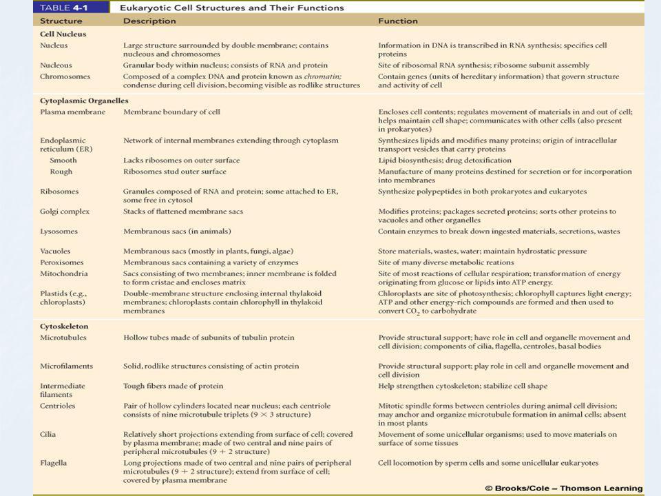
The cell nucleus Contains DNA Bounded by
Nuclear envelope Double membrane perforated with nuclear pores DNA forms chromatin, which is organized into chromosomes Nucleolus RNA synthesis and ribosome assembly

24
The cell nucleus

25
Endoplasmic reticulum (ER)
Network of folded internal membranes in the cytosol Smooth ER Site of lipid synthesis Site of detoxifying enzymes Rough ER Ribosomes manufacture proteins Proteins may be moved into the ER lumen

26
Endoplasmic reticulum (ER)
")
28
Glycoproteins transported to the cis face
Golgi complex Cisternae that process, sort, and modify proteins In animal cells, Golgi complex also manufactures lysosomes Glycoproteins transported to the cis face Golgi modifies carbohydrates and lipids and packages into vesicles; which then may leave the cell

29
Golgi complex

30
Lysosomes break down worn-out cell structures, bacteria, and other substances
Primary lysosomes bud from the Golgi complex Secondary lysosomes form by fusion of a primary lysosome with a vesicle containing ingested material Involved in apoptosis (programmed cell death)
")
31
Lysosomes

32
Peroxisomes metabolize small organic compounds
Transfer hydrogen from various compounds to oxygen, forming hydrogen peroxide Catalase is an enzyme that breaks down hydrogen peroxide Most common in cells that synthesize, store, and degrade lipids

33
Peroxisomes and lysosomes

34
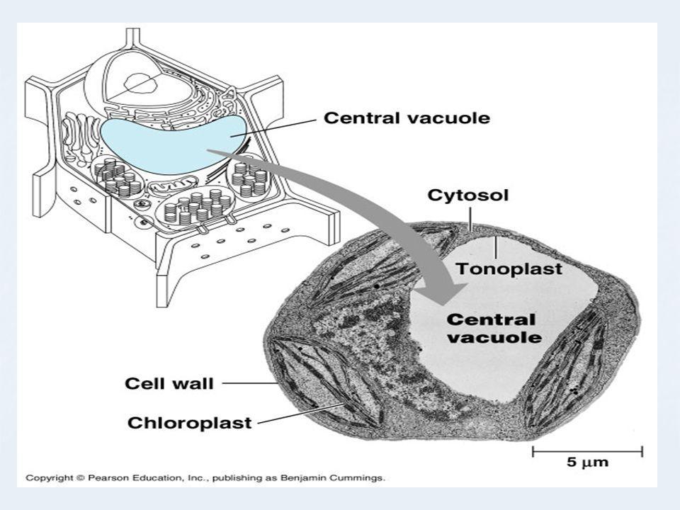
Vacuoles Large, fluid-filled sacs with a variety of functions.
May function in storage of toxins or pigments Plant vacuoles are typically large and allow the cell to increase in size Protist vacuoles may involve digestion or excretion.

37
Mitochondria Sites of aerobic respiration
Organelles enclosed by a double membrane Cristae and matrix contain enzymes for aerobic respiration Nutrients broken down and energy packaged in ATP Carbon dioxide and water by-products

38
Mitochondria

39
Chloroplasts Plastids that carry out photosynthesis
Inner membrane of chloroplast encloses the stroma During photosynthesis, chlorophyll traps light energy Energy converted to chemical energy in ATP; sugars/food for the plant are manufactured.

40
Chloroplast

41
Cellular respiration and photosynthesis

42
Equation for photosynthesis
6H2O + 6CO2 C6H12O6 + 6 O2 If you turn the arrow around, you have the equation for cellular respiration.

43
Internal framework made of
Cytoskeleton Internal framework made of Microtubules Microfilaments Intermediate filaments Provides structural support Involved with transport of materials in the cell

44
The Cytoskeleton

45
Cilia and flagella Thin, movable structures that project from cell surface Function in movement Microtubles anchored in cell by basal body

46
Structure of cilia

47
Cilia and flagella

48
Glycocalyx, cell coat formed by polysaccarides extending from plasma membrane
Many animal cells also surrounded by an extracellular matrix (ECM) Most bacteria, fungi, and plant cell walls made of carbohydrates
 Most bacteria, fungi, and plant cell walls made of carbohydrates.")
49
Extracellular matrix

50
Plant cell walls

Similar presentations


 The McGraw-Hill Companies, Inc.>")


, visible light passes through a specimen and then through glass lenses, which magnify the image The quality of an.>")


