Download presentation
Presentation is loading. Please wait.

1
Differential Diagnosis of Oral and Maxillofacial lesions
General principles of Differential Diagnosis 1. History and examination of the patient 2. The diagnosis sequence 鑑別診斷的基本原則-- 問診與檢查 診斷的步驟 學習病歷的寫法,提供各位病歷寫作時的靈感。熟記關鍵字,寫病歷時才能迅速確實。 王文岑 高雄醫學大學 牙醫學系 高醫大附設醫院S 棟 2 樓 口腔病理影像診斷科 ;

2
學習目標 瞭解主訴的涵義 瞭解問診的內容 熟悉診斷的步驟 瞭解各種檢查方法之使用時機 學習資源及主要圖片引用: 1. Differential diagnosis of oral lesion. Wood, Gooz(Mosby), 5th ed., 1997. 2. 口腔病理科門診臨床記錄

3
Clinical pathology, a study of change
Purpose: overview of significance and application of medicine in the dentistry The dentist does not only treat “teeth in patients “ but patients who have teeth or not”.

4
Possible Procedures Leading to Diagnosis
Clinical pathology Possible Procedures Leading to Diagnosis

5
IN the procedures, you have to do 1. To identified—undetected systemic
disease 2. To identified —taking drug or medicine as clue 3. To modify– treatment planning 4. To protect –medical-dental legal standpoint 5. To communicate –medical consultants 6. To establish –good patient-dentist relation ship 問診的目的:

6
The Diagnostic Sequence
1. Detection and examination of the lesion 2. Examination of the patient Chief complaints Onset and course 3. Re-examination of the lesion 4. Classification of the lesion 5. Listing the possible diagnosis 6. Developing the differential diagnosis 7. Working diagnosis 8. Final diagnosis 診斷操作流程

7
1. RECORDING THE IDENTIFYING DATA 2. HISTORY AND PHYSICAL EXAMINATION
3. CHIEF COMPLAINT 4. PRESENT ILLNESS 5. PAST MEDICAL HISTORY Family history Social history Occupational history Dental history 6. REVIEW OF SYMPTOMS BY SYSTEM 7. PHYSICAL EXAMINATION Radiologic examination 8. DIFFERENTIAL DIAGNOSIS 9. WORKING DIAGNOSIS 10. Medical laboratory studies 11. Dental laboratory studies 12. Biopsy Incisional Excisional Fine-needle aspiration Exfoliationg cytology Toludine blue staining 13. Consultation 14. FINAL DIAGNOSIS 15.TREATMENT PLAN 病歷書寫格式

8
Detection and examination of the patient’s lesion
History taking Inspection Palpation Percussion Aspiration, Auscultation Radiographic examination Laboratory examination

9
History Taking What, where, when, how Past medical history
Chief complaint Present illness Past medical history Family history Social history Occupational history Dental history Review of symptoms by system Physical examination Radiographic examination

10
Chief complaint(s) Pain Soreness Burning sensation Bleeding
Loose teeth Recent occlusal problem Delayed tooth eruption Dry mouth Too much saliva Swelling Bad taste Halitosis Parthesia and anesthesia 病人常用的關鍵字

11
Pain -- Location,sharp or dull, severity, duration, precipitating circumstances Teeth Mucous membrane Salivary gland inflammation or infection Lesions of the jaw bone LN inflammation and /or inflammation TMD, MPD Sinus diseases Ear diseases Psychoses Angina pectoris, neuralgia …

12
-presence of mucosa inflammation or ulcers Burning sensation
Soreness -presence of mucosa inflammation or ulcers Burning sensation -thinning or erosion of the surface epi. Burning mouth syndrome Xerostomic condition Anemia Vitamin deficiencies… Psychosis Neurosis Viral, fungal or chronic bacterial infection

13
口腔灼熱症 (Burning mouth syndrome)
Gender effects : F:M = 3:1 ~ 7:1 Age: Middle-aged and elderly-aged Local and systemic preciptating factors: Hematinic deficiency state Undiagnosed maturity onset diabetes Oral candidal infection Xerostomia Denture design faults Parafunction habits Cancerphobia Allergy Psychological state Drug induced Hypothyroid function 常伴有腸躁症、恐癌症 (cancerphobia) 、看遍各科及名醫
 、看遍各科及名醫.")
14
Vit. B12 defficiency (Pernicious anemia)
口腔黏膜灼熱感、疼痛 After tx. From: Oral pathology dept KMUH

15
Bleeding Gingivitis and periodontal disease
Traumatic incidence, surgery Inflammation Tumors (traumatized tumor or vascular tumors) Diseases associated with deficiencies in hemostasis
 Diseases associated with deficiencies in hemostasis.")
16
Bleeding Periodontitis Hematoma Erythema multiform leukemia
From: Oral pathology dept KMUH

17
Loose teeth -Loss of supporting bone or resorption of roots
Perio. Problem Trauma Pulpoperiapical lesions Normal resorption of primary teeth Benign tumors-root resorption Malignant tumors-supporting bone destruction

18
Recent occlusal problem
-recently teeth don’t bite right or recently teeth are out of line Overcontoured restorations Periodontal disease, periapical abscess Traumatic injury, tooth fracture Tumor or cyst of tooth-bearing regions of the jaws Fibrous dysplasia…

19
Delayed tooth eruption
Malposed eruption or impacted teeth Cyst Odontomas, mesiodens Sclerotic bone Tumors Maldevelopment Generalized delay…anodontia, cleidocranial dysplasia or hypothyroidism

20
Delayed tooth eruption
From: Oral pathology dept KMUH

21
Cleidocranial Dysplasia
From: Oral pathology dept KMUH

22
Cleidocranial Dysplasia
From: Oral pathology dept KMUH

23
Cleidocranial Dysplasia
From: Oral pathology dept KMUH

24
From: Oral pathology dept KMUH

25
Dry mouth Local inflammation Infection and fibrosis of salivary gland
Dehydration state Drug therapy Tranquilizers Diuretics Antihistamines Anticolinergics Autoimmune diseases H &N radiotherapy Chemotherapy Alcoholism Psychosis From: Oral pathology dept KMUH

26
Swelling Too much saliva -may be related to psychosomatic problem
New denture insertion, increased or decreased vertical dimension Swelling Inflammations and infections Cysts Retention phenomena Tumors

27
Bad taste Diabetes Hypertension Medication Uremia Neurogenic disorder
Aging Heavy smoking Poor oral hygiene Dental caries Periodontal disease Dry mouth Intraoral malignancies Diabetes Hypertension Medication Uremia Neurogenic disorder Psychosis

28
Parthesia and anesthesia
Injury to regional nerve Anesthesia needles Jaw bone fracture Surgical procedure Malignancies Medication Sedatives, Tranquilizers, Hypnotics Diabetes Pernicious anemia Acute infection of the jaw bone Psychosis

29
Onset and Courses 1. Masses increase in size just before eating
ex. salivary retention phenomena, sialolithiasis 2. Slow-growing masses (duration of months to years) 1) Reactive hyperplasia 2) Chronic infection 3) Cysts 4) Benign tumors
 1) Reactive hyperplasia. 2) Chronic infection. 3) Cysts. 4) Benign tumors.")
30
(weeks to about 2 months)
3. Moderately rapid-growing masses (weeks to about 2 months) 1) Chronic infection 2) Cysts 3) Malignant tumors
 1) Chronic infection. 2) Cysts. 3) Malignant tumors.")
31
4. Rapidly growing masses (hrs to days)
1) Abscess (painful) 2) Infected cyst (painful) 3) Aneurysm 4) Salivary retention phenomena 5) Hematomas 5. Masses with accompanying fever 1) Infections 2) lymphoma, leukemia
 Abscess (painful) 2) Infected cyst (painful) 3) Aneurysm. 4) Salivary retention phenomena. 5) Hematomas. 5. Masses with accompanying fever. 1) Infections. 2) lymphoma, leukemia.")
32
Inspection Contours Color Surfaces Aspiration

33
Masticatory mucosa vs lining mucosa
Contours Normal & variation Colors Masticatory mucosa vs lining mucosa

34
White -thickening of epithelium or keratin -dense fibrous tissue
ex.: leukoplakia (epi. hyperplasia, hyperkeratosis), VH, OSF From: Oral pathology dept KMUH
, VH, OSF. From: Oral pathology dept KMUH.")
35
Red -thinning of epithelium -inflammation -increased vascularity
ex.: gingivitis, hemangioma Hemangioma From: Oral pathology dept KMUH

36
Yellow -adipose tissue -sebaceous gland ex.: lipoma, Fordyce’s granule
From: Oral pathology dept KMUH

37
Peutz-Jegher’s syndrome
Brownnish, bluish, black -pigmentation, melanin, hemosiderin, heavy metal, pool of clear fluid ex: nevus, amalgam tattoo , hemangioma, mucocele From: Oral pathology dept KMUH Peutz-Jegher’s syndrome mucocele

38
Betel nut chewer’s mucosa
嚼食檳榔者黏膜 From: Oral pathology dept KMUH

39
Normal --smooth & glistening,
Surfaces Normal --smooth & glistening, except dorsal tongue, rugae & attached gingiva From: Oral pathology dept KMUH

40
Pathologic mass may be--
1) Smooth surface -arises beneath epi, originates from mesenchyme ex : benign & early malig. salivary gland tumors, benign & malig. mesenchymal T. ( fibroma, osteoma, hemangioma, myoma…), cellulitis, mucocele…
 Smooth surface. -arises beneath epi, originates from mesenchyme. ex : benign & early malig. salivary gland tumors, benign & malig. mesenchymal T. ( fibroma, osteoma, hemangioma, myoma…), cellulitis, mucocele…")
41
Ex: papilloma, VH , V.ca, ulcerative & exophytic SCC
2) Rough surface -except due to trauma, infection and malig., originates in the epithelium Ex: papilloma, VH , V.ca, ulcerative & exophytic SCC polypoid or papillomatous mass pedunculated
 Rough surface. -except due to trauma, infection and malig., originates in the epithelium. Ex: papilloma, VH , V.ca, ulcerative & exophytic SCC. polypoid or papillomatous mass. pedunculated.")
42
From: Oral pathology dept KMUH

43
-granular cell tumor, lymphangioma
3)Pebbly surface -granular cell tumor, lymphangioma 4)Flat & raised entities -Hyperplasia (cell number↑) & hypertrophy (cell size↑) papule, nodule From: Oral pathology dept KMUH
Pebbly surface. -granular cell tumor, lymphangioma. 4)Flat & raised entities. -Hyperplasia (cell number↑) & hypertrophy (cell size↑) papule, nodule. From: Oral pathology dept KMUH.")
44
irritation fibroma Mixed tumor From: Oral pathology dept KMUH

45
Palpation Surface temperature Anatomic regions & planes involved
--A third eye of clinical examination Surface temperature Anatomic regions & planes involved Mobility Extent Size & shape Consistency Fluctuance & emptiability Painless, tender or painful Unilateral or bilateral Solitary or multiple

46
Surface temperature Temperature↑, → Inflamed or infected → Vascular problems, ex. aneurysm, AV shunts

47
Anatomic regions & planes involved
Locates a firm mass, superficial or deep Difficult if swelling or painful From: Oral pathology dept KMUH

48
Mobility 1. free movable 2. fixed to skin but not to the underlying tissue 3. free movable to the skin but fixed to the underlying tissue

49
4. bound to both skin or mucosa and to the underlying tissue
1) fibrosis after a previous inflammation 2) malignancy from skin or mucosa invade to underlying tissue 3) malignancy from deeper tissue invade to surface epithelium 4) malignancy from loose CT to both the superficial & the deeper layers
 fibrosis after a previous inflammation. 2) malignancy from skin or mucosa. invade to underlying tissue. 3) malignancy from deeper tissue invade. to surface epithelium. 4) malignancy from loose CT to both the. superficial & the deeper layers.")
50
well defined, moderate defined or poor defined depend on :
Extent Border of a mass : well defined, moderate defined or poor defined depend on : -Border of the mass -Consistency of surrounding tissue -Thickness of overlying tissue -Sturdiness of underlying tissue

51
Fluctuance & emptiability
Size & Shape Fluctuance & emptiability Fluid contented lesion Cyst, mucocele, ranuna, hemangioma Ranuna From: Oral pathology dept KMUH

52
Consistency Soft: vein, loose CT, glandular tissue
Cheesy: sebaceous cyst, epidermoid cyst Rubbery: relaxed muscle, glandular tissue with capsule, arteries Firm: fibrous tissue, tensed muscle, large nerve Bony hard: bone, cartilage, tooth structure

53
Torus palatini or exostosis
From: Oral pathology dept KMUH

54
Painless, tender or painful
1.inflammation-- mechanical trauma or infection 2.painful tumors--some neural tumors 3.sensory nerve encroachment Tenderness low-grade inflammation & internal pressure, chronic infection

55
Unilateral or bilateral
Solitary or multiple Solitary : a local benign or early malignancy Multiple : systemic, disseminated diseases or syndrome

56
Lichen planus From: Oral pathology dept KMUH

57
Aspiration --Investigate the fluid contents of the lesions Cyst Tumor
Pus Sticky, clear, viscous fluid

58
Radiographic examination
Intraoral From: Oral pathology dept KMUH

59
Extraoral radiographies
From: Oral pathology dept KMUH

60
artifact From: Oral pathology dept KMUH

61
Re-examination of the lesion
Re-evaluate his origin findings or detailed observation Classification of the lesion Soft tissue origin ? Bone orign? Subclassified: soft tissuewhite, exophytic, ulcerative, etc or bone lesion periapical, cystic like, radiolucency, multiple separate, etc.

62
Listing the possible diagnosis
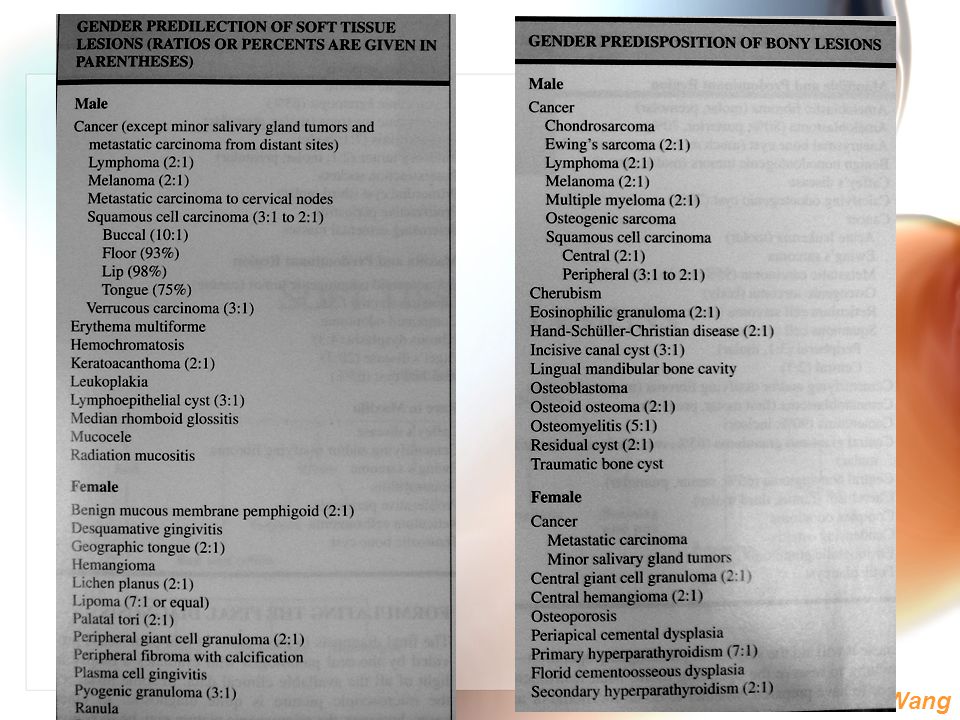
Clinically and /or radiographically Developing the possible diagnosis Sign—symptoms—statistical knowledge relate to the incidence of each disease entity—in order of their relative frequency of occurrence Age, gender, race, country of origin, anatomic location

65
Two or more lesions present
1. Lesions are related a. Lesion A and lesion B are identical (ex. 2 aphthous ulcers) b. Lesion B is secondary to lesion A (ex. metastatic tumor and primary) c. Lesion A and lesion B are both secondary to a third lesion, which may be occult (ex. metastatic tumors and primary) d. Lesion A and lesion B are manifestation of a systemic disease (ex. infections, Langerhan’s cell disease. Disseminated malignancy) e. Lesion A and lesion B form part of a syndrome (ex. caf’e-au-late spots and multiple neurofibromatosis in von Recklinghausen’s disease) 2. Lesions are completely unrelated to each other and occur together only by chance
 b. Lesion B is secondary to lesion A (ex. metastatic tumor and primary) c. Lesion A and lesion B are both secondary to a third lesion, which may be occult (ex. metastatic tumors and primary) d. Lesion A and lesion B are manifestation of a systemic disease (ex. infections, Langerhan’s cell disease. Disseminated malignancy) e. Lesion A and lesion B form part of a syndrome (ex. caf’e-au-late spots and multiple neurofibromatosis in von Recklinghausen’s disease) 2. Lesions are completely unrelated to each other and occur together only by chance.")
66
Developing the working diagnosis
(=opertational diagnosis, fentative diagnosis, clinical impression) Further exam. the lesion, more definitive questions to expand the history, additional tests —reevaluating all the assembled pertinent data Formulating the finial diagnosis Biopsy---microscopic examination
 Further exam. the lesion, more definitive questions to expand the history, additional tests —reevaluating all the assembled pertinent data. Formulating the finial diagnosis. Biopsy---microscopic examination.")
67
- Artifact: improper fixation, freezing, curling og the specimen
Biopsy - Artifact: improper fixation, freezing, curling og the specimen - Specimen should be identified with : patient’s name, clinician’s name, location of the lesion, patient history Exfoliating cytology Toludine blue staining

68
Excisional biopsy when lesion <=1cm, does not necessitate a major surgical procedure

69
Incisional biopsy too large to excision, may require multiple tissue samples Most suspect area, should be relatively large and deep and include the junction with surrounding normal tissue Necrotic tissue, electrosurgery should be avoided as possible

70
Punch biopsies: used on surface oral tissue (trismus patient)
Wedge-shaped biopsies: used for vesiculoerosive disease Fine-needle aspiration(fine-needle aspiration FNA, aspiration biopsy): gauge.
: gauge.")
71
Exfoliative cytology Fungal or viral disease or malignant-appearing cells Not used in smooth-surfaced exophytic lesion, homogeneous leukoplakia, submucosa lesions, unulcerated pigmented lesions, verruca vulgaris, papilloma

72
Toludine blue staining
Stained with 1 % Toludine blue, then washed or rinsed with 1% acetic solution Toludine blue is an acidophilic metachromatic nuclear dye, selectively stains acidic tissue esp. DNA and RNA (affinity DNA>RNA). False positive 8-10%, false negative 6-7%
. False positive 8-10%, false negative 6-7%")
73
提醒 病歷是個人一生健康的重要記錄,需受同儕一再檢視。病患及法定之第三人均有權調閱影印,務求正確及詳實的記載,以示尊重與負責。
避免自創之簡寫及縮寫,字跡應力求不潦草,不要有情緒性字眼或加註,可減少日後不必要的困擾。 病歷如需修改,應劃線修正後於旁蓋章,不可塗毀或使用立可白。

74
我們都在學習中不知不覺地同時傳承,與您共勉~
結語 為減少忙中有錯,除了在病歷上,下筆小心之外,如何在忙碌(亂)中能保持冷靜、有耐心而不冷漠是保護病人也保護自己的重要條件,是習醫生涯中非常重要的功課。 從病歷記載中可以看出各個醫師的個性。您希望為您看病的醫師是哪一型? 我們都在學習中不知不覺地同時傳承,與您共勉~
中能保持冷靜、有耐心而不冷漠是保護病人也保護自己的重要條件,是習醫生涯中非常重要的功課。 從病歷記載中可以看出各個醫師的個性。您希望為您看病的醫師是哪一型 我們都在學習中不知不覺地同時傳承,與您共勉~")
75
Summary 1. RECORDING THE IDENTIFYING DATA
2. HISTORY AND PHYSICAL EXAMINATION 3. CHIEF COMPLAINT 4. PRESENT ILLNESS 5. PAST MEDICAL HISTORY Family history Social history Occupational history Dental history 6. REVIEW OF SYMPTOMS BY SYSTEM 7. PHYSICAL EXAMINATION Radiologic examination 8. DIFFERENTIAL DIAGNOSIS 9. WORKING DIAGNOSIS Summary 10. Medical laboratory studies 11. Dental laboratory studies 12. Biopsy Incisional Excisional Fine-needle aspiration Exfoliationg cytology Toludine blue staining 13. Consultation 14. FINAL DIAGNOSIS 15.TREATMENT PLAN 病歷書寫格式

Similar presentations


>")


:實驗法(實驗設計) (第七章)>")
與研究問題.>")


 來判斷是否為場景變換,以方便使用者來 找出所要的片段。>")


>")

 ? 或是一個必要的需要 (demanding needs)?>")
 樣本必須是隨機樣本 (random sample) ,才能代表母體 Sample mean 是一隨機變數,隨著每一次抽出來的 樣本值不同,它的值也不同,但會有規律性 為了要知道估計的精確性,必需要知道樣本平均數.>")
