Download presentation
Presentation is loading. Please wait.

1
Chapter 18 Lecture Slides
Copyright © The McGraw-Hill Companies, Inc. Permission required for reproduction or display.

2
Functions 1. Excretion 2. Blood volume and blood pressure control
3. pH regulation 4. Concentration of solutes 5. Vitamin D production 6. Red blood cell concentration

5
Components of Urinary System
2 kidneys 2 ureters 1 urinary bladder 1 urethra

6
Figure 18.2a

7
Kidney Characteristics
Shape and size: - bean shaped - weighs 5 oz. (bar of soap or size of fist) Location: between 12th thoracic and 3rd lumbar vertebra
 Location: between 12th thoracic and 3rd lumbar vertebra.")
9
Kidney Structures Renal capsule:
- connective tissue around each kidney - protects and acts as a barrier Hilum: - indentation - contains renal artery, veins, nerves, ureter

10
Renal sinus: contains renal pelvis, blood vessels, fat Renal cortex: outer portion Renal medulla: inner portion

11
Renal pyramid: junction between cortex and medulla Calyx: tip of pyramids Renal pelvis: - where calyces join - narrows to form ureter

14
Nephron What is it? - functional unit of kidneys
- over 1 million/kidney

16
Components of Nephron Renal corpuscle:
structure that contains a Bowman’s capsule and glomerulus Bowman’s capsule: - enlarged end of nephron - opens into proximal tubule - contains podocytes (specialized cells around glomerular capillaries) Glomerulus: contains capillaries wrapped around it
 Glomerulus: contains capillaries wrapped around it.")
18
Filtration membrane: - in renal corpuscle - includes glomerular capillaries, podocytes, basement membrane Filtrate: fluid that passes across filtration membrane Proximal tubule: where filtrate passes first

19
Loop of Henle: - contains descending and ascending loops - water and solutes pass through thin walls by diffusion Distal tubule: structure between Loop of Henle and collecting duct Collecting duct: - empties into calyces - carry fluid from cortex through medulla

20
Figure 18.5a

21
Figure 18.4

22
Flow of Filtrate through Nephron
Renal corpuscle Proximal tubule Descending loop of Henle Ascending loop of Henle Distal tubule Collecting duct Calyx Renal pelvis Ureter

23
Blood Flow through Kidneys
Renal artery Interlobal artery Arcuate artery Interlobular artery Afferent arteriole Glomerulus Efferent ateriole Peritubular capillaries Vasa recta Interlobular vein Arcuate vein Interlobar vein

26
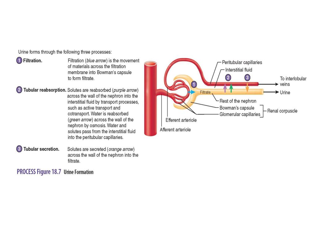
Urine Formation-Filtration
Movement of water, ions, small molecules through filtration membrane into Bowman’s capsule 19% of plasma becomes filtrate 180 Liters of filtrate are produced by the nephrons each day 1% of filtrate (1.8 L) become urine rest is reabsorbed
 become urine rest is reabsorbed.")
27
Only small molecules are able to pass through filtration membrane
Formation of filtrate depends on filtration pressure Filtration pressure forces fluid across filtration membrane Filtration pressure is influenced by blood pressure

30
Urine Production-Reabsorption
99% of filtrate is reabsorbed and reenters circulation Proximal tubule is primary site for reabsorption of solutes and water Descending Loop of Henle concentrates filtrate Reabsorption of water and solutes from distal tubule and collecting duct is controlled by hormones

35
Urine Production-Secretion
Water, small ions, by products of metabolism, drugs, urea are found in urine

36
Ureters, Urinary Bladder, Urethra
small tubes that carry urine from renal pelvis of kidney to bladder Urinary bladder: - in pelvic cavity - stores urine - can hold a few ml to a max. of 1000 ml Urethra: - tube that exits bladder - carries urine from urinary bladder to outside of body

38
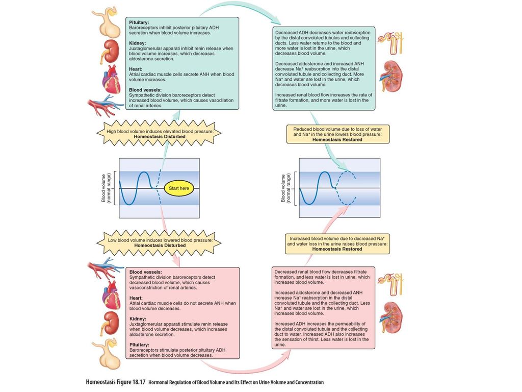
Renin-Angiotensin-Aldosterone Mechanism
Renin acts on angtiotensinogen to produce angiotensin I Angiotensin-converting enzyme converts angiotensin I to angtiotensin II Angiotensin II causes vasoconstriction Angiotensin II acts on adrenal cortex to release aldosterone Aldosterone increases rate of active transport of Na+ in distal tubules and collecting duct Volume of water in urine decreases

40
Antidiuretic Hormone Mechanism
ADH is secreted by posterior pituitary gland ADH acts of kidneys and they absorb more water (decrease urine volume) Result is maintain blood volume and blood pressure
 Result is maintain blood volume and blood pressure.")
42
Atrial Natriuretic Hormone
ANH is secreted from cardiac muscle to right atrium of heart when blood pressure increases ANH acts on kidneys to decrease Na+ reabsorption Sodium ions remain in nephron to become urine Increased loss of sodium and water reduced blood volume and blood pressure

45
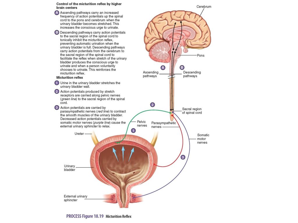
Urine Movement Micturition reflex:
activated by stretch of urinary bladder wall Action potentials are conducted from bladder to spinal cord through pelvic nerves Parasympathetic action potentials cause bladder to contract Stretching of bladder stimulates sensory neurons to inform brain person needs to urinate

47
Thirst Regulation Water intake is controlled by hypothalamus in thirst center Conc. of blood increase thirst center responds by initiating sensation of thirst When water is consumed, conc. of blood decreases and sensation of thirst decreases

49
Regulation of Acid-Base Balance
Buffers Chemicals resist change in pH of a sol’n Buffers in body contain salts of weak acids or bases that combine with H+ - 3 classes of buffers: proteins, phosphate buffer, bicarbonate buffer

50
Respiratory System Responds rapidly to change in pH Increased resp. rate raise pH due to rate of carbon dioxide elimination being increased Reduced respiratory rate reduced pH due to rate of carbon dioxide elimination being reduced

51
Kidneys Nephrons secrete H+ into urine and directly regulate pH of body fluids More H+ if pH is decreasing and less H+ if pH is increasing

53
Acidosis and Alkalosis
Acidosis occurs when pH of blood falls below 7.35 2 types are resp. acidosis and metabolic acidosis Alkalosis occurs when pH of blood increases above 7.45

Similar presentations


 System>")


