Download presentation
Presentation is loading. Please wait.

1
Body Cavities & Membranes

2
2 1.6: Organization of the Human Body Body cavities Thoracic cavity Abdominopelvic cavity Abdominal cavity Diaphragm Pelvic cavity Cranial cavity Vertebral canal (a) Thoracic cavity Abdominopelvic cavity Abdominal cavity Pelvic cavity Right pleural cavity Mediastinum Left pleural cavity Pericardial cavity Diaphragm Vertebral canal Cranial cavity Thoracic cavity (b) Copyright © The McGraw-Hill Companies, Inc. Permission required for reproduction or display.

3
Body Cavities 1) Cranial cavity – contains the brain 2) Vertebral cavity – spinal cord 3) Thoracic cavity (pleural cavity) – contains the lungs and mediastinum. Mediastinum – region between the lungs. Contains the heart, esophagus, trachea and thymus. Divides the thoracic cavity into left and compartments (L & R pleural cavity). (Contains pericardial cavity surrounding the heart) Diaphragm – broad, thin muscle that divides the thoracic cavity from the abdominopelvic cavity. 3
. (Contains pericardial cavity surrounding the heart) Diaphragm – broad, thin muscle that divides the thoracic cavity from the abdominopelvic cavity. 3.")
4
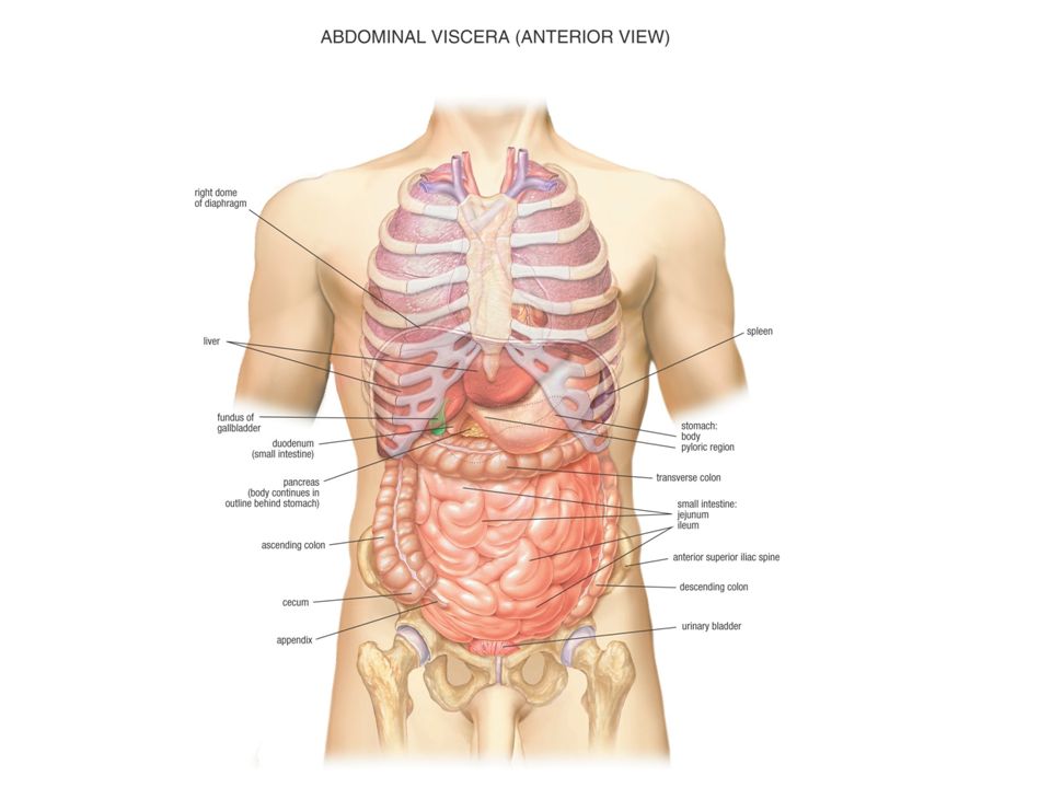
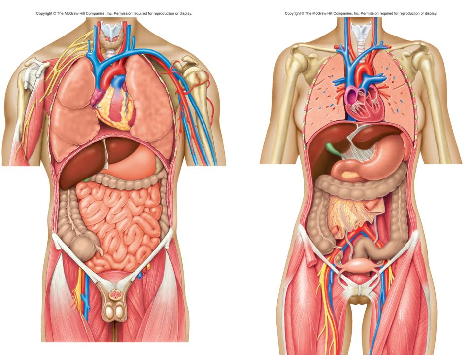
Body Cavities 4) Abdominopelvic cavity (Peritoneal cavity) – extends from the diaphragm to the floor of the pelvis. It has two divisions: Abdominal cavity – contains the stomach, liver, gall bladder, spleen, small and large intestine, kidneys and ureters. Pelvic cavity – area enclosed by pelvic bone. Contains bladder, terminal end of large intestine, internal reproductive organs. 4

7
7 What are the four main body cavities? Cranial cavity, vertebral canal, thoracic cavity, abdominopelvic cavity What are the divisions of the thoracic cavity? Left and right pleural cavity and the pericardial cavity

8
Membranes Serous membranes – thin membranes that line the walls of the thoracic and abdominopelvic cavities and fold back to cover the organs within the cavities. Secrete a slippery, watery fluid (serous fluid) that lubricates the surface of the walls and organs to moisturize and reduce friction. Visceral layer – covers the surface of an organ (viscera) Parietal layer – covers the walls of the cavity 8
 that lubricates the surface of the walls and organs to moisturize and reduce friction. Visceral layer – covers the surface of an organ (viscera) Parietal layer – covers the walls of the cavity 8.")
9
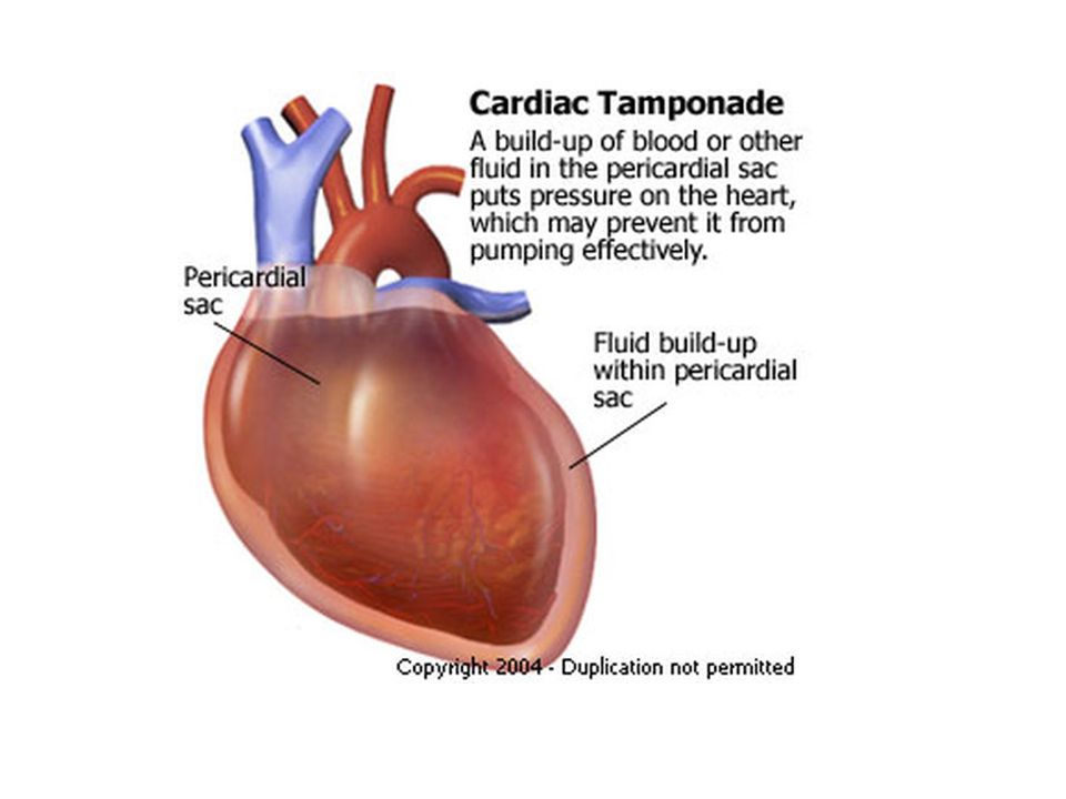
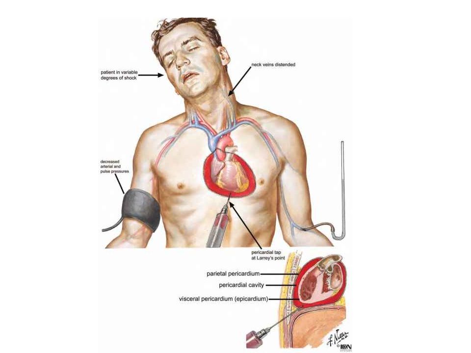
Thoracic Membranes Thoracic membranes = Pleural membranes Visceral pleura – covers the surface of the lungs Parietal pleura – covers the walls of the thoracic cavity Heart membranes have their own special name - pericardium: Visceral pericardium – surface of the heart Parietal pericardium – outer membrane surrounding the heart 9

10
Abdominopelvic Membranes (Peritoneal Membranes) Visceral peritoneum – covers the surface of an organ (viscera) in the abdominopelvic cavity Parietal peritoneum – covers the walls of the abdominopelvic cavity 10
 Visceral peritoneum – covers the surface of an organ (viscera) in the abdominopelvic cavity Parietal peritoneum – covers the walls of the abdominopelvic cavity 10")
11
Two words to name a membrane: 1 st :_______________ 1) Visceral – covers an organ 2) Parietal – covers the walls of a cavity Heart has 2 membranes: Inner membrane – surface of heart = visceral Outer membrane = parietal 2 nd :________________ 1) pleura – thoracic cavity 2) peritoneum – abdominopelvic cavity 3) pericardium - heart
 Visceral – covers an organ 2) Parietal – covers the walls of a cavity Heart has 2 membranes: Inner membrane – surface of heart = visceral Outer membrane = parietal 2 nd :________________ 1) pleura – thoracic cavity 2) peritoneum – abdominopelvic cavity 3) pericardium - heart")
12
Membranes What are the membranes that line the walls of cavities called? Parietal membranes What are the membranes that line organs called? Visceral membranes 12

13
Membranes Membranes in the thoracic cavity = ___________ Pleura Membranes that cover the heart = _________ Pericardium Membranes in the abdominopelvic cavity = _______ Peritoneum 13

14
Name the following… The membrane lining the walls of the Thoracic cavity Parietal pleura The membrane covering the lungs Visceral pleura The membrane lining the walls of the abdominopelvic cavity Parietal peritoneum 14

15
15 Name the membrane covering the organs in the abdominopelvic cavity Visceral peritoneum The outer membrane surrounding the heart Parietal pericardium The inner membrane surrounding the heart Visceral pericardium

16
16 Thoracic Serous Membranes Vertebra Aorta Esophagus Right lung Visceral pleura Pleural cavity Parietal pleura Sternum Plane of section Spinal cord Mediastinum Left lung Rib Left ventricle of heart Visceral pericardium Pericardial cavity Parietal pericardium Anterior Azygos v. Right atrium of heart Right ventricle of heart Fibrous pericardium Copyright © The McGraw-Hill Companies, Inc. Permission required for reproduction or display.

17
17 Abdominal Serous Membranes Vertebra Right kidney Pancreas Large intestine Liver Gallbladder Duodenum Peritoneal cavity Parietal peritoneum Plane of section Left kidney Spinal cord Spleen Rib Small intestine Large intestine Stomach Anterior Visceral peritoneum Costal cartilage Aorta Inferior vena cava Copyright © The McGraw-Hill Companies, Inc. Permission required for reproduction or display.

18
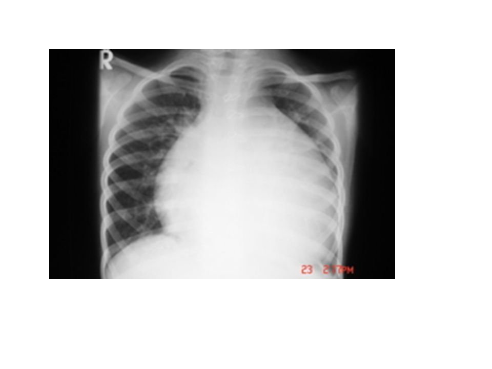
Normal Chest Radiograph 18

22
22

23
23

24
24

Similar presentations