Download presentation
Presentation is loading. Please wait.

1
MEDIASTINUM EDITED BY: DR. NIVIN SHARAF MD LMCC

2
OBJECTIVES By the end of this lecture the students should be able to: Define mediastinum. Enlist the divisions of mediastinum. Describe the boundaries and contents of mediastinum.

3
MEDIASTINUM Central compartment of the thoracic cavity Covered by mediastinal pleura Contents: all thoracic viscera EXCEPT lungs Extent: Superior - thoracic inlet Inferior - diaphragm Anterior - sternum & costal cartilages Posterior- bodies of thoracic vertebrae

4
MEDIASTINUM Surrounded by blood and lymphatic vessels Lymph nodes, nerves and adipose tissues Looseness of structures enable mediastinum to accommodate changes in movement, volume & pressure in the thoracic cavity

5
Divisions of the Mediastinum SUPERIOR MEDIASTINUM Superior - thoracic inlet Inferior - transverse thoracic plane Anterior - sternal angle Posterior - IV disc T4 & T5 INFERIOR MEDIASTINUM Superior – transverse thoracic plane Inferior - diaphragm

6
What can you spot here?

7
Divisions of the Mediastinum INFERIOR MEDIASTINUM a. ANTERIOR MEDIASTINUM - contains thymus remnant, lymph nodes & fats - contains thymus remnant, lymph nodes & fats b. MIDDLE MEDIASTINUM - contains the heart & great vessels - contains the heart & great vessels c. POSTERIOR MEDIASTINUM - contains esophagus, great vessels,vagus nerves & symphathetic trunks - contains esophagus, great vessels,vagus nerves & symphathetic trunks

8
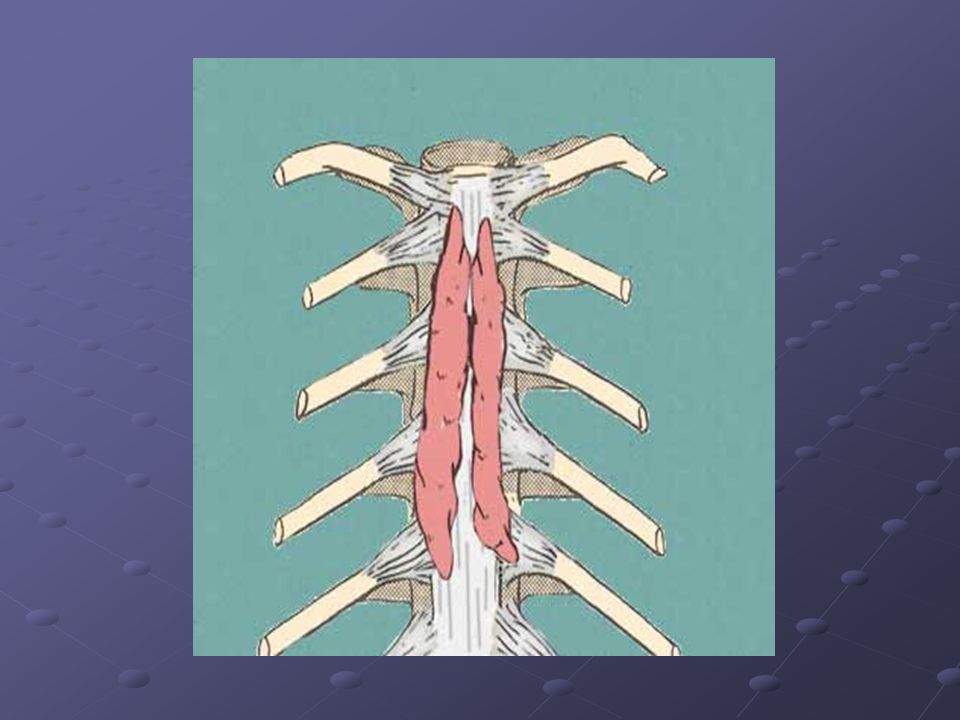
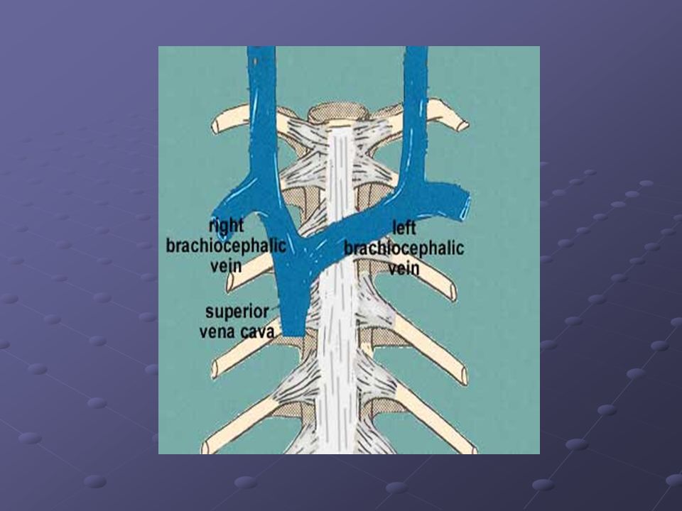
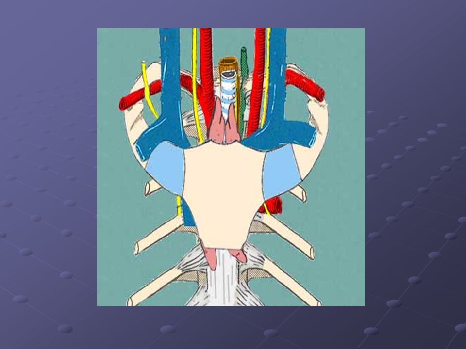
Superior Mediastinum Contents: (Anterior – Posterior) 1. Thymus gland - primary lymphoid organ located behind manubrium - puberty undergoes gradual involution 2. Great Vessels Brachiocephalic Veins Superior Vena Cava - formed at level of 1 st right costal cartilage - enters right atrium at level of 3 rd right costal cartilage

11
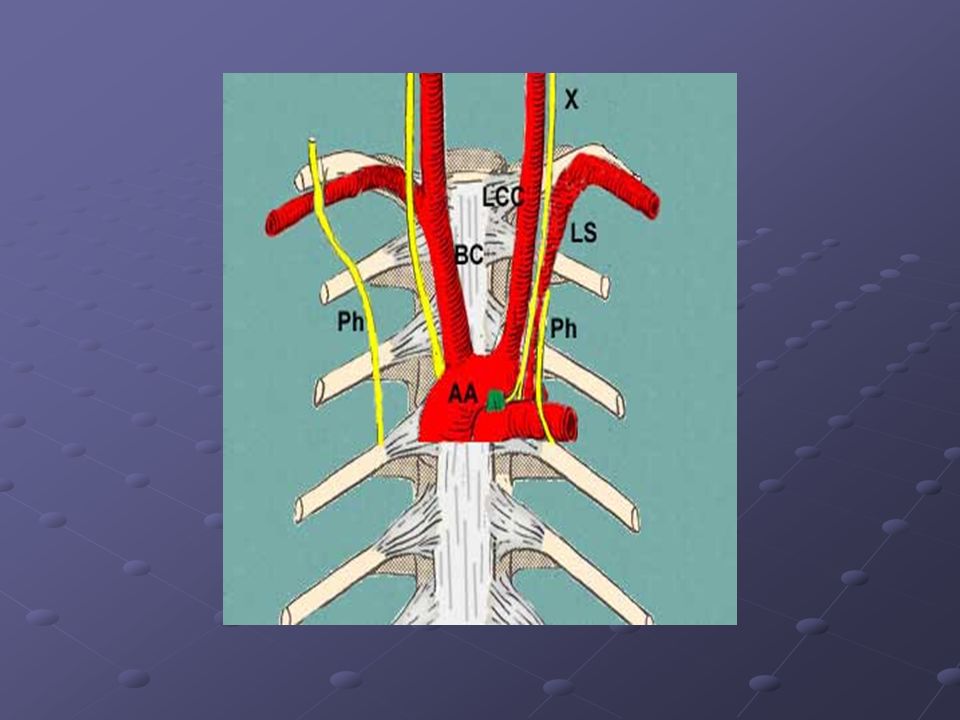
Superior Mediastinum Contents: Arch of the Aorta - starts behind 2 nd right SC joint - ends at 2 nd left SC joint Branches: Brachiocephalic Trunk Left Common Carotid Artery Left Subclavian Artery

12
Aortic Arch Passes upwards from the sternal angle behind the manubrium, backwards and to the left of the 4 th Thoracic vertebra BRANCHES SUPPLY UL, HEAD, and NECK

13
Branches

14
Relations

15
Relations Cont.

17
Ligamentum arteriosum Ligamentum arteriosum Aortic Arch is connected inferiorly to the left pulmonary artery by the Ligamentum arteriosum” fibrous remnant of ductus arteriosus”

18
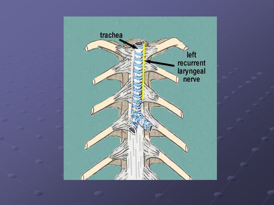
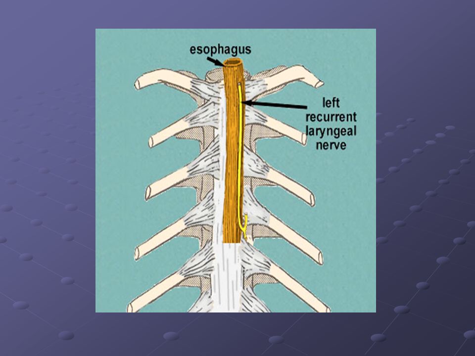
Superior Mediastinum Contents: 3. Nerves Vagus & Phrenic Nerves Cardiac Plexus of Nerves Left Recurrent Laryngeal Nerve 4. Trachea 5. Esophagus 6. Thoracic Duct 7. Prevertebral Muscles

24
Anterior Mediastinum Smallest subdivision of the Inferior Mediastinum Boundaries: Anterior: body of sternum & trans thoracis muscles Posterior: pericardium Contents: Loose CT (Sternopericardial Ligament) Adipose tissue Adipose tissue Lymphatic Vessels & lymph nodes Lymphatic Vessels & lymph nodes Branches of Internal Thoracic Vessels Branches of Internal Thoracic Vessels
 Adipose tissue Adipose tissue Lymphatic Vessels & lymph nodes Lymphatic Vessels & lymph nodes Branches of Internal Thoracic Vessels Branches of Internal Thoracic Vessels")
25
Posterior Mediastinum Boundaries: Anterior- Pericardium & Diaphragm Posterior- T5 to T12 vertebrae Contents: Thoracic AortaEsophagus & Plexus Thoracic DuctThoracic Sympathetic Trunks Post Mediastinal LNThoracic Splanchnic Nerve Azygos & Hemiazygos Veins

26
Thoracic Aorta Branches: 1. Bronchial Arteries5. Esophageal Arteries 2. Pericardial Arteries6. Mediastinal Arteries 3. Post. Intercostal Arteries7. Subcostal Arteries 4. Superior Phrenic Arteries

27
Esophagus Course: Superior to Posterior Mediastinum Located behind: Arch of Aorta Pericardium & Left Atrium Pericardium & Left Atrium Enters Esophageal Hiatus of the Diaphragm at level of T10 Anatomic Impressions or “Constrictions”: 1. Crossing with Aortic Arch 2. Crossing with Left Main Bronchus 3. Diaphragmatic Hiatus

28
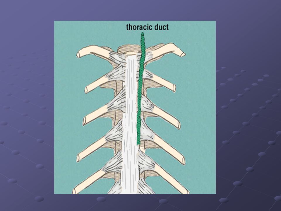
Thoracic Duct Largest lymphatic channel in the body Originates from Cisterna Chyle in the abdomen & passes thru aortic hiatus of diaphragm at level of T12 Relations: Posterior: bodies of inferior 7 thoracic vertebrae Anterior : Esophagus Anterior : Esophagus Left: Thoracic Aorta Left: Thoracic Aorta Right : Azygos Vein Right : Azygos Vein Conveys lymph from: Lower extremitiesLeft side of thorax Pelvic CavityLeft side of H & N Abdominal CavityLeft upper limb

29
Lymph Nodes of the Posterior Mediastinum Posterior Mediastinal Lymph Nodes - receives lymph from esophagus, posterior aspect of the pericardium & diaphragm & middle posterior ICS

30
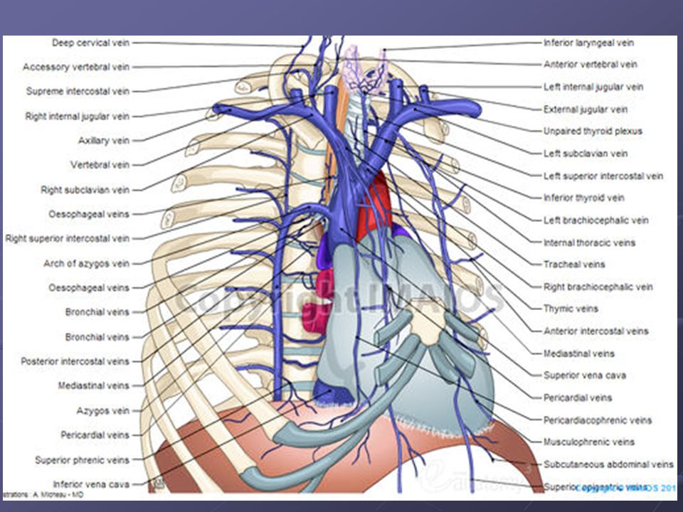
Azygos Venous System of the Posterior Mediastinum Drains the back & thoracoabdominal walls and the mediastinal viscera Azygos Vein - forms a collateral pathway b/w SVC & IVC - passes to the right side of inferior 8 thoracic vertebrae - arches over the root of the right lung to enter the SVC - receives posterior intercostal veins, mediastinal, esophageal & bronchial veins

31
Azygos Venous System of Posterior Mediastinum Hemiazygos Vein - arises on left side of the vertebral column to level of T9 - receives the inferior 3 PIV, inferior esophageal veins, small mediastinal veins Accessory Hemiazygos Vein - starts at medial end of 4 th or 5 th ICS - descends on left of VC from T5 thru T8 - crosses to the right to join the Azygos Vein - receives 4 th thru 8 th IC Veins - communicates with Superior IC Vein which drains 1 st thru 3 rd ICS

33
Nerves of Posterior Mediastinum Thoracic Sympathetic Trunks - lie against heads of ribs in superior thorax costovertebral joints in midthorax sides of vertebral bodies in lower thorax Lower Thoracic Splanchnic Nerves (Greater, Lesser and Least SN) - presynaptic fibers from 5 th thru 12 th sympathetic ganglia - sympathetic innervation for most abdominal viscera
 - presynaptic fibers from 5 th thru 12 th sympathetic ganglia - sympathetic innervation for most abdominal viscera")
34
References Illustrated Clinical Anatomy by:Peter Abrahams Clinical Anatomy by Systems for Richard S Snell Pages Gray’s Anatomy for students

Similar presentations


 in the diaphragm. Give the.>")


 THE THORACIC WALL Posteriorly by the thoracic part of the vertebral column Posteriorly by the thoracic.>")

