Download presentation
Presentation is loading. Please wait.

1
Slacker’s Guide to Thoracic Gross Anatomy
Prepared by Mike Ori

2
Caveat Emptor As with all slackers guides, I have omitted, condensed, and misunderstood information so don’t complain if I got something wrong, after all, you’re a slacker. Also, it goes without saying that the faculty has not vetted the information in any way. Always refer to their notes and lectures for the last word.

3
CT and Such You’re probably best off working on an interactive system try CPR Radiograph key from Slice-o lecture is important

4
Structures from labs Veins Azygous vein (c) Hemiazygous vein (c) Posterior intercostal veins (c) Pulmonary vein (c) Nerves Intercostal nerves (c) Phrenic nerve (c) Viscera Lungs Horizontal fissure (right only) (c) Inferior lobe (left and right) (c) Middle lobe (right only) (c) Oblique fissure (left and right) (c) Superior lobe (left and right) (c) Pleura Parietal pleura (c) Visceral pleura (c) Primary (main) bronchi (c,r) Secondary (lobar) bronchi (c) Superior lobar bronchi (left and right) (c) Middle lobar bronchus (right only) (c) Inferior lobar bronchi (left and right) (c) Trachea (c,r) Viscera Esophagus (c, r) Thoracic duct (c) Trachea (c,r) Bones Ribs (b, r) Sternal Body (b,r) Manubrium (b) Sternal angle (b) Xiphisternum (xiphoid process) (b) Muscles External intercostal (c) Internal intercostal (c) Diaphragm (c) Arteries Pulmonary artery (c) Subclavian artery (c,r) Internal thoracic artery (c) Anterior intercostals arteries (c) Thoracic (descending) aorta (c,r) Posterior intercostal arteries (c) Arteries and Veins Ascending aorta (c,r) Aortic Arch (c,r) Brachiocephalic artery (c,r) Right subclavian artery (c) Right common carotid artery (c) Left common carotid artery (c,r) Left subclavian artery (c,r) Descending aorta (c,r) Posterior intercostal arteries (c) Inferior vena cava (c,r) Pulmonary trunk (c,r) Left pulmonary artery (c) Right pulmonary artery (c) Superior vena cava (c,r) Azygous vein (c) Hemiazygous vein (c) Posterior intercostal veins (c) Brachiocephalic veins (c) Nerves Phrenic nerve (c) Thoracic sympathetic chain (c) Greater splanchnic nerve (c) Vagus nerve (c) Anterior vagal trunk (c) Posterior vagal trunk (c) Esophageal plexus (c) Left recurrent laryngeal nerve (c) These are set out exactly as found in the labs so there are duplicates
 Nerves. Intercostal nerves (c) Phrenic nerve (c) Viscera. Lungs. Horizontal fissure (right only) (c) Inferior lobe (left and right) (c) Middle lobe (right only) (c) Oblique fissure (left and right) (c) Superior lobe (left and right) (c) Pleura. Parietal pleura (c) Visceral pleura (c) Primary (main) bronchi (c,r) Secondary (lobar) bronchi (c) Superior lobar bronchi (left and right) (c) Middle lobar bronchus (right only) (c) Inferior lobar bronchi (left and right) (c) Trachea (c,r) Viscera. Esophagus (c, r) Thoracic duct (c) Trachea (c,r) Bones. Ribs (b, r) Sternal Body (b,r) Manubrium (b) Sternal angle (b) Xiphisternum (xiphoid process) (b) Muscles. External intercostal (c) Internal intercostal (c) Diaphragm (c) Arteries. Pulmonary artery (c) Subclavian artery (c,r) Internal thoracic artery (c) Anterior intercostals arteries (c) Thoracic (descending) aorta (c,r) Posterior intercostal arteries (c) Arteries and Veins. Ascending aorta (c,r) Aortic Arch (c,r) Brachiocephalic artery (c,r) Right subclavian artery (c) Right common carotid artery (c) Left common carotid artery (c,r) Left subclavian artery (c,r) Descending aorta (c,r) Posterior intercostal arteries (c) Inferior vena cava (c,r) Pulmonary trunk (c,r) Left pulmonary artery (c) Right pulmonary artery (c) Superior vena cava (c,r) Azygous vein (c) Hemiazygous vein (c) Posterior intercostal veins (c) Brachiocephalic veins (c) Nerves. Phrenic nerve (c) Thoracic sympathetic chain (c) Greater splanchnic nerve (c) Vagus nerve (c) Anterior vagal trunk (c) Posterior vagal trunk (c) Esophageal plexus (c) Left recurrent laryngeal nerve (c) These are set out exactly as found in the labs so there are duplicates.")
5
Structures from Labs Muscles Papillary muscles
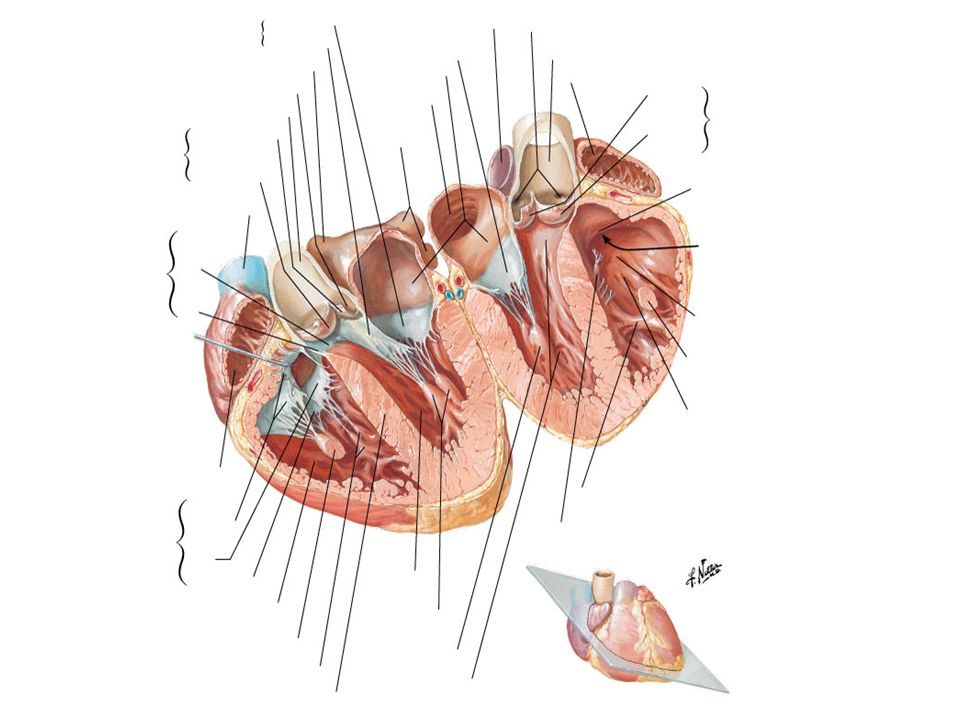
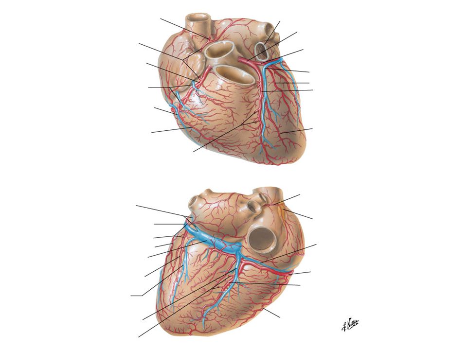
Viscera Anterior interventricular sulcus Chordae tendineae Coronary sulcus Fossa ovalis Interventricular septum Left atrium Openings of pulmonary veins Left auricle Left ventricle Aortic valve Mitral (bicuspid) valve Pericardial sac Fibrous pericardium Parietal (serous) pericardium Visceral (serous) pericardium = epicardium Posterior interventricular sulcus Right atrium Crista terminalis Opening of coronary sinus Opening of inferior vena cava Opening of superior vena cava Right auricle Right ventricle Moderator band Pulmonary valve Tricuspid valve Muscles Papillary muscles Anterior papillary muscle – both right and left ventricles Posterior papillary muscle – both right and left ventricles Septal papillary muscle – right ventricle only Pectinate muscle (right atrium) Trabeculae carnae (ventricles) Vessels Aorta Left coronary artery Anterior interventricular artery Circumflex artery Right coronary artery Marginal artery Posterior interventricular artery Coronary sinus Great cardiac vein Middle cardiac vein Small cardiac vein Ligamentum arteriosum Pulmonary trunk Pulmonary arteries Pumlonary veins
 valve. Pericardial sac. Fibrous pericardium. Parietal (serous) pericardium. Visceral (serous) pericardium = epicardium. Posterior interventricular sulcus. Right atrium. Crista terminalis. Opening of coronary sinus. Opening of inferior vena cava. Opening of superior vena cava. Right auricle. Right ventricle. Moderator band. Pulmonary valve. Tricuspid valve. Muscles. Papillary muscles. Anterior papillary muscle – both right and left ventricles. Posterior papillary muscle – both right and left ventricles. Septal papillary muscle – right ventricle only. Pectinate muscle (right atrium) Trabeculae carnae (ventricles) Vessels. Aorta. Left coronary artery. Anterior interventricular artery. Circumflex artery. Right coronary artery. Marginal artery. Posterior interventricular artery. Coronary sinus. Great cardiac vein. Middle cardiac vein. Small cardiac vein. Ligamentum arteriosum. Pulmonary trunk. Pulmonary arteries. Pumlonary veins.")
6
Heart X-Ray

11
Manubrium Sternal angle Sternal body Xyphoid process

13
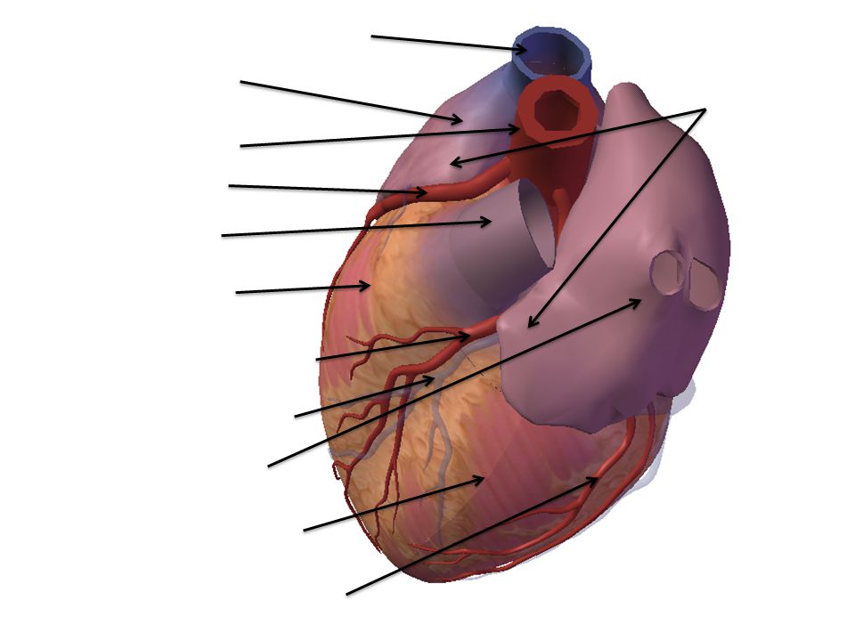
Pulmonary trunk Auricle of LA and RA Superior vena cava Aorta Right atrium Right Coronary Artery Right Ventricle Anterior interventricular artery Left ventricle Anterior interventricular Sulcus

15
Superior vena cava Right atrium Auricle of LA and RA Aorta Right Coronary Artery Pulmonary trunk Right Ventricle Anterior interventricular artery Great cardiac vein Left atrium Left ventricle Circumflex artery

17
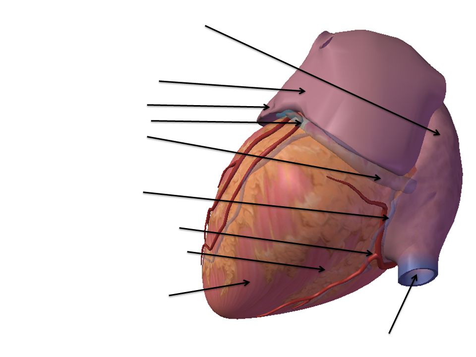
Right atrium Left atrium Auricle of Left Atrium Great Cardiac Vein Coronary sinus Middle cardiac vein Posterior interventricular artery Posterior interventricular sulcus Left ventricle Inferior vena cava

19
Superior vena cava Right atrium Aorta Right Coronary Artery Pulmonary trunk Right Ventricle Left coronary artery Anterior interventricular artery Great cardiac vein Left ventricle Left atrium Circumflex artery Mitral valve

21
Marginal artery Right atrium Small cardiac vein Right coronary artery Inferior vena cava Coronary Sinus Posterior interventricular artery Middle cardiac vein Left ventricle Right Ventricle

37
Manubrium Superior Vena Cava Intercostal Neurovascular bundle Internal Thoracic A/V Sternal body Xyphoid process

39
Sternal Angle Manubrium Intercostal A V Post Ant Internal Thoracic A/V Sternal body Intercostal nerve

41
Aortic Arch Right superior lobe Left superior lobe Internal thoracic A/V Right middle lobe Cardiac Notch Left inferior lobe Right inferior lobe Lingula of Left Lung Diaphragm

43
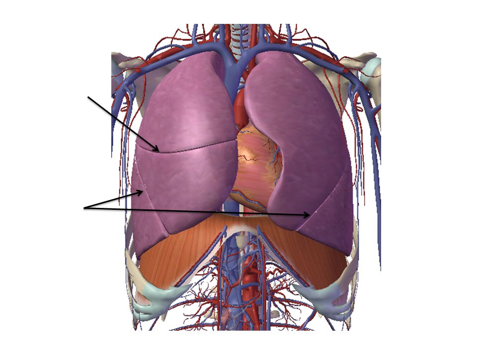
Horizontal fissure Oblique Fissure

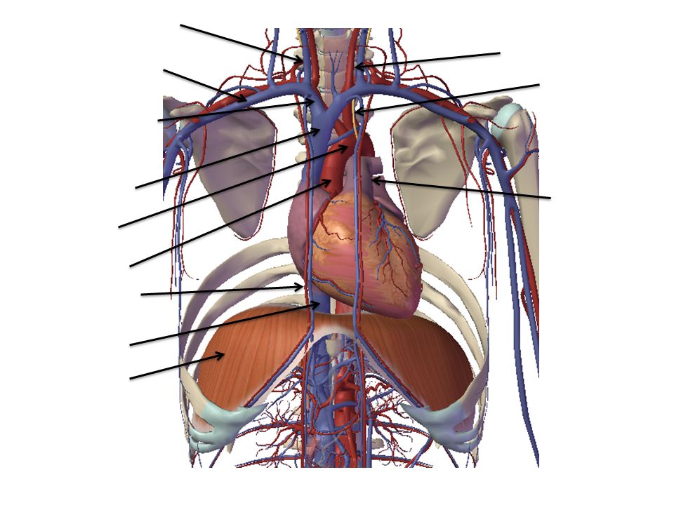
45
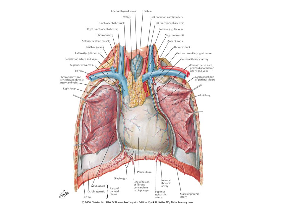
Internal Jugular V Common Carotid A Subclavian A/V Left Vagus N (CNX) Brachiocephalic v Sup Vena Cava Pulmonary trunk Aortic Arch Ascending Aorta Int thoracic A/V Inf Vena Cava Diaphragm

47
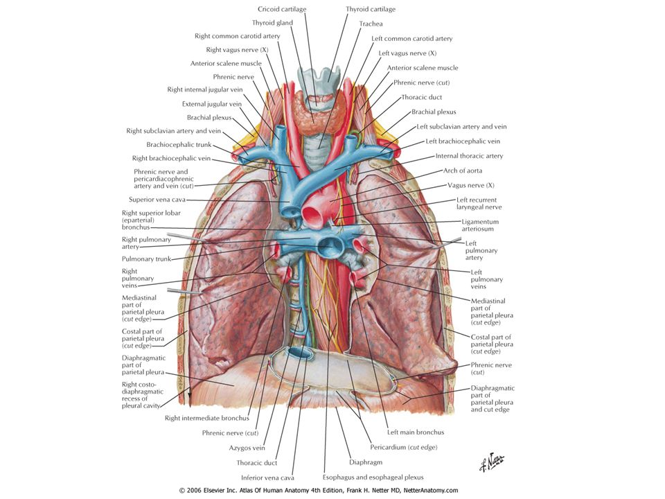
Larynx Trachea External Jugular V Common Carotid A Internal Jugular V Left Vagus N (CNX) Subclavian A/V Brachiocephalic V Sup Vena Cava Pulmonary trunk Aortic Arch Pulmonary A/V Ascending Aorta Descending Aorta Hemiazygos V Esophagus Azygos V Inf Vena Cava Diaphragm

49
Larynx Trachea External Jugular V Vagus N (CNX) Recurrent Laryngeal of CNX Common Carotid A. Subclavian A. Brachiocephalic A. Sup Vena Cava Aortic Arch Ascending Aorta Pulmonary trunk Esophagus

51
Larynx External Jugular V Recurrent Laryngeal of CNX Vagus N (CNX) Common Carotid A. Subclavian A. Brachiocephalic A. Hemiazygos V azygos V Esophagus Descending Aorta Inf Vena Cava

53
Posterior

57
Note lack of cartilage on posterior trachea

60
Posterior

64
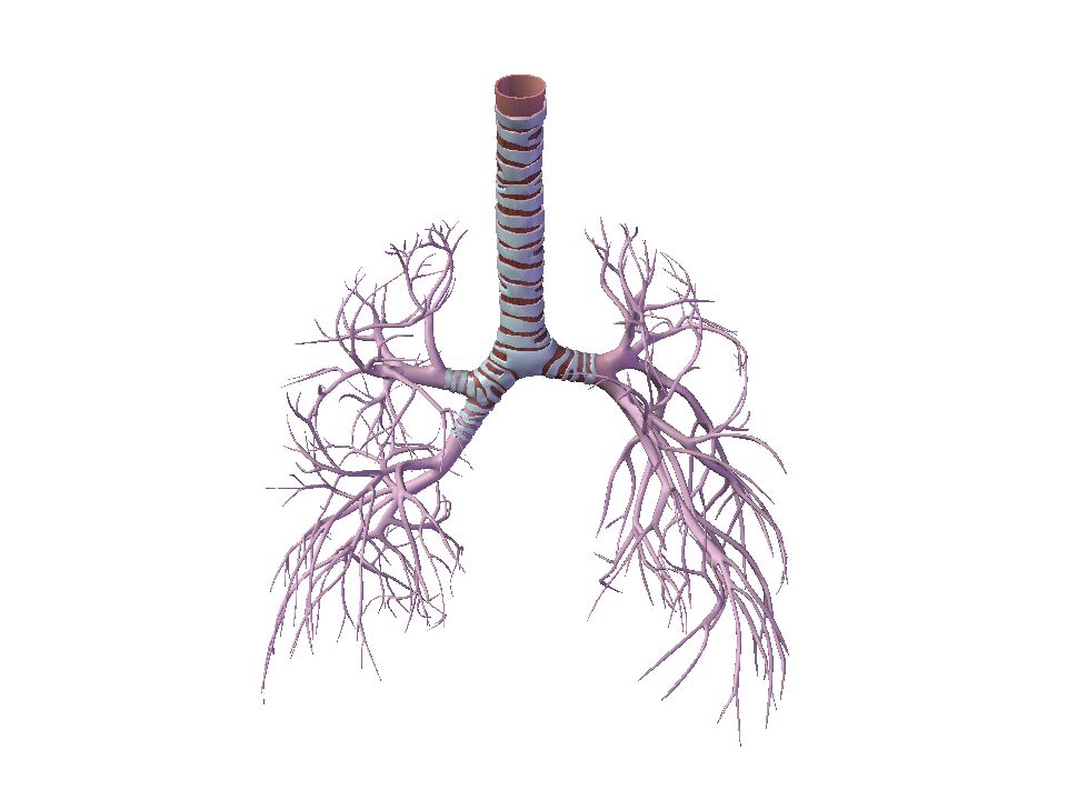
Larynx Trachea Primary (main) Bronchus Tertiary bronchus Secondary (lobar) bronchus
 Bronchus Tertiary bronchus Secondary (lobar) bronchus")
65
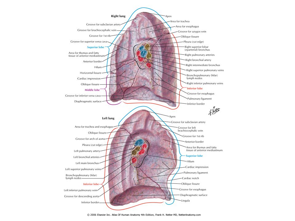
Right lung hilar view

66
Right lung hilar view Right superior lobe Primary bronchus (Be careful with the split here, could be 2o) Pulmonary artery Horizontal fissure Oblique fissure Pulmonary vein (ant and inf. most) Right inferior lobe Right middle lobe
 Right inferior lobe. Right middle lobe.")
67
Left lung hilar view

68
Left lung hilar view Pulmonary artery Primary bronchus (Be careful with the split here, could be 2o) Left superior lobe Oblique fissure Pulmonary Vein (ant and inf. most) Left inferior lobe
 Left inferior lobe.")
70
Renal pelvis Renal Cortex Medullary pyramids Calyces

71
Just because it could do this.

73
Kidney Ureter Uterus The model system didn’t have male anatomy so picture a walnut just below the bladder. Urinary bladder Pelvic floor muscles Urethra

74
References www.visiblebody.com
Netter’s Atlas of Human Anatomy ( Dr A’s lecture notes Dr C’s slides

Similar presentations


 Coronary sulcus>")

4Lateral border of descending aorta5Main pulmonary artery 6Azygo- esophageal line7Posterior.>")
