Download presentation
Presentation is loading. Please wait.

1
Mediastinum Dr.Hassan Shaibah

2
Chest Cavity Pleurae & lungs Mediastinum pleurae & lungs

3
Chest Cavity Mediastinum pleurae & lungs pleurae & lungs

4
Mediastinum

5
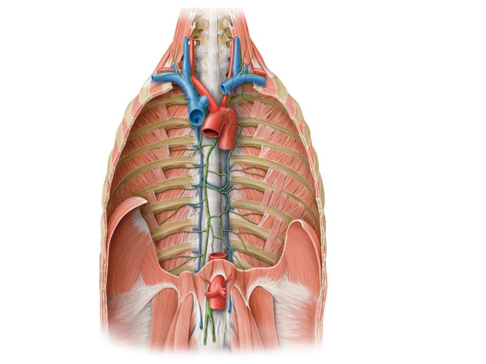
Mediastinum Extends superiorly to the thoracic outlet ,root of the neck &inferiorly to the diaphragm. extends anteriorly to the sternum & posteriorly to the vertebral column. It contains : thymus, trachea, thoracic duct ,the heart esophagus, large blood vessels, lymph nodes, vagus & phrenic nerves, & sympathetic trunks.

6
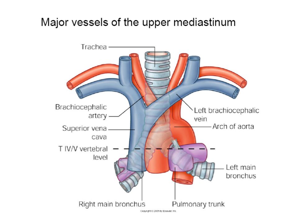
trachea phrenic& vagus nerves large blood vessels thymus Heart Some Contents Mediastinum

7
esophagus trachea large blood vessels phrenic& vagus nerves Some Contents Mediastinum

8
The mediastinum is divided by an imaginary plane passing from sternal angle to the Intervertebral discs T4 & T5 into: superior mediastinum inferior mediastinum

9
Mediastinum Divsion 4 superior sternal angle 5 inferior

10
The inferior mediastinum
subdivided into: anterior mediastinum, a space between the pericardium and the sternum Middle mediastinum pericardium and heart posterior mediastinum, between “pericardium &vertebral column”

11
MIDDLE Anterior Posterior * *inferior mediastinumm superior

12
Superior Mediastinum is bounded: front by manubrium sterni
behind by first 4 thoracic vertebrae. It contains: (a) Thymus, (b) large veins, (c) large arteries, (d) trachea, (e) esophagus and thoracic duct, (f) nerves
 Thymus, (b) large veins, (c) large arteries, (d) trachea, (e) esophagus and thoracic duct, (f) nerves.")
13
1 manubrium 4

14
RT & LT brachiocephalic v.
nerves RT & LT brachiocephalic v. LT common carotid a. Brachiocephalic Trunk thymus

15
Inferior Mediastinum Bondries behind lower 8 thoracic vertebrae
front Body of sternum 5 behind lower 8 thoracic vertebrae 12

16
Inferior Mediastinum It contains: (a) Thymus, (b) heart within the pericardium. (c) phrenic nerves (d) esophagus and thoracic duct, (e) descending aorta (f)Azygous venous system (g) sympathetic trunks
 Thymus, (b) heart within the pericardium. (c) phrenic nerves (d) esophagus and thoracic duct, (e) descending aorta (f)Azygous venous system (g) sympathetic trunks")
17
2)heart within the pericardium
1)Thymus 3)esophagus 2)heart within the pericardium
Thymus. 3)esophagus. 2)heart within the pericardium.")
18
4)phrenic nerves
phrenic nerves")
19
5)Descending aorta
Descending aorta")
20
6)Thoracic duct
Thoracic duct")
21
Sympathetic trunks

22
Anterior mediastinum, a space between the pericardium and the sternum
Middle mediastinum pericardium and heart will be discussed with Cardiovascular block

23
Posterior Mediastinum
Boundaries: . Sup. transverse thoracic plane 5 Ant. pericardium Post. bodies of the vertebral column 12 Inf. diaphragm laterally the pleura (on either side)
")
24
Contents 1)Descending aorta
Descending aorta")
25
2)Azygos venous system 3)Thoracic duct
Azygos venous system 3)Thoracic duct")
26
Sympathetic trunks

27
9)vagus nerve Vagal plexus
vagus nerve Vagal plexus")
28
Contents artery Veins nerves esophagus thoracic duct
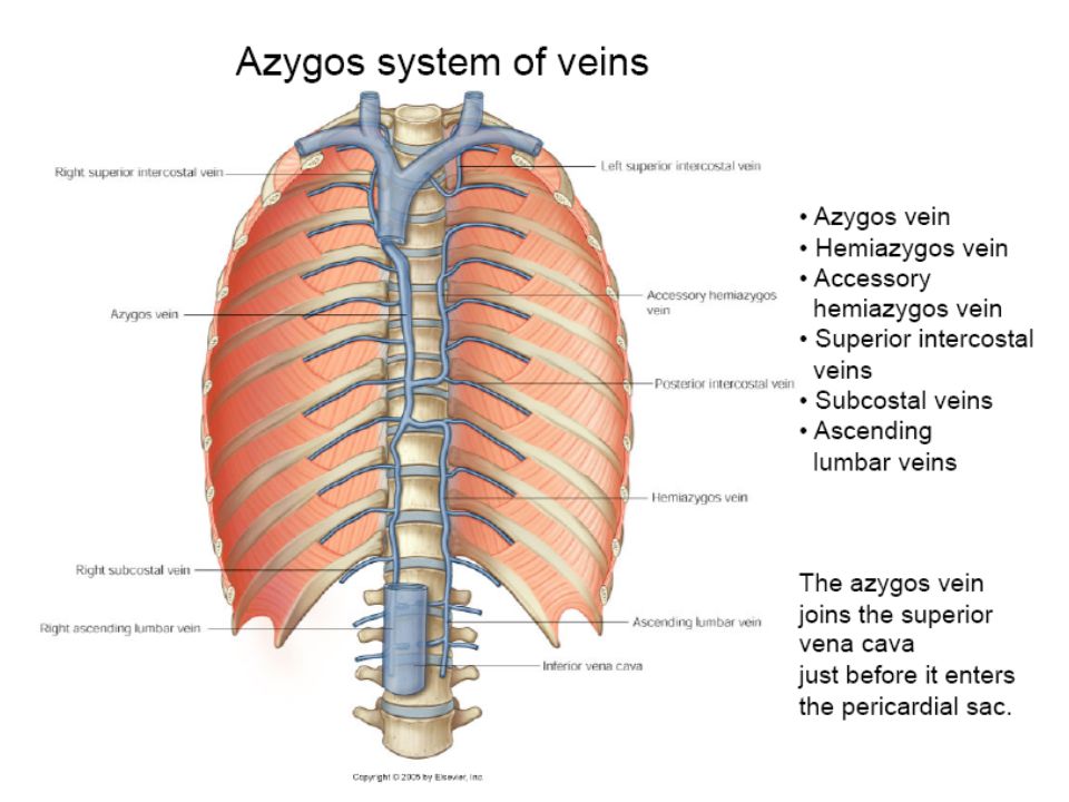
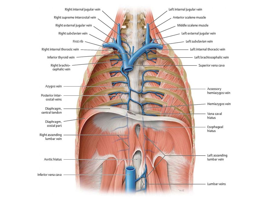
descending thoracic aorta Veins azygos vein the sup. & inf. hemiazygos vein nerves vagus nerve Sympathetic trunks esophagus thoracic duct

30
2)superior hemiazygos v. 3)Inferior hemiazygos v.
Azygos Venous system: consist of: 2)superior hemiazygos v. azygos v. 3)Inferior hemiazygos v.
superior hemiazygos v. azygos v. 3)Inferior hemiazygos v.")
31
They drain blood from: posterior intercostal spaces posterior abdominal wall pericardium diaphragm bronchi esophagus.

32
Azygos Vein formed by union of:
SVC T5 Azygos V. 1)right subcostal v. 2)right ascending lumbar v. IVC
right subcostal v. 2)right ascending lumbar v. IVC.")
33
8 lower right posterior intercostal v.
Tributaries: Superior hemiazygos veins. RT superior intercostal v. 8 lower right posterior intercostal v. Inferior hemiazygos veins Mediastinal veins.

34
1)Azygos Vein formed by union of the right ascending lumbar vein and the right subcostal vein. b. It ascends through aortic opening in the diaphragm on the right side of the aorta to the level of the fifth thoracic vertebra. Arch over the root of the right lung to empty into the SVC e. The azygos vein tributaries are: a. The 8 lower right posterior intercostal veins. b. The right superior intercostal vein. c. The superior and inferior hemiazygos veins. d. Mediastinal veins.

35
Inferior Hemiazygos Vein
It is formed by the union of the left ascending lumbar vein & left subcostal vein. It ascends through the left crus of the diaphragm at T8. turns to the right and joins the azygos vein. It receives as tributaries some lower left intercostal veins and mediastinal veins.

36
Superior Hemiazygos Vein
It is formed by the union of the 4 to the 8 intercostal veins. It joins the azygos vein at the level of the T7.

37
Mediastinum lymph Lymph nodes draining the lungs, mediastinal structures empty into the : bronchomediastinal trunks & thoracic duct.

38
left Brachiocephalic v.
Thoracic Duct begins in the abdomen as a dilated sac (cisterna chyli) Asend to the root of the neck to empty into beginning of the left Brachiocephalic vein cisterna chyli
 Asend to the root of the neck to empty into beginning of the left Brachiocephalic vein. cisterna chyli.")
39
3) broncho-mediastinal lymph trunks.
At the root of the neck, the thoracic duct receives: . 1)left jugular trunk 2)LT Subclavian trunk 3) broncho-mediastinal lymph trunks.
left jugular trunk. 2)LT. Subclavian trunk. 3) broncho-mediastinal lymph trunks.")
41
The thoracic duct carries lymph from:
i. The lower limbs. ii. The pelvic cavity. iii. The abdominal cavity. vi. The left side of the thorax. v. The left side of the head, neck. vi. The left arm.

43
3)bronchomediastinal trunks
Right Lymphatic Duct formed by: It opens into beginning Right brachiocephalic vein. 1)RT jugular 2)RT subclavian 3)bronchomediastinal trunks
RT. jugular. 2)RT. subclavian. 3)bronchomediastinal trunks.")
45
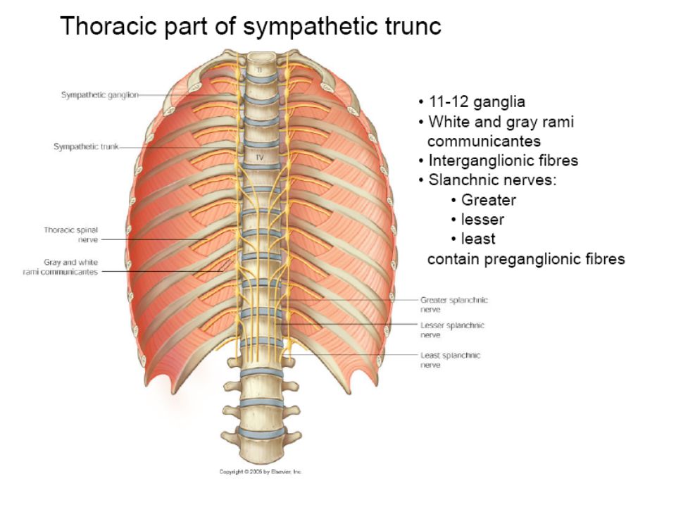
Thoracic Part of the Sympathetic Trunk 1
Thoracic Part of the Sympathetic Trunk 1. continuous above with the cervical and below with the lumbar parts of the sympathetic trunk. 2. It is the most laterally placed structure in the mediastinum. 3. It runs downward on the heads of the ribs. 4. It leaves the thorax on the side of the body of the T12 by passing behind the medial arcuate ligament.

46
5.. The sympathetic trunk has 12 (often only 11) segmentally arranged ganglia, each with white and gray ramus communicans passing to the corresponding spinal nerve. 6. The first ganglion is often fused with the inferior cervical ganglion to form the stellate ganglion.
 segmentally arranged ganglia, each with white and gray ramus communicans passing to the corresponding spinal nerve. 6. The first ganglion is often fused with the inferior cervical ganglion to form the stellate ganglion.")
59
Thymus 1. The thymus is a flattened, bilobed structure. 2. It lies between the sternum & pericardium in the anterior mediastinum. 3. In newborn infant, it reaches its largest size so it may extend up through the superior mediastinum in front of the great vessels into the root of the neck. 4. It continues to grow until puberty but thereafter undergoes involution

60
5. It has a pink, lobulated appearance.
6. It is the site for development of T (thymic) lymphocytes. Blood Supply The blood supply of the thymus is from the inferior thyroid & internal thoracic arteries.
 lymphocytes. Blood Supply. The blood supply of the thymus is from the inferior thyroid & internal thoracic arteries.")
61
Large Veins of the Thorax
1. Brachiocephalic Veins: The right brachiocephalic vein is formed by union of right subclavian & right internal jugular veins. begins posterior to the sternoclavicular joint of the right clavicle, and descends almost vertically to join the left brachiocephalic vein, forming superior vena cava Its tributaries vertebral, internal thoracic, inferior thyroid sometimes the first right posterior intercostal veins.

62
b. The left brachiocephalic vein.
It is formed by the union of the left subclavian and the right internal jugular veins. begins posterior to the sternoclavicular joint of left clavicle * It passes obliquely behind the manubrium sterni and in front of the large branches of the aortic arch. * sternal end of the first right costal cartilage it joins the right brachiocephalic vein to form the superior vena cava Its tributaries vertebral, internal thoracic, inferior thyroid, superior intercostal, sometimes the first left posterior intercostal, thymic and pericardial veins.

63
2. Superior Vena Cava: It contains all the venous blood from head and neck and both upper limbs. It is formed by the union of the two brachiocephalic veins. It passes downward to end in the right atrium of the heart. The azygos vein joins the posterior aspect of the superior vena cava just before it enters the pericardium.

Similar presentations


 in the diaphragm. Give the.>")


 THE THORACIC WALL Posteriorly by the thoracic part of the vertebral column Posteriorly by the thoracic.>")