Download presentation
Presentation is loading. Please wait.

1
Foot& ankle deformity Most of those occur due to: Congenital defects. Muscle imbalance. Ligament laxity. Joint instability

2
the foot postures are: Equinous foot: the foot is planter flexed at the ankle. Calcaneus foot: the foot is dorsiflexed at the ankle. Varus foot; is like an inverted foot with the sole facing medially. Valgus foot; is like an everted foot with the sole facing laterally. Flat foot (pes planus): is the condition where there is flattening of the medial longitudinal arch of the foot and this is usually associated with valgus deformity. Pes cavus: it is the deformity where there is increased medial arch of the foot and this is usually associated with varus deformity and claw toes. Clawing of the toes: is the deformity where there is abnormally maintained hyperextension of the metatarso-phalangial joints and flexion of the interphalangial joints. Hallux valgus (Hallux is the big toe): is an abnormal lateral deviation of the big toe. Talipes: it means any deformity where the foot is no more in plantigrade position, its usually congenital and the most frequently seen in practice is the cogenital talipes eqinovarus.
: is the condition where there is flattening of the medial longitudinal arch of the foot and this is usually associated with valgus deformity. Pes cavus: it is the deformity where there is increased medial arch of the foot and this is usually associated with varus deformity and claw toes. Clawing of the toes: is the deformity where there is abnormally maintained hyperextension of the metatarso-phalangial joints and flexion of the interphalangial joints. Hallux valgus (Hallux is the big toe): is an abnormal lateral deviation of the big toe. Talipes: it means any deformity where the foot is no more in plantigrade position, its usually congenital and the most frequently seen in practice is the cogenital talipes eqinovarus..")
3
Cogenital talipes eqinovarus: Common deformity of polygenic pattern, boys affected twice more often than girls, It’s bilateral in one third of the cases. This deformity may occur in cases of myelomeningocele and in arthrogryposis. Pathology: The talus and calcanium are pointing downwards. equines position, while the naviculum and the whole forefoot are shifted medially (adducted) and medially rotated (varus position), there is subluxation of the talo-navicular joint. talipes equinovarus. The soft tissues of the medial side of the foot are short and underdeveloped or contracted. If the condition is not treated early there will be chronic irreversible changes of bone and soft tissue that will be permanent. Clinical features: At birth the foot looks abnormally in equines, adduction and varus. Sometimes the deformity due to intrauterine malposition and is flexible, in these cases and in normal feet we can do gentile manipulation of the foot with dorsiflexion and eversion and the lateral aspect of the foot can touch the front of the leg. While in real congenital eqinovarus the deformity is fixed, rigid and cannot be corrected by manipulation. Always look for associated abnormalities as CDH, arthrogryposis and spina bifida.
 and medially rotated (varus position), there is subluxation of the talo-navicular joint. talipes equinovarus. The soft tissues of the medial side of the foot are short and underdeveloped or contracted. If the condition is not treated early there will be chronic irreversible changes of bone and soft tissue that will be permanent. Clinical features: At birth the foot looks abnormally in equines, adduction and varus. Sometimes the deformity due to intrauterine malposition and is flexible, in these cases and in normal feet we can do gentile manipulation of the foot with dorsiflexion and eversion and the lateral aspect of the foot can touch the front of the leg. While in real congenital eqinovarus the deformity is fixed, rigid and cannot be corrected by manipulation. Always look for associated abnormalities as CDH, arthrogryposis and spina bifida..")
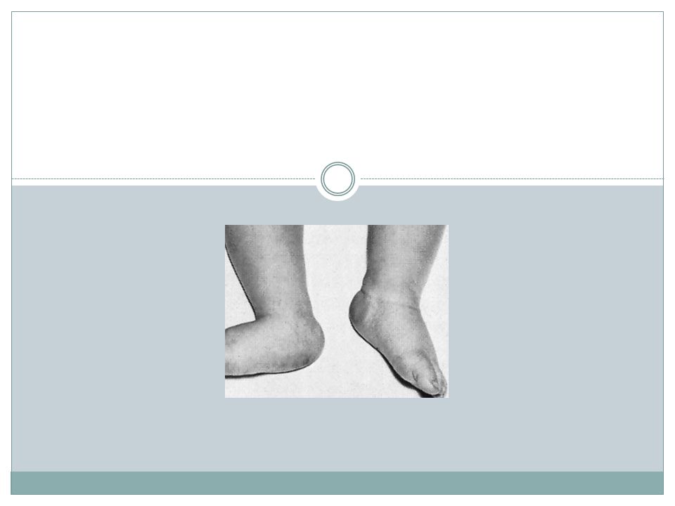
4
Club foot

5
X-ray: Lines can be drawn through the long axis of the talus parallel to its medial border & aline passing in the longtudinal axis of calcanium parallel to its latral border should de 20-40 digree

6
Treatment: The aims of the treatment are: Correct the deformity early. To correct the deformity fully. To hold the foot in the corrected position until it stop growing. Correction can be achieved by one of the followings; 1- Stretching and splintage: This is useful for simple cases its started in the 2nd Or 3rd day of life and useful until the age of three months where the bone and soft tissues still viscoelastic and allow correction. Here the foot is frequently, gently and gradually manipulated into the normal position weekly, and every time the position is held in by gently well-molded POP cast above the knee. The correction must be gentle, gradual and frequent every week to avoid forceful correction and traumatic foot injuries. We must start to correct the foot adduction first then varus and lastly the equinus. The role here is to correct and over correct the foot position.

7
2- Operative surgical correction: This is left for those who fail to respond to stretching and splintage and for cases that are resistant from the start. It’s usually done after the age of three months or according to foot size and body weight. Here Achilles tendon is elongated, all the shortened medial soft tissues are released and the long plantiflexors are elongated putting the foot in the normal position and this position held in POP cast. Here we start to correct equines then varus and lastly the forefoot adduction. The role here is to correct only and never overcorrect. The corrected foot position after stretching or operation must be held continuously by splints until the child start walking where the splints are only used at night until skeletal maturity to prevent recurrence. For children with failed correction and those who present between 5-10 years the correction is usually surgical and needs in addition to soft tissue release bone reshaping and possible tendon transfer. For children above the age of 10 years wedge tarsectomy or triple arthrodesis is indicated and the prognosis is poor.

9
Flat foot (pes planus): It’s the condition where there is flattening of the medial longitudinal arch of the foot, its usually associated valgus foot (flat everted foot). Flat foot can be primary, secondary or rigid. primary flat foot: Normally the foot is flat early in life in young infants and gradually corrects with early development, if persist it can be any of the below. Familial or idiopathic this is usually bilateral. secondary flat foot : Generalized ligament laxity. Valgus knee. Tight tendo-achellis with mild equinus foot. External rotation and valgus foot (Charlie Chaplin look). Overweight. Pregnancy with increased weight and ligament Laxity. In most of the above conditions the flat foot is said to be simple or flexible and on examination when we dorsiflex the big toe the medial arch of the foot can be restored. The patient or family notices the deformity or the abnormal shoe wear. The foot is examined with the patient standing and best from behind; patient should walk and tiptoe to see the state of the deformity and its correction.
. Overweight. Pregnancy with increased weight and ligament Laxity. In most of the above conditions the flat foot is said to be simple or flexible and on examination when we dorsiflex the big toe the medial arch of the foot can be restored. The patient or family notices the deformity or the abnormal shoe wear. The foot is examined with the patient standing and best from behind; patient should walk and tiptoe to see the state of the deformity and its correction..")
10
X-ray : may exclude bone abnormality. Treatment: Young infants with physiological flat foot needs no treatment, most younger children may need inner shoe raise, in adolescents we may use simple medial arch support inside the shoe, all to avoid later painful foot and prevent permanent secondary changes of bone and soft tissues.

12
Other causes of stiff or rigid flat foot are: Congenital vertical talus. Tarsal coalition (spasmodic flat foot). Neuromuscular disorders and muscle imbalance. Rheumatoid foot. Congenital vertical talus: Its rare and uncommon, it present at birth. the foot is convex on the sole (like the bottom of the rocker), the talus is vertically placed with dislocation of the talonavicular joint, the whole forefoot is dorsiflexed and valgus at the tarso- metatarsal region. Any attempt to correct this rigid deformity by manipulation is very difficult, it usually needs surgery and prognosis is poor. Spasmodic flat foot: This is a condition that occurs in adolescents where the foot at rest looks normal but after walking or exercise it get painful with spasm of the peronial muscles so the foot go into valgus and get flattened.
. Neuromuscular disorders and muscle imbalance. Rheumatoid foot. Congenital vertical talus: Its rare and uncommon, it present at birth. the foot is convex on the sole (like the bottom of the rocker), the talus is vertically placed with dislocation of the talonavicular joint, the whole forefoot is dorsiflexed and valgus at the tarso- metatarsal region. Any attempt to correct this rigid deformity by manipulation is very difficult, it usually needs surgery and prognosis is poor. Spasmodic flat foot: This is a condition that occurs in adolescents where the foot at rest looks normal but after walking or exercise it get painful with spasm of the peronial muscles so the foot go into valgus and get flattened..")
13
Sometimes its idiopathic although tarsal fusion (coalition) by a bony or cartilage bar has been commonly blamed. X-ray, CTscan or MRI may show the abnormal coalition. The condition usually relieved by resting the foot in a cast or splint, if there is abnormal bar this may need surgical interference.

14
Pes cavus (increased or high medial arch of the foot): The medial arch is higher than normal with varus foot and finger clawing the main Causes are: Primary idiopathic, usually familial and bilateral. Neuromuscular conditions like cerebral palsy, peroneal muscle dystrophy or friedreich’s ataxia This deformity of the foot with the clawing of the toes puts the body weight on the metatarsal heads that projects down into the sole of the foot and usually there is an overlying skin callosities due to friction with the shoe. In the mobile flexible early deformity the foot shape can be restored if the metatarsal heads pushed up by the examiner’s finger, as the arch gets normal and the clawing of the toes corrected. Later the deformity if untreated gets fixed and painful. Treatment: In cases of painless mobile deformity no treatment is needed apart from special shoe wear. In severe deformities which is still mobile the foot shape can be improved and weight bearing on metatarsal heads can be decreased by rebalancing surgery correcting the clawing by tendon transfer so the long toe flexors are released and transferred from the planter to the dorsal aspect of the toes and fixed on the extensor expansion so it will correct the hyperextension and put the toes straight. For fixed deformities no much can be done, if special shoe wear is not enough complex bone surgeries and arthrodesis can be done, operations must always delayed after the age of 16 years.

Similar presentations


 Dr. Mazloumi MD Associate Professor Pediatric Orthopedic Surgeon.>")


