Download presentation
Presentation is loading. Please wait.

1
Female Reproductive System

2
Components of the female reproductive system
(1) a pair of ovaries (2) a pair of oviducts (3) a uterus (4) a vagina (5) the external genitalia
 a pair of ovaries. (2) a pair of oviducts. (3) a uterus. (4) a vagina. (5) the external genitalia.")
3
Ovary Functions: ► 1. produce ova
► 2. secrete hormones – estrogens + progesterone Histology: ● consists of a cortical and medullar regions (not sharply delineated) ● the surface of the ovary is covered by a simple cuboidal epithelium – the germinal epithelium ● under the germinal epithelium – tunica albuginea – layer of dense irregular connective tissue
 ● the surface of the ovary is covered by a simple cuboidal epithelium – the germinal epithelium. ● under the germinal epithelium – tunica albuginea – layer of dense irregular connective tissue.")
4
A. CORTICAL REGION (CORTEX):
○ the stroma composed from connective tissue – cells respond to hormonal stimuli ○ contains numerous ovarian follicles in various stages of development ○ ovarian follicle consists of an oocyte surrounded by one or more layers of follicular cells – the granulosa cells ● Development of ovarian follicles: 1. Primordial follicles 2. Primary follicles 2.1. Unilaminar primary follicles 2.2. Multilaminar primary follicles 3. Secondary (antral) follicles 4. Mature (Graafian) follicles . A. MEDULLARY REGION (MEDULLA): ○ contains a rich vascular bed within a cellular loose connective tissue
 follicles. 4. Mature (Graafian) follicles . A. MEDULLARY REGION (MEDULLA): ○ contains a rich vascular bed within a cellular loose connective tissue.")
5
PRIMORDIAL FOLLICLES ● at the time of birth 400.000 - 1.000.000
At puberty: 40,000 ● only few hundred maturate ● arrested in prophase of meiosis I ● Composition : a. primary oocyte b. single layer of flat follicular cells ● surrounded by basement membrane

6
GROWING FOLLICLES: GROWING FOLLICLES:
● Beginning in puberty with the release of (FSH) from the pituitary, a small group of primordial follicles each month begins a process of follicular growth. ● distinct changes in morphology of oocyte, follicular cells and stroma arround growing follicle
 from the pituitary, a small group of primordial follicles each month begins a process of follicular growth. ● distinct changes in morphology of oocyte, follicular cells and stroma arround growing follicle.")
7
GROWING FOLLICLES 2. PRIMARY FOLLICLES
2.1. Unilaminar primary follicle ● follicular cells form single layer of cuboidal cells ● primary oocyte follicular cells proliferate by mitosis and form a stratified follicular epithelium, or granulosa layer → multilaminar primary follicle

8
Primary multilaminar follicle
Avascular part: ● primary oocyte ● granulosa layer → stratified follicular epithelium that communicate by gap junctions ● a thick coat – the zona pellucida composed of glycoproteins, surround the oocyte Filopodia of follicular cells and microvilli of the oocyte penetrate the zona pellucida, allowing communication between these cells via gap junctions Vascularized part: ● stromal tissue around differentiates into two layers: - Theca folliculi interna – cellular layer - Theca folliculi externa- fibrous layer ● theca folliculi interna cells – production of steroid hormones

9
SECONDARY FOLLICLES liquor folliculi – fluid accumulate in the spaces between granulosa cells (plasma exudate with hormons)
")
10
MATURE GRAAFIAN FOLLICLE
● about 2.5 cm in diameter ● protrudes deep into cortical tissue and extends over the surface of the organ ● the granulosa cells that form the first layer around the oocyte and are in close contact with zona pellucida – become elongated and form the corona radiata ● a hillock called cumulus oophorus, carrying an oocyte – positioned off center ● antrum folliculi with follicular fluid ● oocyte prior to ovulation complete first meiotic division forming secondary oocyte ● usually only one dominant follicle undergoes ovulation

12
CORPUS LUTEUM (CL) ● after ovulation, the granulosa cells and those of theca interna that remain in the ovary form a temporary endocrine gland called the corpus luteum under the infleuence of LH ● CL is localized in the cortical region of the ovary and persists 14 days after ovulation ● secretes hormones → progesteron + estrogens + relaxin ● the granulosa cells make up 80% of the parenchyma of the CL and are now called – granulosa lutein cells ● the cells of the theca interna form – theca lutein cells Remnants from its degeneration and regression are phagocytosed by macrophages, after which fibroblasts invade the area and produce a scar of dense connective tissue called corpus albicans (white body)
")
15
ATRETIC FOLLICLE ● the most ovarian follicles undergo the process – atresia → follicular cells + oocytes die and are disposed of by phagocytic cells (MQ) ● the atresia can take place during any stages in the development of a follicle ● this process is histologically characterized by: a. stop of mitosis in the granulosa cells b. detachment of granulosa cells from the BM c. death of the oocyte it is most prominent just after birth, when levels of maternal hormones decline rapidly, and during both puberty and pregnancy, when qualitative and quantitative hormonal changes occur again.
 ● the atresia can take place during any stages in the development of a follicle. ● this process is histologically characterized by: a. stop of mitosis in the granulosa cells. b. detachment of granulosa cells from the BM. c. death of the oocyte. it is most prominent just after birth, when levels of maternal hormones decline rapidly, and during both puberty and pregnancy, when qualitative and quantitative hormonal changes occur again.")
16
OVIDUCT (FALLOPIANE TUBE)
the wall is composed of 3 main layers : 1) Tunica mucosa ● forms longitudinal mucosal folds (mainly in ampulla → labyrinth) ● Epithelium– simple columnar epithelium 1.) CILIATED CELLS –possess many cilia- transport of the ovum and embryo 2.) SECRETORY CELLS (Peg Cells) – secrete a nutrient rich medium – nutrition of spermatozoa and preimplantation embryo ● lamina propria – loose connective tissue (is richly vascularized!)
 Tunica mucosa. ● forms longitudinal mucosal folds (mainly in ampulla → labyrinth) ● Epithelium– simple columnar epithelium. 1.) CILIATED CELLS –possess many cilia- transport of the ovum and embryo. 2.) SECRETORY CELLS (Peg Cells) – secrete a nutrient rich medium – nutrition of spermatozoa and preimplantation embryo. ● lamina propria – loose connective tissue (is richly vascularized!)")
18
OVIDUCT (FALLOPIANE TUBE)
(B) Tunica muscularis ● consists of smooth muscle cells disposed as: 1) INNER CIRCULAR (SPIRAL) LAYER 2) OUTER LONGITUDINAL LAYER (C) Tunica serosa ● composed of visceral peritoneum (mesothelium + submesothelial layer)
 Tunica muscularis. ● consists of smooth muscle cells disposed as: 1) INNER CIRCULAR (SPIRAL) LAYER. 2) OUTER LONGITUDINAL LAYER. (C) Tunica serosa. ● composed of visceral peritoneum (mesothelium + submesothelial layer)")
19
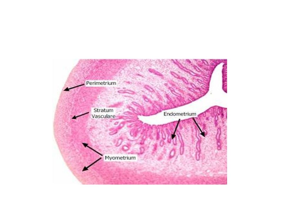
UTERUS the uterus is a thick-walled organ, wall consists of 3 layers:
A – tunica mucosa – ENDOMETRIUM B – tunica muscularis – MYOMETRIUM C – tunica serosa – PERIMETRIUM * PARAMETRIUM – dense regular C.T. of the broad ligament

21
ENDOMETRIUM ● consists of lamina epithelialis and lamina propria
● epithelial lining – simple columnar epithelium containing secretory and ciliated cells ● lamina propria – loose connective tissue with many stellate fibroblasts, contains abundant amorphous ground substance→ uterine glands – simple tubular glands (covered by simple columnar epithelial cells)
")
22
Layers of endometrium:
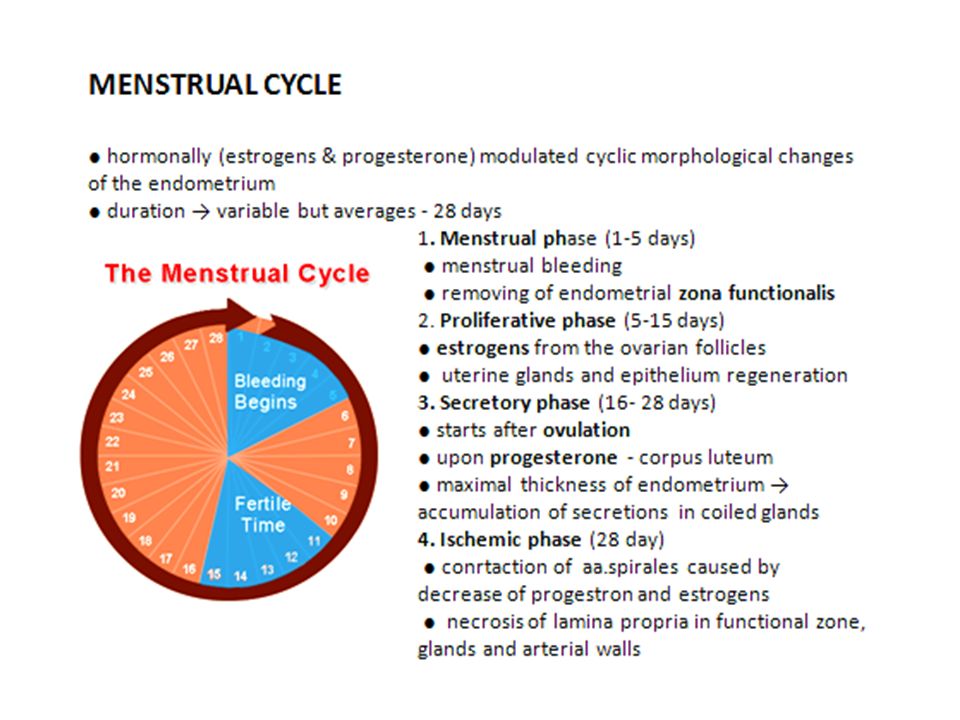
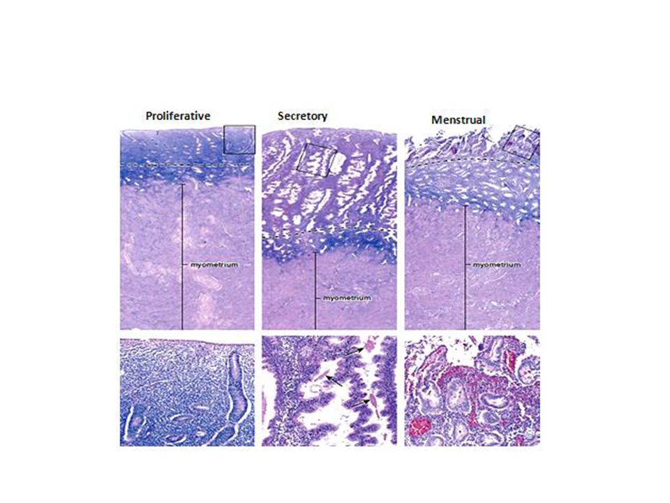
1. Zona functionalis ○ superficial layer contains more spongy and less cellular lamina propria, richer in ground substance, most of the length of the glands, as well as the surface epithelium ○ exhibit dramatic changes during menstrual cycle every month as a result of hormonal changes ○ shed during menstruation ! 2. Zona basalis ○ basal layer adjacent to the myometrium contains highly cellular lamina propria and the deep basal ends of the uterine glands. ○ undergoes little changes during the menstrual cycle ○ not shed during menstruation ! ○ provides a new epithelium and lamina propria for the renewal of the endometrium!

23
ENDOMETRIAL BLOOD SUPPLY
○ the endometrium has a unique dual blood supply ○ the uterine artery distributes blood to arcuate arteries in the middle layer of the myometrium ○ from these vessels, 2 sets of arteries arise to supply blood to the endometrium: (1) STRAIGHT (BASAL) ARTERIES → which supply the zona basalis (2) COILED (SPIRAL) ARTERIES → which bring blood to the zona functionalis and undrrgo pronounced changes during menstrual cycle (progesterone-sensitive) many dilated, thin-walled vessels called vascular lacunae.
 STRAIGHT (BASAL) ARTERIES → which supply the zona basalis. (2) COILED (SPIRAL) ARTERIES → which bring blood to the zona functionalis and undrrgo pronounced changes during menstrual cycle (progesterone-sensitive) many dilated, thin-walled vessels called vascular lacunae.")
24
MYOMETRIUM ● thick muscular layer (4 poorly defined layers)
● composed of bundles of smooth muscle cells separated by connective tissue ● inner and outer layers are longitudinal, thick middle circular ● the middle layers contain the larger blood vessels (arcuate arteries)- stratum vasculare ● myometrium thicknes during pregnancy because of the hypertrophy and hyperplasia of individual smooth muscle cells During this growth, many of the smooth muscle cells also actively synthesize collagen, strengthening the uterine wall. After pregnancy, uterine smooth muscle cells shrink and many undergo apoptosis, with removal of unneeded collagen, and the uterus returns almost to its prepregnancy size
- stratum vasculare. ● myometrium thicknes during pregnancy because of the hypertrophy and hyperplasia of individual smooth muscle cells. During this growth, many of the smooth muscle cells also actively synthesize collagen, strengthening the uterine wall. After pregnancy, uterine smooth muscle cells shrink and many undergo apoptosis, with removal of unneeded collagen, and the uterus returns almost to its prepregnancy size.")
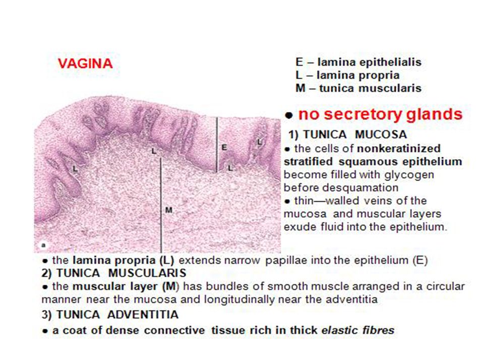
27
Cervix In comparison to the body, the myometrium of the cervix is less muscular and contains proportionally more connective tissue, much of it elastic fibers. In contrast to the body, the mucosa of the cervix does not undergo cyclical changes. External to the cervical canal (the ecotcervix or vaginal portion) the mucosa is lined by stratified squamous epithelium. Internally, a mucous secreting simple columnar epithelium lines the cervical canal and forms the cervical glands. The mucous secretion of the cervical glands varies with the menstrual cycle, being more copious and less viscous at ovulation (to promote fertilization). Blockage of the lumen of a cervical gland results in the formation of a Nabothian cyst (or more simply, dilated cervical gland); these cysts are of no pathological consequence. Before parturition the cervix dilates greatly and softens due to intense collagenolytic activity in the stroma.
 the mucosa is lined by stratified squamous epithelium. Internally, a mucous secreting simple columnar epithelium lines the cervical canal and forms the cervical glands. The mucous secretion of the cervical glands varies with the menstrual cycle, being more copious and less viscous at ovulation (to promote fertilization). Blockage of the lumen of a cervical gland results in the formation of a Nabothian cyst (or more simply, dilated cervical gland); these cysts are of no pathological consequence. Before parturition the cervix dilates greatly and softens due to intense collagenolytic activity in the stroma.")
29
Mammary gland The mammary glands of the breasts develop embryologically as invaginations of surface ectoderm resembling highly modified apocrine sweat glands Each mammary gland consists of 15–25 lobes of the compound tubuloalveolar Each lobe, separated from the others by dense connective tissue with much adipose tissue, is a separate gland with its own excretory lactiferous duct. These ducts, each 2–4.5 cm long, emerge independently in the nipple, which has 15–25 pore-like openings, each about 0.5 mm in diameter.

30
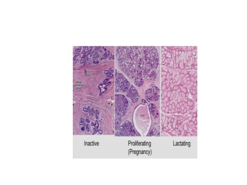
Mammary gland. the sequence of changes that occur in the duct system and secretory units before, during, and after pregnancy and lactation. (1) Before pregnancy, the gland is inactive, with small ducts and only a few small secretory alveoli. (2) Alveoli develop and begin to grow early in a pregnancy. (3) By mid-pregnancy, the alveoli and ducts have become large and have dilated lumens. (4) At parturition and during the time of lactation, the alveoli are greatly dilated and maximally active in production of milk components. (5) After weaning, the alveoli and ducts regress with apoptotic cell death
 Before pregnancy, the gland is inactive, with small ducts and only a few small secretory alveoli. (2) Alveoli develop and begin to grow early in a pregnancy. (3) By mid-pregnancy, the alveoli and ducts have become large and have dilated lumens. (4) At parturition and during the time of lactation, the alveoli are greatly dilated and maximally active in production of milk components. (5) After weaning, the alveoli and ducts regress with apoptotic cell death.")
32
Mammary gland

Similar presentations