Download presentation
Presentation is loading. Please wait.

1
Reproductive System Continued Exercises 42 and 43

2
Reproduction is the process by which: new individuals of a species are produced the genetic material is passed on from generation to generation. Organisms that reproduce sexually produce specialized reproductive cells called gametes: ova (ovum) - eggs sperm (spermatozoa)
 - eggs sperm (spermatozoa).")
3
Meiosis in the male Spermatogenesis takes about 65-70 days. Begins with spermatogonium – 2n First division is through mitosis – one daughter cell remains near basement membrane of the seminiferous tubule, the other forms sperm – primary spermatocyte (2n) Sperm production stops here until puberty.
 Sperm production stops here until puberty..")
4
First phase of meiosis the primary spermatocyte enlarges, and duplicates its DNA. It then goes through two nuclear divisions, resulting in 4 cells with n chromosomes. During first division homologous pairs of chromosomes line up in the center of the cell – synapsis. Crossing-over may occur and increase genetic variability by recombination of genes.

7
During the first meiotic division, the centromeres do not break, and one member of each pair (two identical chromosomes linked together) is pulled to opposite sides of the cell. Secondary spermatocytes. The nucleus reforms, but the DNA is not duplicated. The second meiotic division proceeds just like mitosis, and we end with 4 haploid spermatids.

13
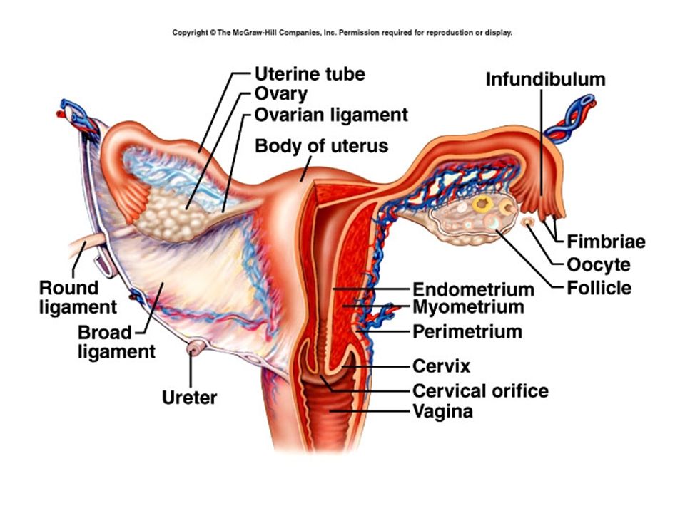
Female Reproductive System Ovaries – gonads – produce oocytes and hormones Uterine (Fallopian) tubes or oviducts Uterus Vagina Vulva (female external reproductive organs)
 tubes or oviducts Uterus Vagina Vulva (female external reproductive organs)")
15
Ovaries Located in upper pelvic cavity, on either side of uterus Held in place by a number of ligaments Descend from abdominal cavity to pelvic brim

18
Each ovary has a hilus – blood vessels and nerves enter Several layers: –Germinal epithelium – simple squamous cells –Tunica albuginea- white capsule of C. T. –Stroma – connective tissue, can be divided into: Medulla – loose connective tissue, blood vessels, nerves Cortex - contains ovarian follicles – consist of oocytes at various stages of development

19
Single layer – follicular cells Several layers – granulosa cells Mature or Graafian follicle is a large, fluid-filled follicle that will rupture and release a secondary oocyte in process called ovulation. Corpus luteum is the remnant of a ruptured follicle – produces estrogen, progesterone and relaxin until degenerates into the corpus albicans.

21
Oogenesis During fetal development, germ cells differentiate into oogonia millions of germ cells. Many degenerate, but a few develop into primary oocytes that enter Prophase I of meiosis before birth – stop there. At birth, about 1 million oogonia and primary oocytes in each ovary About 400 mature over a woman’s lifetime.

22
Primordial follicle – primary oocyte and single layer of follicular cells Under influence of FSH, become primary follicles which have several layers of cells. Layer of glycoprotein, the zona pellucida, separates oocyte from the granulosa cells. Ovarian cells outside follicle form two layers: –Inner vascular layer (theca interna) that secretes hormones –Outer fibrous layer (theca externa) – connective tissue.
 that secretes hormones –Outer fibrous layer (theca externa) – connective tissue..")
23
Granulosa cells begin to secrete fluid, forms a cavity called the antrum. Now called a secondary follicle. After puberty, each month one secondary follicle resumes meiosis. Meiosis I results in two unequal cells – secondary oocyte and a polar body. Begins to divide again but stops at metaphase II.

25
At ovulation, secondary oocyte and polar body are released. If not fertilized, degenerates. If penetrated by sperm, meiosis resumes, forming ovum and another polar body. Nuclei of ovum and sperm unite to form a zygote. (2n or diploid) So, meiosis results in ONE OVUM and a polar body (which degenerates)
 So, meiosis results in ONE OVUM and a polar body (which degenerates).")
28
Uterine (Fallopian) Tubes Extend laterally from uterus Open end is funnel shaped – infundibulum Many finger-like projections – fimbriae Local currents produced by movement of fimbriae draws ovum into uterine tube. Uterine tube is lined with ciliated columnar epithelium, which move ovum along. Peristaltic movements also propel ovum toward uterus.

29
The secondary oocyte is usually fertilized in the outer one-third of the uterine tube, although fertilization may occur in the abdominopelvic cavity. Fertilization can occur up to about 24 hours after ovulation. If oocyte is fertilized, it will reach the uterus in about 7 days.

31
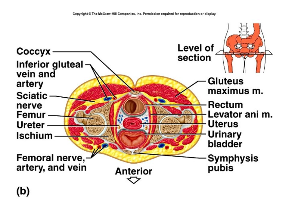
The Uterus Is an organ about the size and shape of an inverted pear. Uterus can be divided into the fundus, body and cervix.

33
Layers of uterus : perimetrium, myometrium and endometrium Perimetrium – a part of visceral peritoneum Myometrium – bulk of uterus – three layers of muscle that contract under influence of oxytocin during labor. Endometrium – highly vascular mucosa –Stratum functionalis – shed during menstruation –Stratum basalis – deeper, permanent layer, gives rise to new stratum functionalis after each cycle.

34
The Vagina Passageway for sperm and menstrual flow Receptacle for penis during intercourse Inferior portion of birth canal Capable of considerable distention (stretching) Mucosa is continuous with that of uterus and consists of nonkeratinized stratified squamous epithelium. Contains large stores of glycogen which decomposes to organic acids – lower pH = less susceptible to infection & less hospitable to sperm

36
The Vulva External genitals of female Mons pubis – pad of adipose tissue which cushions pubic symphysis Labia majora (labium majus) – folds of skin analogous to the scrotum –Adipose tissue, sebaceous glands, sweat glands Labia minora (labium minus) – folds of skin with many blood vessels and sebaceous (oil) glands
 – folds of skin analogous to the scrotum –Adipose tissue, sebaceous glands, sweat glands Labia minora (labium minus) – folds of skin with many blood vessels and sebaceous (oil) glands")
37
Clitoris – homologous to the penis, also composed of two corpora cavernosa and a glans. Vestibule – between the labia minora –External urethral orifice –Openings for ducts of paraurethral glands : secrete mucus and are homologous to prostate –Beside vaginal orifice are the vestibular (Bartholin’s) glands – produce mucus during arousal – homologous to bulbourethral glands
 glands – produce mucus during arousal – homologous to bulbourethral glands.")
Similar presentations


 Gametogenesis Process of gamete formation with the reduction by half.>")


