Download presentation
Presentation is loading. Please wait.

1
Descriptive Histology CLS 222 Mrs. Saida Almashharawi

3
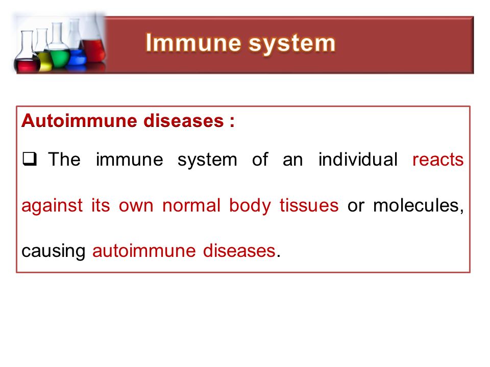
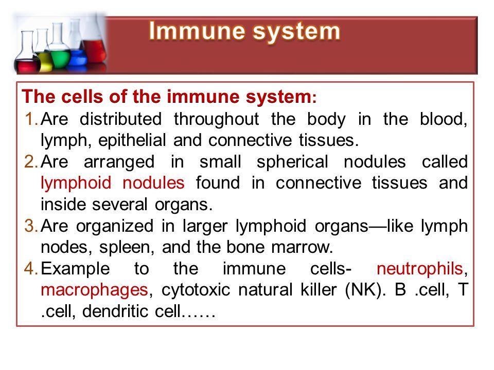
The immune and lymphatic systems are two closely related organ systems that share several organs and physiological functions. The immune system is our body’s defense against infectious pathogenic viruses, bacteria, and fungi as well as parasite and protists. The immune system works to keep these harmful agents out of the body and attacks those that manage to enter

7
The lymphatic system is part of the circulatory system, comprising a network of lymphatic vessels that carry a clear fluid called lymph (from Latin lympha meaning water), lymph is a fluid containing infection- fighting white blood cells, throughout the body. The lymph has a number of functions, including:- 1- Helps the body to get rid of toxins, waste and other unwanted materials 2-Removal of interstitial fluid ( extracellular fluid of tissue).
..")
10
Primary lymphoid organs Secondary lymphoid organs Secondary lymphoid organs Bone marrow Thymus Lymph node Spleen Tonsils

12
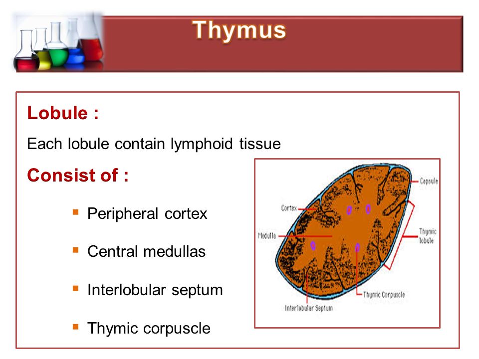
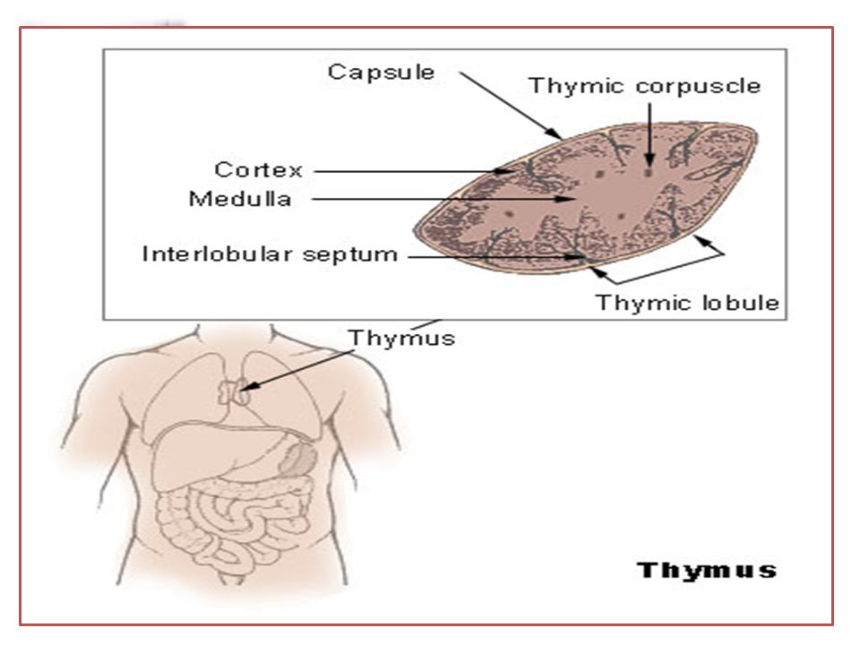
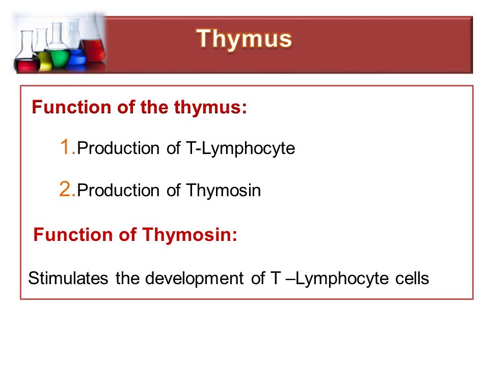
primary lymphoid organ, composed of two identical lobes, located anatomically in front of the heart and behind the sternum(is a long flat bony plate shaped like a capital "T" located anteriorly to the heart in the center of the chest) This small organ stores immature lymphocytes (specialized white blood cells) and prepares them to become active T cells, which help destroy infected or cancerous cells.
 This small organ stores immature lymphocytes (specialized white blood cells) and prepares them to become active T cells, which help destroy infected or cancerous cells.")
15
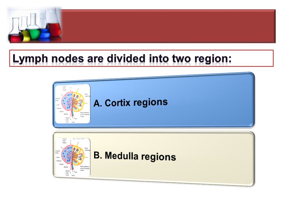
Cortex: The cortical portion is mainly composed of lymphocytes, supported by a network of finely- branched epithelial reticular cells, which is continuous with a similar network in the medullary portion. The cortex is the location of the earliest events in thymocyte development

16
Medulla : In the medullary portion, the reticulum is coarser than in the cortex, the lymphoid cells are relatively fewer in number. The medulla is the location of the latter events in thymocyte development. Both cortex and medulla contain small lymphocyte (Called in this particular location as thymocytes. No lymphatic nodules are present in the thymus.

21
Descriptive Histology CLS 222 Mrs. Saida Almashharawi

22
Revision to lymphatic organs Structure & functions of lymph node Structure, location, types of tonsils Structures& functions of spleen

23
Primary lymphoid organs Secondary lymphoid organs Secondary lymphoid organs Bone marrow Thymus Lymph node Spleen Tonsils

25
lymph node Tonsils Spleen

26
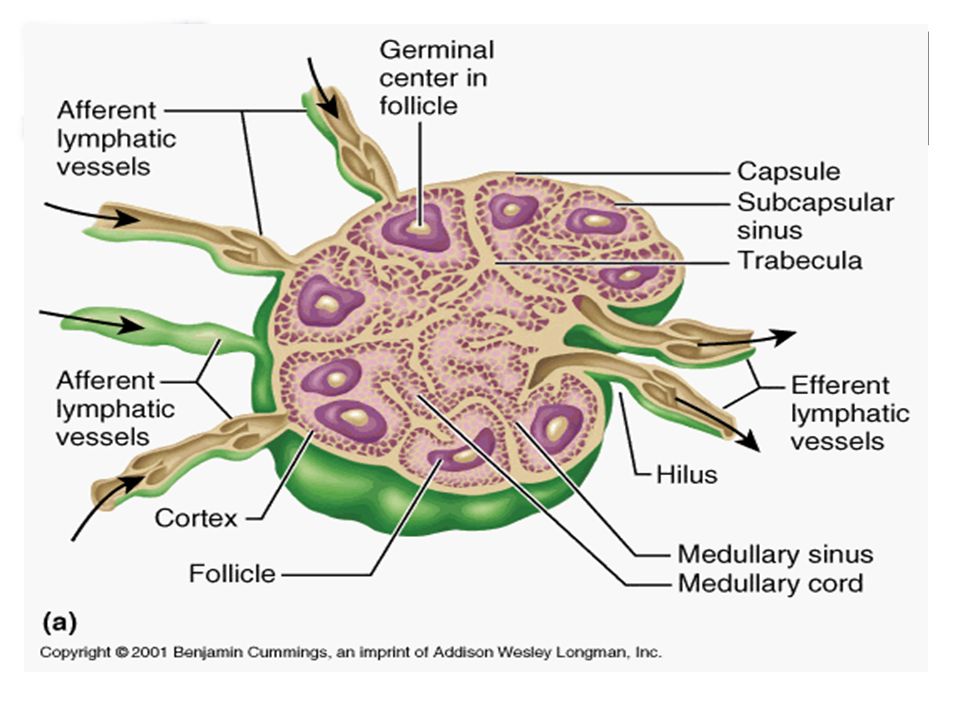
1.Lymph nodes are bean-shaped, encapsulated structures 2. generally 2–10 mm in diameter 3.distributed throughout the body along the course of the lymphatic vessels, in the chest, neck, pelvis, axilla (armpit), groin region, and in association with the blood vessels of the intestines
, groin region, and in association with the blood vessels of the intestines.")
28
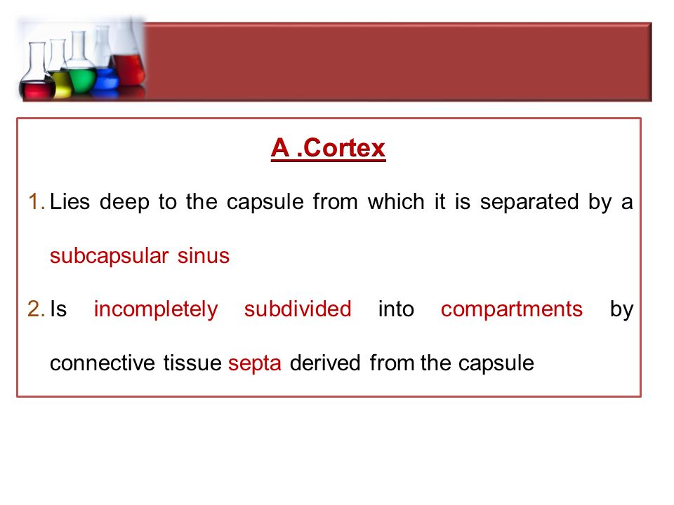
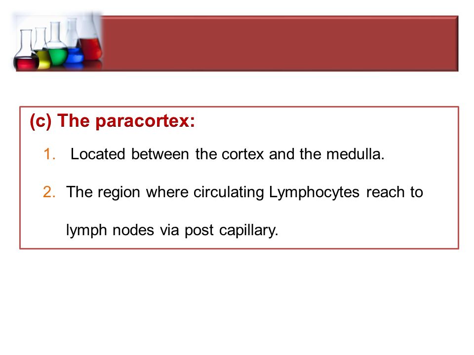
The lymph node consists of lymphoid follicles in an outer portion called the "cortex.“, and the inner portion called the "medulla," which is surrounded by the cortex on all sides except for a portion known as the "hilum.“ The hilum presents as a depression on the surface of the lymph node, which makes the spherical lymph node, bean-shaped or ovoid. The efferent lymph vessel, arteries and veins supplying the lymph node with blood enter and exit through the hilum.

38
1.Lymph nodes have the primary function of producing lymphocytes which help protect the body against microorganisms and harmful foreign particles. 2. Lymph nodes normally swell when they are actively responding to infection as they fill with the pathogenic cells they filter from the lymph.

40
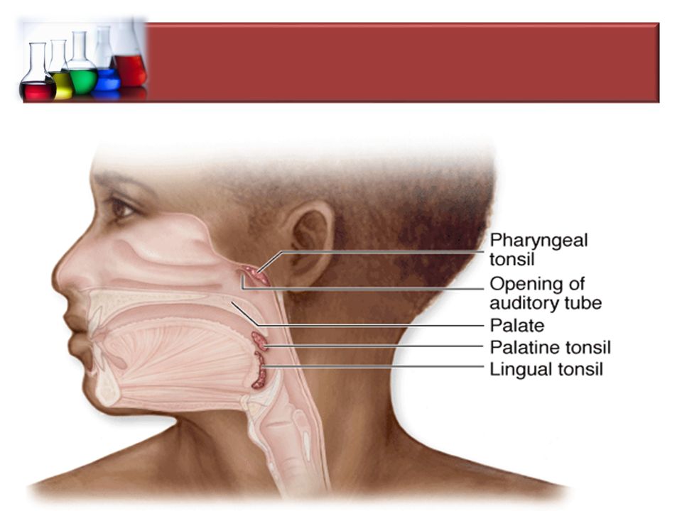
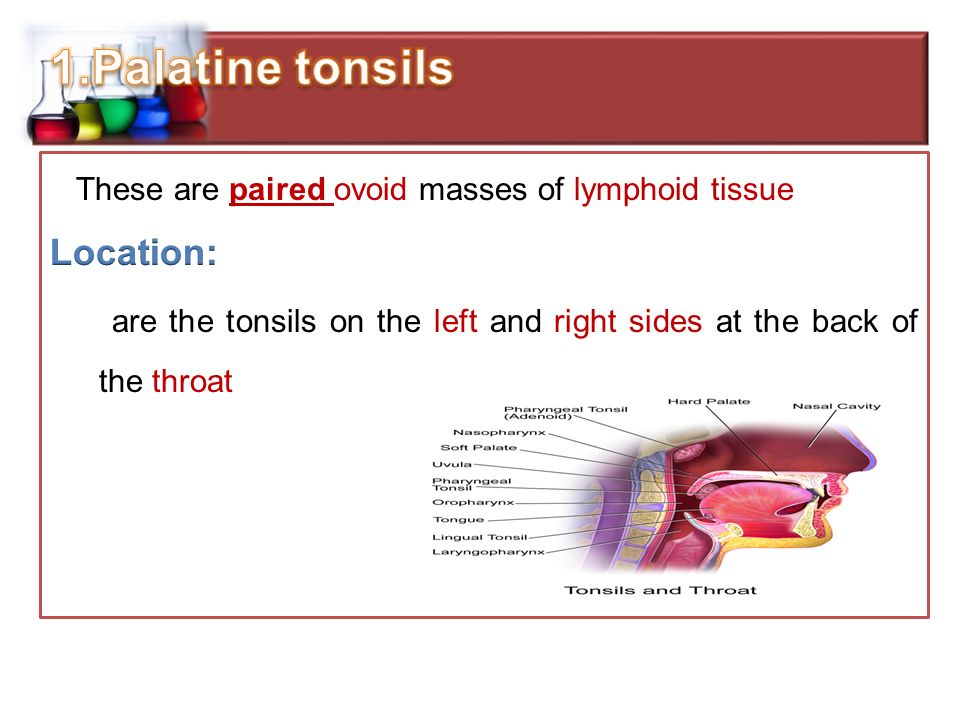
These lymphoid masses are named according to their location as : 1.Palatine tonsils 2.Pharyngeal tonsil 3.Lingual tonsil

49
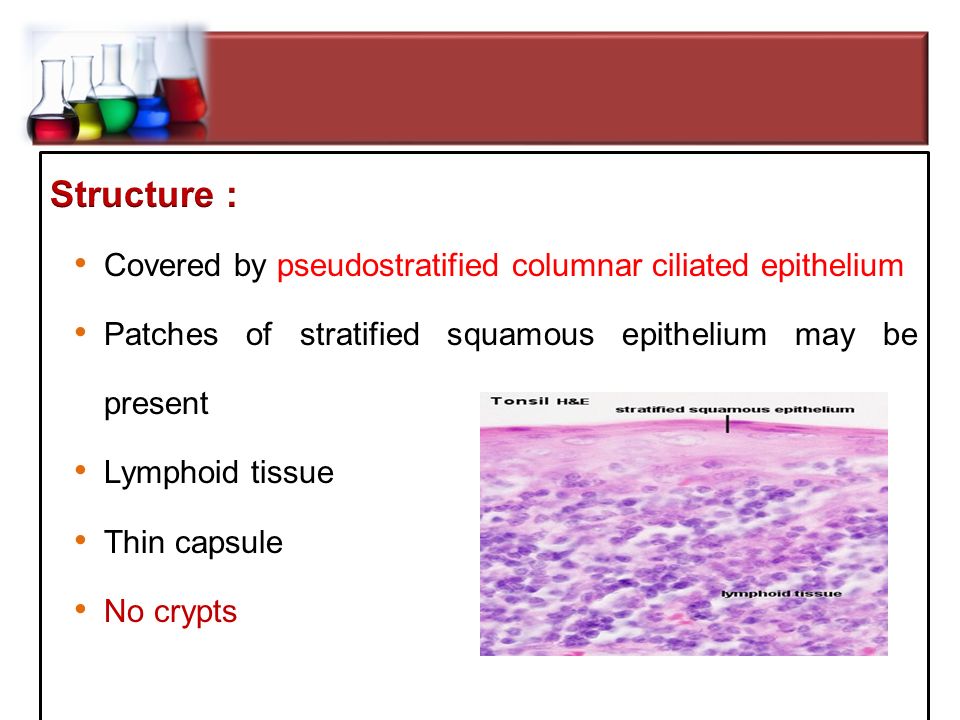
TonsilLocationEpitheliumCrypts Pharyngea l In the midline on the roof of the nasopharynx Ciliated pseudostratified columnar epithelium, with some patches of stratified squamous epithelium observed Numerous folds of pharyngeal epithelium, not true crypts Lingual Posterior third of tongue Stratified squamous non-keratinising Multiple monocryptic 'units' PalatinePaired at oropharynx Stratified squamous non-keratinising 10-30 deep and sometimes branching crypts (Polycryptic)
")
Similar presentations


 -mast cells -interdigitating.>")


 from the lamina propria. Absorb.>")

