Download presentation
Presentation is loading. Please wait.

1
Histological Structure of Lymphoid Organs
DR RANIA GABR

2
Objectives Understand the location of lymphatic organs.
Discuss the microscopic features of Lymph Node. Discuss the microscopic features of Spleen. Discuss the microscopic features of Thymus. Discuss the microscopic features of Tonsils

3
Lymphoid Tissue Lymphoid tissue is connective tissue chch by rich supply of lymphocytes. It is found either Free in regular CT 2-Surrounded by capsules, forming the “lymphoid organs” Very little cytoplasm so stain dark blue with H&E. Rich network of reticular fibrils produced by fibroblasts.

4
Lymphoid System Basics
Two main tissue architecture types: Diffuse: uniform appearance Follicular: consists of lymphoid follicles Two types of lymphoid tissues: Encapsulated: connective tissue capsule spleen, thymus, lymph nodes Unencapsulated (or partly encapsulated) Tonsils, Peyer’s patches, lymphoid nodules in GI tract, respiratory tract, urinary & reproductive tracts
 Tonsils, Peyer’s patches, lymphoid nodules in GI tract, respiratory tract, urinary & reproductive tracts.")
5
2 Types of Lymphoid Organs
Central (primary) lymphoid organ: where lymphoid cells undergo maturation T cells in thymus B cells in bone marrow Peripheral (secondary) lymphoid organ: where functional lymphocytes go including: 1- lymph nodes spleen, 3- Peyer’s patches, lymphoid nodules of GI and other tracts
 lymphoid organ: where lymphoid cells undergo maturation. T cells in thymus. B cells in bone marrow. Peripheral (secondary) lymphoid organ: where functional lymphocytes go including: 1- lymph nodes 2- spleen, 3- Peyer’s patches, 3- lymphoid nodules of GI and other tracts.")
7
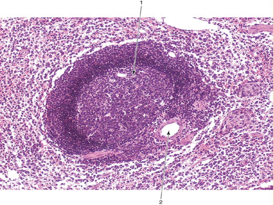
Lymphoid Follicles Nodules of densely packed lymphocytes located in all peripheral lymphoid tissues. Most lymphocytes are B cells. Two distinct areas: 1- Mantle – darker stained, mainly small, resting lymphocytes 2- Germinal center – (defines “secondary” or “reactive” lymphoid follicles): lighter stained, larger, activated B cells
: lighter stained, larger, activated B cells.")
8
Lymph follicle: Mantle = cap (dark) Germinal center (light)
 Germinal center (light)")
10
Lymph Nodes Present throughout the body, along lymph vessels
Numerous in axilla, groin, cervical area and thoracic/abdominal mesenteries Filter lymph before it returns to vasculature Hilum: concave side, arteries, nerves enter; veins and efferent lymph vessels leave the organ Afferent lymph vessels enter convex surface

12
Covered by a capsule which extends to form Trabeculae.
Divided into outer cortex and inner medulla. OUTER CORTEX contains: Lymphatic nodules with germinal center INNER MEDULA contains: Medullary Cords and Medullary Sinus

13
Medullary cords Are branched, cordlike extensions of lymphoid tissue arising from the paracortex. They contain primarily B- lymphocytes and often plasma cells and macrophages. Medullary cords are separated by dilated spaces , frequently bridged by reticular cells and fibers , called Medullary sinuses They contain lymph , lymphocytes, macrophages, sometimes granulocytes if the lymph node is draining an infected organ

15
Lymph node CT --- Connective tissue C Cortex M Medulla P Paracortex LN --- Lymph Node T Trabeculae MS --- Medullary Sinus MC --- Medullary Cords

16
Spleen It is an encapsulated lymphoid organ
The capsule extends to form Trabeculae which contain the trabecular arteries and trabecular veins. It consists of stroma and parenchyma. The parenchyma of the spleen consists of: 1.White Pulp: lymphoid follicles or nodules with germinal centre. A Central artery passes through lymphatic nodules.

17
Spleen

18
2.Red Pulp: surrounds the white pulp and is also divided to splenic cords and splenic sinuses. Splenic Cords: Known as Splenic Cords of Billroth They consist of all WBCs, macrophages, RBCs ,B & T Lymphocytes. Splenic Sinuses: Full of blood because the spleen acts as a filter of blood. Splenic sinuses are lined by endothelium. The stroma of the spleen is similar to that of the lymph node with a capsule, trabeculae & reticular fibers

19
The Spleen

20
Spleen

21
Splenic Cords and Splenic Sinusoids

22
Open and Closed Circulation in Spleen

23
Thymus The thymus is a primary lymphoid organ in that it supplies other lymphoid organs and tissues with T-lymphocytes. The thymus is enclosed by a thin C.T capsule from which numerous septa extend into the thymus subdividing the 2 lobes into numerous lobules.

24
Each lobule is divided into:
Cortex : darker peripheral zone with densely packed lymphocytes (No lymphatic nodules). Medulla: lighter central zone with fewer lymphocytes but more epithelial reticular cells.
. Medulla: lighter central zone with fewer lymphocytes but more epithelial reticular cells.")
25
Thymus Medulla also contains Thymic (Hassall’s) Corpuscles.
Thymic (Hassall’s) Corpuscles are oval structures consisting of round whorls of flattened epithelial reticular cells. Thymus
 Corpuscles are oval structures consisting of round whorls of flattened epithelial reticular cells. Thymus.")
26
Thymus

27
Palatine Tonsil Surface of the Tonsil is covered by Stratified Squamous nonkeratinized epithelium. Tonsil is invaginated by deep grooves called Tonsillar Crypts. Below epithelium, lymphatic nodules are present in the connective tissue. Dense connective tissue underlies the palatine tonsil and forms capsule.

28
Palatine Tonsil

Similar presentations


 System Color Textbook of Histology, 3rd ed. Gartner & Hiatt Copyright.>")

 -mast cells -interdigitating.>")


 from the lamina propria. Absorb.>")

