Download presentation
Presentation is loading. Please wait.

1
KCP 784 경희대학교병원 병리과 박재영

2
Clinical History F/54 20여 일간 지속된 폐경 후 질 출혈을 주소로 내원 2010년 자궁경부 생검에서 Mild dysplasia (CIN 1) 진단 이 후 추적 검사(Pap smear)에서 특이 소견 없음 내원 시 시행한 자궁경부 세포진 검사
 진단 이 후 추적 검사(Pap smear)에서 특이 소견 없음 내원 시 시행한 자궁경부 세포진 검사")
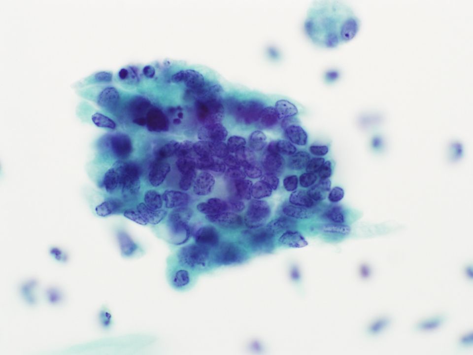
13
Cytologic findings Background Moderate cellularity Inflammatory background No diathesis Cell arrangement Mainly 3D clusters with smooth border Rare single cell Irregular arrangement No strips, rosettes No SIL Slightly increased nucleus High N/C ratio Irregular nuclear membrane Nuclear size variation Prominent single nucleoli Mild to moderate hyperchromasia Fine to granular chromatin Scant to moderate cytoplasm Vacuolated cytoplasm Intracytoplasmic inflammatory cells Atypical Glandular Lesion Endometrial vs Endocervical

14
Endocervical cells vs Endometrial cells Endocervical cellsEndometrial cells CellularityAbundantSparse CellsLarger Tall columnar Preserved Smaller Rounder Degenerated GroupsFlat 2-D sheets, honeycomb Crowded groups, rosettes Feathering Crowded, 3-D Balls Molded groups Nuclei Shape Size Multinucleation Chromatin Nucleoli Elongated Larger (2-5x intermediate) Common Coarse, darker Multiple, larger Round Smaller (<3x intermediate) Rare Finer, paler Single, smaller Cytoplasm Texture Stain PMNs Abundant cytoplasm Granular More acidophilic Uncommon Scant cytoplasm Vacuolar More basophilic More common
 Common Coarse, darker Multiple, larger Round Smaller (<3x intermediate) Rare Finer, paler Single, smaller Cytoplasm Texture Stain PMNs Abundant cytoplasm Granular More acidophilic Uncommon Scant cytoplasm Vacuolar More basophilic More common")
15
Endocervical cells vs Endometrial cells Endocervical cellsEndometrial cells CellularityAbundantSparse CellsLarger Tall columnar Preserved Smaller Rounder Degenerated GroupsFlat 2-D sheets, honeycomb Crowded groups, rosettes Feathering Crowded, 3-D Balls Molded groups Nuclei Shape Size Multinucleation Chromatin Nucleoli Elongated Larger (2-5x intermediate) Common Coarse, darker Multiple, larger Round Smaller (<3x intermediate) Rare Finer, paler Single, smaller Cytoplasm Texture Stain PMNs Abundant cytoplasm Granular More acidophilic Uncommon Scant cytoplasm Vacuolar More basophilic More common
 Common Coarse, darker Multiple, larger Round Smaller (<3x intermediate) Rare Finer, paler Single, smaller Cytoplasm Texture Stain PMNs Abundant cytoplasm Granular More acidophilic Uncommon Scant cytoplasm Vacuolar More basophilic More common")
16
Differential diagnosis Atypical endometrial cells Chronic endometritis Associated with IUD, PID etc. Endometrial polyp Endometrial hyperplasia Well-differentiated endometrial carcinoma

17
Differential diagnosis Atypical endometrial cellsFavor endometrial carcinoma Small 3D clusters (5 to 20 cells)3D cluster Small to moderate in cell sizeClusters of cells with smooth border Scant to moderate cytoplasmVariation in nuclear size Mildly to moderately enlarged nucleiLarge nuclei (>X2 intermediate) Slightly hyperchromatic nucleiChromatic clearing With of without nucleoliProminent nucleoli Cytoplasm may be vacuolatedCytoplasmic vacuolation Nuclear irregularity Intracytoplasmic neutrophils Tumor diathesis CAP TODAY, January 2013, Cytopathology and More Acta Cytol 2007;51:735–741 Acta Cytol 2002;46:458–464
3D cluster Small to moderate in cell sizeClusters of cells with smooth border Scant to moderate cytoplasmVariation in nuclear size Mildly to moderately enlarged nucleiLarge nuclei (>X2 intermediate) Slightly hyperchromatic nucleiChromatic clearing With of without nucleoliProminent nucleoli Cytoplasm may be vacuolatedCytoplasmic vacuolation Nuclear irregularity Intracytoplasmic neutrophils Tumor diathesis CAP TODAY, January 2013, Cytopathology and More Acta Cytol 2007;51:735–741 Acta Cytol 2002;46:458–464")
18
Differential diagnosis Atypical endometrial cellsFavor endometrial carcinoma Small 3D clusters (5 to 20 cells)3D cluster Small to moderate in cell sizeClusters of cells with smooth border Scant to moderate cytoplasmVariation in nuclear size Mildly to moderately enlarged nucleiLarge nuclei (>X2 intermediate) Slightly hyperchromatic nucleiChromatic clearing With of without nucleoliProminent nucleoli Cytoplasm may be vacuolatedCytoplasmic vacuolation Nuclear irregularity Intracytoplasmic neutrophils Tumor diathesis CAP TODAY, January 2013, Cytopathology and More Acta Cytol 2007;51:735–741 Acta Cytol 2002;46:458–464
3D cluster Small to moderate in cell sizeClusters of cells with smooth border Scant to moderate cytoplasmVariation in nuclear size Mildly to moderately enlarged nucleiLarge nuclei (>X2 intermediate) Slightly hyperchromatic nucleiChromatic clearing With of without nucleoliProminent nucleoli Cytoplasm may be vacuolatedCytoplasmic vacuolation Nuclear irregularity Intracytoplasmic neutrophils Tumor diathesis CAP TODAY, January 2013, Cytopathology and More Acta Cytol 2007;51:735–741 Acta Cytol 2002;46:458–464")
19
Diagnosis Uterine Cervix, Pap smear; Adenocarcinoma, endometrial favor well-differentiated endometrioid type

Similar presentations


 Reactive endocervical cells B) HSIL C) LSIL D) Squamous metaplasia E) ASCUS 1.>")


