Download presentation
Presentation is loading. Please wait.

1
Inflammation of FGT 2

2
Vaginitis Definition: inflammation of the vagina Types: Bacterial vaginitis Parasitic vaginitis Atrophic vaginitis Fungal vaginitis Viral vaginitis

3
Bacterial vaginitis Haemophilis vaginitis: – Most common cause of non-specific vaginitis – Diagnosis: presence of Small polymorphic G-ve aerobic cocco-bacilli Few inflammatory cells – Lactobacilli are usually present

4
E. coli vaginitis: – Suppurative vaginitis in young girls or post- menopausal women due to atrophic changes – Infected epithelial cells show common cellular changes with large number of neutrophils neutrophils

5
Actinomycosis : Causative organism s: G+ve branched filamentus bacteria. Causes: Increased pH Decreased body resistance Injury to epithelium Diagnosis : Amorphus clusters of small filaments with large number of neutrophils. Histocytes, lymphocytes, few giant cells and necrotic debris. Most common in women with prolonged intrauterine device

6
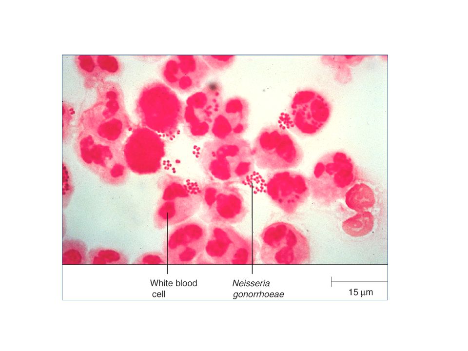
Gonococcal vaginitis: – It is venereal infection caused by G-ve diplococci (Neisseria gonorrhea). – Clinically: Suppurative vaginal discharge involve urethra, and vagina. – Diagnosis: By the presence of diplococci inside or outside neutrophils or histocytes.

8
TB of female genital tract: – Common in endometrium and fallopian tube. – Rare sites are: vagina and cervix. – Diagnosis: Typical Langhan`s giant cells, lymphocytes and cluster of epithelial cells. Presence of Acid fast mycobacterial bacilli.

9
Granuloma Inguinal: Venereal G-ve encapsulated coccobacilli ( Donovania granulumatis ) Diagnosis: Scraping margins of ulcero-granulomatous regions of moist skin, surface genitals, rectal, urethral, and inguinal areas intra-cytoplasmic donovania bodies (bean-shaped organisms) of large cystic macrophages with eccentric nuclei. Large number of neutrophils, cellular debris, and granular protein deposit in the background.

11
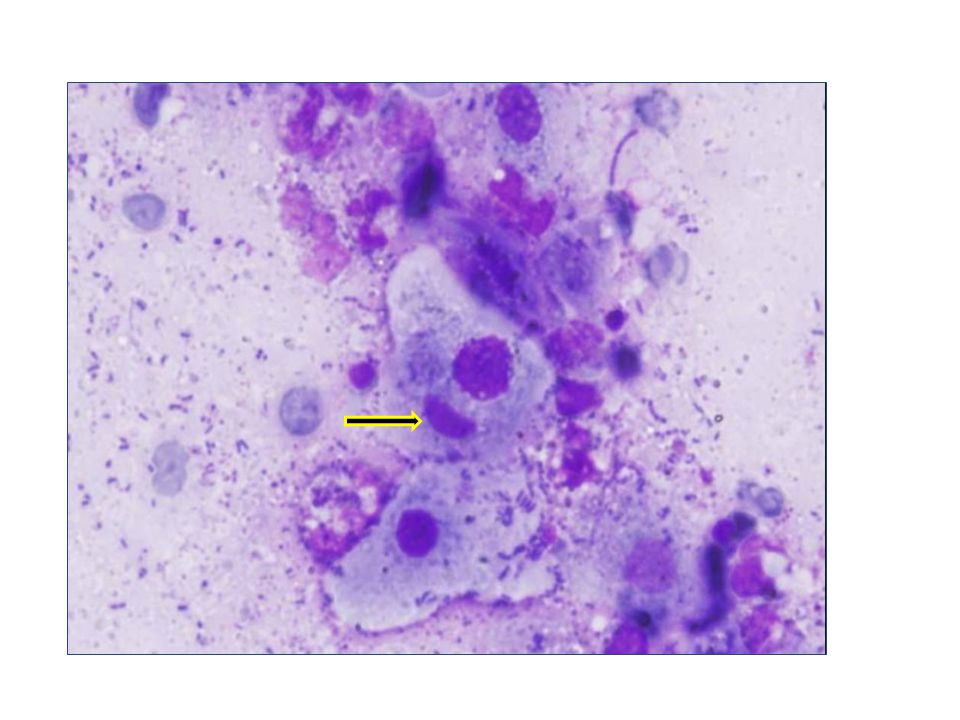
Parasitic (Protozoal) vaginitis Trichomonas vaginalis: – Causative organisms: Flagellated parasite, oval or pear shaped with active amoeboid movements. – Predisposing factors: pH of vagina, tissue injury – Clinically: is associated with greenish-yellow discharge, itching, soreness, and offensive odour.

12
Diagnosis by vaginal smear: Fresh specimen of vaginal smear without delay of examination shows motile parasite.

13
Diagnosis by vaginal smear: – Fixed and stained smear field show large cells, greenish grey in colour, with red granular cytoplasm, and dark shadow nuclei, surrounded by large number of neutrophils. – Cytoplasm of epithelial cells show polychromasia, red in colour, irregular membrane, with hyperkeratosis, pyknosis, and karyorrhexis. – Hypertrophy of intermediate and basal cells.

14
Trichomonas

15
Fungal vaginitis Moniliasis or Candida albicans: – It is the most common cause of acute and chronic vaginitis – Commonly seen with pregnancy – Causative organisms: yeast like cells – Clinically: milky white vaginal discharge – Diagnosis: red long bodies (hyphae), with pear shaped rounded bodies (budding yeast)
, with pear shaped rounded bodies (budding yeast)")
16
Candida spores- hyphae sporeshyphae

17
Atrophic (senile) vaginitis Definition: it is a condition result from combination of three factors: – Lack of estrogenic stimulation with thin vaginal wall. – Secondary bacterial infection due to loss protective epithelium and increase pH ------- expose less resistant parabasal cells to infection. – Vaginal pH become alkaline

18
Clinically: postmenopausal bleeding with yellow bloody discharge. Diagnosis: cytological smear contain – Increased number of parabasal cells – Parabasal cells vary in shape and size – Degenerative changes are seen in many parabasal cells – Inflammation of endocervical canal with some stripped nuclei may be involved

19
Etiology Menpopause: due to lack of estrogen. Dramatic decline in the circulating estrogen levels occurs at menopause. Pelvic irradiation or chemotherapy Oophorectomy Anti-estrogns: e.g. Tamoxifen, Danazol. Immediately after delivery or breast feeding Anorexic women & women who have recently lost a significant body weight Idiopathic.

20
Endometritis Definition: inflammation of the endometrium. Types: Acute endometritis Chronic endometritis – Chronic non-specific endometritis (Puerperal endometritis) – Chronic specific endometritis: TB; actinomycosis, …. Atrophic endometritis
 – Chronic specific endometritis: TB; actinomycosis, …. Atrophic endometritis.")
21
Causes: – Bacterial invasion of endometrial cavity – Retained gestational contents – Endometrial polyp – Fibromyoma – Secondary to introduction of foreign objects: – Criminal abortion – Contaminated intrauterine device

22
Puerperal endometritis – Causes: Following bacterial invasion in abortion or labour Clinically: – Vaginal bleeding – Offensive vaginal discharge – Fresh or clotted blood with endometrial cells Diagnostic endometrial smear: – Large number of acute and chronic inflammatory cells – Abundant fragmented cytoplasm and nuclei – Increase number of histocyte and multinucleated giant cells

23
Atrophic endometritis Causes: – Sever deficiency in estrogenic hormones in postmenopausal women associated with secondary bacterial infection.

24
Diagnostic endometrial aspiration: – Few endometrial cells – Fresh or clotted blood – Increased number of lymphocytes and plasma cells – Moderate amount of cellular debris and protein deposits – Metaplastic epithelium usually parabasal cells; with semi-keratinized cytoplasm.

25
Viral inflammation of FGT

26
General considerations: – Most of the viruses infecting oral mucosa have the ability to infect the vagina. – Routine vaginal smears are usually useful for detecting the various typical viral cellular changes.

27
There arte non-specific structural changes of infected cells commonly seen with viral infection; such as: 1. Hypertrophy of the cytoplasm, or of the nucleus, or of both. 2. Changes of the normal granular cytoplasm into hyaline pink cytoplasm. Viral cytoplasmic changes

28
3. Enlargement of the nucleolus followed by its distortion and disappearance or lysis. 4. Single or multiple intracytoplasmic or intranuclear inclusion bodies surrounded by prominent halo (clear space). 5. Late, balloning cytoplasm and nuclear degenerative vacolization.
. 5. Late, balloning cytoplasm and nuclear degenerative vacolization..")
29
Herpes simplex virus (HSV) Gross lesion: Vesicle Ulcer Occult Site of lesion: Cervix Vagina Valva
 Gross lesion: Vesicle Ulcer Occult Site of lesion: Cervix Vagina Valva")
30
Clinical findings It is transmitted during sexual intercourse. Early: Fever, enlarged regional lymph nodes, painful lesion, and thin watery discharge. In pregnant women, the infection could transmit to the baby during delivery. It could cause abortion It may play a role in cervical dysplasia or carcinoma

31
Diagnostic scraping of exfoliated infected cells in vaginal smear True hypertrophy of the cytoplasm, or of the nucleus, or of both. An irregular perinuclear halo (early changes). Homogenous dense pink hyaline cytoplasm, or more basophilic blue cytoplasm.
. Homogenous dense pink hyaline cytoplasm, or more basophilic blue cytoplasm..")
32
Nuclear and cytoplasmic enlargement

33
Multinucleated giant cells with characteristic molding (crowded nuclei without overlapping) with variation in size and shape. Change of nuclear chromatin distribution: loss of uniform granularity of the chromatin marginal deep basophilic chromatin clumps adherent to the inner surface of the nuclear membrane, with central amorphus esinophilic zone.

34
Multinucleated giant cell

35
– Intranuclear acidophilic inclusion red bodies with halo or ground glass appearance of nuclei. – Balloning cytoplasm with multiple vacolization, or nuclear degeneration indicating irreversible cell injury.

36
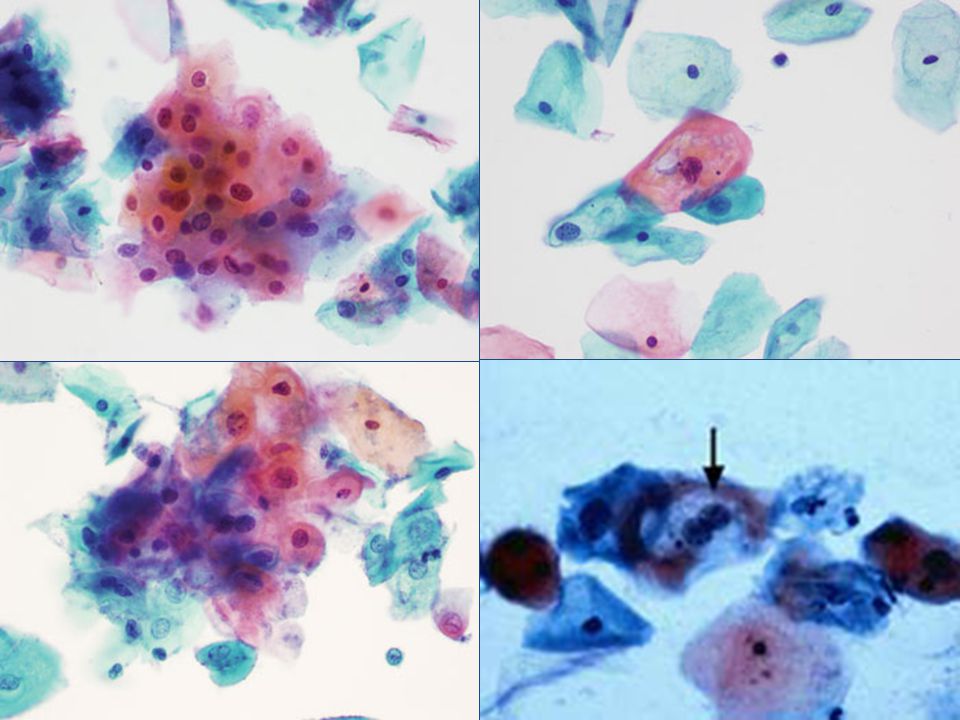
Human Papilloma Virus (HPV) HPV is one of the most common sexually transmitted viral infection of FGT. HPV has a role in the pathogenesis of cervical dysplasia and/or carcinoma. HPV involves skin and mucous membrane of vulva, vagina, and cervix.

37
Diagnostic cytological scraping smears for HPV Moderate increase number of inflammatory cells (acute and chronic). Altered squamous epithelial cells: Koilocytes Dyskeratocyte and dyskeratosis Atypical parabasal cells and dyskeratocytes

38
Koilocytic cells Koilos = cavity = clearing space Single, clusters, or sheet of parabasal and intermediate squamous cells showing nuclear and cytoplasmic changes; charactrestic for HPV infection. Koilocytic cells have characteristic well defined zone of perinuclear halo with darkly stained peripheral zone of cytoplasm adjacent to distinct irregular cytoplasmic membrane.

39
Nuclei of koilocytic cells are central, round or irregular enlarged. Some cells are binucleated or multinucleated with variable coarse granular chromatin without visible inclusion bodies. Paoilloma type virus particles may be demonstrated in perinuclear zone of cytoplasm.

41
Dyskeratocytes Dyskeratocytic cells are single or clustres of small oval or spindle shaped squamous cells with small round pyknotic nuclei and refractile pink cytoplasm without perinuclear halo. The presence of dyskeratotic cells in cytological smear is strongly suggestive of HPV.

43
Atypical parabasal and dyskeratotic cells Parabasal squmous cells and dyskeratotic cells showing moderate nuclear atypia. It is precancerous lesion for cervical carcinoma.

44
Thank you

Similar presentations


>")


>")

