Download presentation
Presentation is loading. Please wait.

1
بسم الله الرحمن الرحيم

2
Objectives 1- To know the definition and value of identification of living and dead persons. 1-To know how to identify living persons 2- To know how to identify dead bodies. 3- To know the characters useful in identification of dead persons 4-To know how to identify age ,sex ,stature ,race, time pass since death ,and cause of death from the skeletal remains.

3
Identification Definition:- The application of forensic science and technology to personalize someone through specific criteria that individualizes him than others throughout his life. The medicolegal expert may be asked to examine for identification a living , a dead person or even parts of dead bodies or collection of bones.

4
Medicolegal importance of identification
I-Civil cases:- 1- In unconscious patients admitted to hospital. 2-Amnesia( memory loss”). 3- Unknown persons and those reported to be lost. 4- Persons having no authentic records when facing any legal situation. 5- Inheritance. 6- Military services.
. 3- Unknown persons and those reported to be lost. 4- Persons having no authentic records when facing any legal situation. 5- Inheritance. 6- Military services.")
5
7- Emigration at exit ports
7- Emigration at exit ports. 8- Controversy about a prominent , important persons . II- In criminal cases:- 1- In criminals who impersonate others. 2- Offenders below age of 18 having no official records of age to be tried in the related court eg:- juvenile court.

6
Identification of living
Primarily a matter for police investigation. Methods of identification:- 1-Personal impression (visual identification) Basis of identification parades but is notoriously unreliable. Witness in court may be asked to point out the accused. Identikit is a composite picture of a person from a witness account.
 Basis of identification parades but is notoriously unreliable. Witness in court may be asked to point out the accused. Identikit is a composite picture of a person from a witness account.")
7
Personal impression depends on features such as hair, moustache and beard, all of which can be changed easily. Plastic surgery can easily alter other features.

8
2-Photography More useful in identifying the living than the dead
2-Photography More useful in identifying the living than the dead. 3-Handwriting Possible for experts to identify a person . Methods used include photographic enlargement, analysis of ink, analysis of paper.

9
4-Fingerprints (Dactylography) How fingerprints are produced: friction skin (i.e. palms and soles) have a ridge pattern. Sweat glands open through minute openings on the summits of the ridges. The sweat contains fat. When the skin is applied to a smooth surface a greasy impression is left behind.
 have a ridge pattern. Sweat glands open through minute openings on the summits of the ridges. The sweat contains fat. When the skin is applied to a smooth surface a greasy impression is left behind.")
10
Principles of fingerprint identification: Fingerprint patterns are unique (1 in 64 billion chance of 2 prints being identical). FBI has over 100 million records, no two of which are alike. Fingerprint pattern of an individual remains unchanged throughout life. Reversible atrophy occurs in certain diseases (eg; dermatitis).
.")
11
Permanent impairment occurs in leprosy and after exposure to radiation
Permanent impairment occurs in leprosy and after exposure to radiation. Attempts to mutilate fingerprints are sometimes made. If only the epidermis is destroyed there is no alteration in ridge pattern. If dermis is destroyed additional points of identification are created. 6-DNA:- This is forensically useful in the living (strong evidence of involvement in assault, rape, disputed paternity)
")
13
Identification of the dead
Primary Physical Characteristics :- (characteristics which are very difficult for a person to change during life. Some of these characteristics will appear to alter post mortem): Sex - usually obvious. Age - external appearances, internal degenerative disease, bones, joints.
: Sex - usually obvious. Age - external appearances, internal degenerative disease, bones, joints.")
14
Height or stature: - N.B. height of corpse differs from that in life by up to 2-3 cm, due to joint and muscle relaxation. Race DNA (unique to every individual) except monozygous (identical) twins. DNA comparisons allow for definitive identification of an individual
 except monozygous (identical) twins. DNA comparisons allow for definitive identification of an individual.")
15
This is forensically useful in dead (DNA survives in bone for many years, comparison of DNA with family members) as well as in living. Samples may be taken from blood of dead bodies (on EDTA),or from spleen, or organs or from bone marrow( without fixation).
,or from spleen, or organs or from bone marrow( without fixation).")
16
Secondary physical characters:-
(characteristics which can change during life, Some of these characteristics will appear to alter post mortem): Skin :-Color in undamaged unputrified bodies only (alters post mortem) Eyes - :-Iris color can be useful in fresh corpse only, but color can alter PM.
: Skin :-Color in undamaged unputrified bodies only (alters post mortem) Eyes - :-Iris color can be useful in fresh corpse only, but color can alter PM.")
17
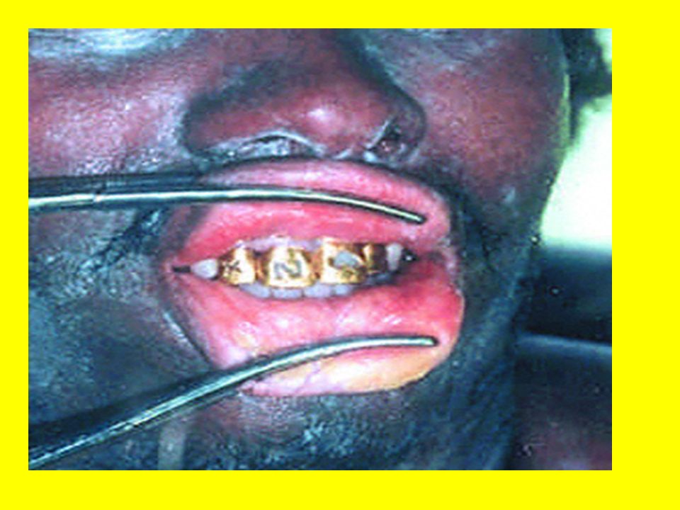
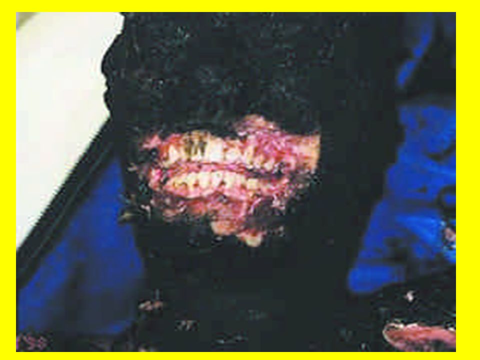
Teeth :- very resistant and bear much useful information.
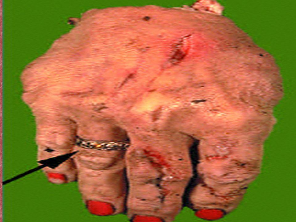
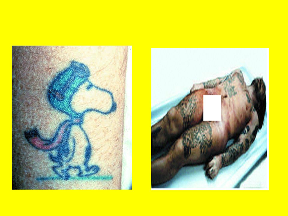
Hair color and structure - color, style, length, beard/moustache give an idea about sex , race, type of crime and identification of the victim or assailant Scars - surgical procedures and prostheses Tattooing - seen even if the body is putrefied. Fingerprints :- from the palm or sole

18
External peculiarities - circumcision, moles, warts.
Deformities Clothing and other objects (as:-Jewelers ) Occupational stigmata :-( eg:-shoe maker kyphosis ,and sternum, blue scars of coal miners).
 Occupational stigmata :-( eg:-shoe maker kyphosis ,and sternum, blue scars of coal miners).")
21
Identification of decomposed or skeletal reminants
1- Are they really bones? (wood, stones) 2-Are the remains human or animal? 3-Are they human? 4-How many bodies?
 2-Are the remains human or animal 3-Are they human 4-How many bodies")
22
5-Time pass since death. - recent or ancient (e. g
5-Time pass since death? - recent or ancient (e.g. construction or digging at an old burial site) 6-Cause of death? 7-Sex? 8-Age? 9-Race? 10-Stature?
 6-Cause of death 7-Sex 8-Age 9-Race 10-Stature")
23
Identification of sex I- In intact bodies:- Straight forward in intact bodies except in adrenogenital syndrome and hermaphrodites. In mutilated/dismembered or charred bodies the internal sex organs, especially the uterus, cervix and prostate are resilient.

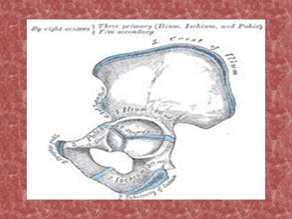
24
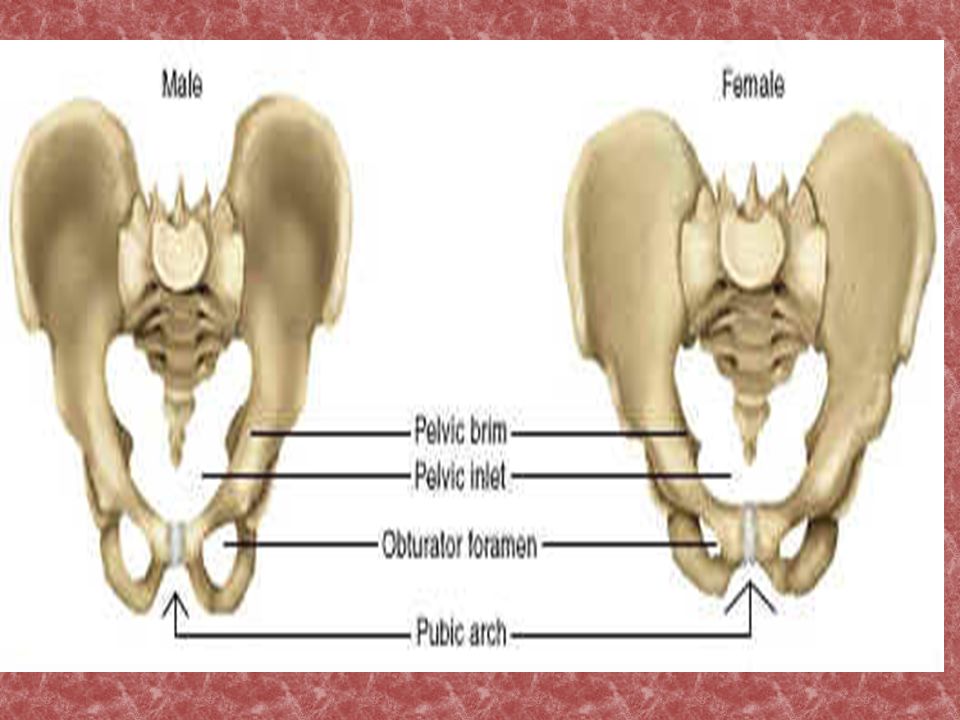
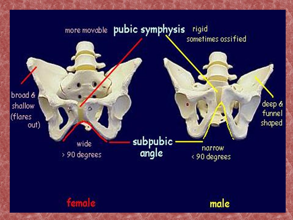
II- In skeleton remnant: Based on appearance of pelvis, skull, sternum, long bones. Pelvis is the most important bone for sex determination. Subjective and objective differences are seen between male and female pelvis.

25
The differences between male and female pelvis
MALE FEMALE Pelvis as a whole Thick, heavy, markedly Smoother, lighter, more muscular spacious Brim Heart shaped Circular or elliptical Body of pubis Triangular shape Quadrangular Sub pubic arch Inverted V shaped Inverted U shaped Greater sciatic notch Deep and narrow Broad and shallow Sacro iliac joint Large Small Sacrum Long and narrow Short and wide

28
III-Sex determination from the skull :-
Supra orbital ridges Mastoid process condylar process

29
Race determination from bones
The skull is the only reliable bone. Cheekbones (Zygomatic arches): determine facial width. More prominent in Mongoloids. Width between eyes greater in mongoloids. Nasal openings: Wider and flatter in Negroids. Narrow in Caucasians.
: determine facial width. More prominent in Mongoloids. Width between eyes greater in mongoloids. Nasal openings: Wider and flatter in Negroids. Narrow in Caucasians.")
30
Determination of stature from bones
Long bone length (femur, tibia, humerus) is proportional to height. Tables are used. Fairly reliable up to the age of epiphyseal fusion. There are sex, race, nutrition and personal variations to consider.
 is proportional to height. Tables are used. Fairly reliable up to the age of epiphyseal fusion. There are sex, race, nutrition and personal variations to consider.")
31
Age determination from skeleton
Sex has to be taken into account as bone development and epiphyseal fusion is different between the sexes. Epiphyseal fusion :- The pattern of fusion of bone ends (epiphysis) to bone shaft (metaphysis) in each bone indicates age. Charts & tables are used. Cranial suture fusion is less reliable. Arthritic changes, osteophytes and osteoporosis give further clues.
 to bone shaft (metaphysis) in each bone indicates age. Charts & tables are used. Cranial suture fusion is less reliable. Arthritic changes, osteophytes and osteoporosis give further clues.")
32
Identification of age The age can be determined from :- 1- Skull including mandible and teeth. 2- Appearance of ossific centers. 3- Union of epiphysis.

33
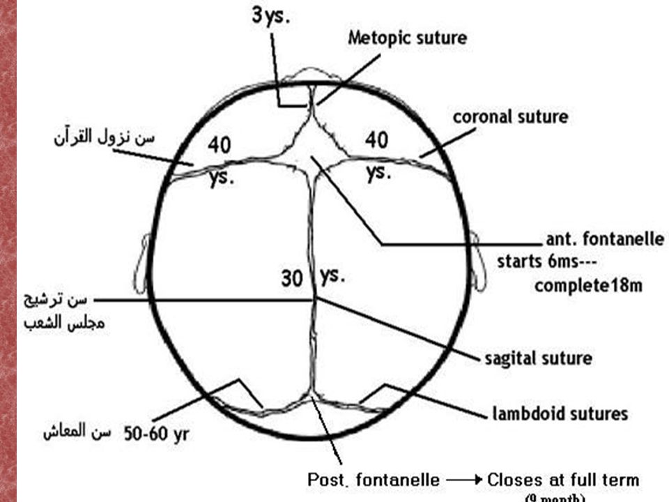
1- Skull A- Dimensions :- In the full term infants , the circumference is 13 inches,length is 5 inches and width is 4 inches. B- Fontanells:- The posterior fontanel is closed at full term while the anterior fontanel can admit the tips of the middle 3 fingers and closed at 18 months after birth.

34
C- Sutures:- - Frontal suturre closes at 3 years ( 30 years in negroid). - Basioccipital –basisphenoidal sutures closes at 23 years. Saggital sutures closes at 30 years. Coronal sutures closes at 40 years. Lamboid sutures at 50 years.

36
D-Age detection from teeth:- A- temporary teeth:-
Tooth Eruption Time Lower Central Incisors ( CI ) Upper central incisors(UI) 6-8th month 9-12 month Lateral Incisors ( LI ) 18-24th month(10) Canine ( C ) 18-24th month(18) Temporal Molar 1 (TM1) 12-24th month Temporal Molar 2 (TM2) 24-36th month
 Upper central incisors(UI) 6-8th month month. Lateral Incisors ( LI ) 18-24th month(10) Canine ( C ) 18-24th month(18) Temporal Molar 1 (TM1) 12-24th month. Temporal Molar 2 (TM2) 24-36th month.")
37
b- Permanent teeth :- Tooth E.T Central Incisors ( CI ) 6-7TH year
Lateral Incisors ( LI ) 7-8th year Canine ( C ) 9-11th year Premolar 1 (PM1) 9-12th year Premolar 2 (PM2) Molar 1 (M1) 5-7th year Molar 2 (M2) 10-13th year Molar3 (M3) 18-25th year
 7-8th year. Canine ( C ) 9-11th year. Premolar 1 (PM1) 9-12th year. Premolar 2 (PM2) Molar 1 (M1) 5-7th year. Molar 2 (M2) 10-13th year. Molar3 (M3) 18-25th year.")
38
TEMPORARY TEETH PERMANENT TEETH
- small, narrow, light & delicate - china white crown - constricted neck - serrated edges - - big, broad, heavy & strong - ivory white crown - less constricted - not serrated

39
Bite marks Caused by front teeth with gap at either side
May be contusion, abrasion & laceration Swab of bite marks & victims saliva should be taken Matched by photographic method & casts

41
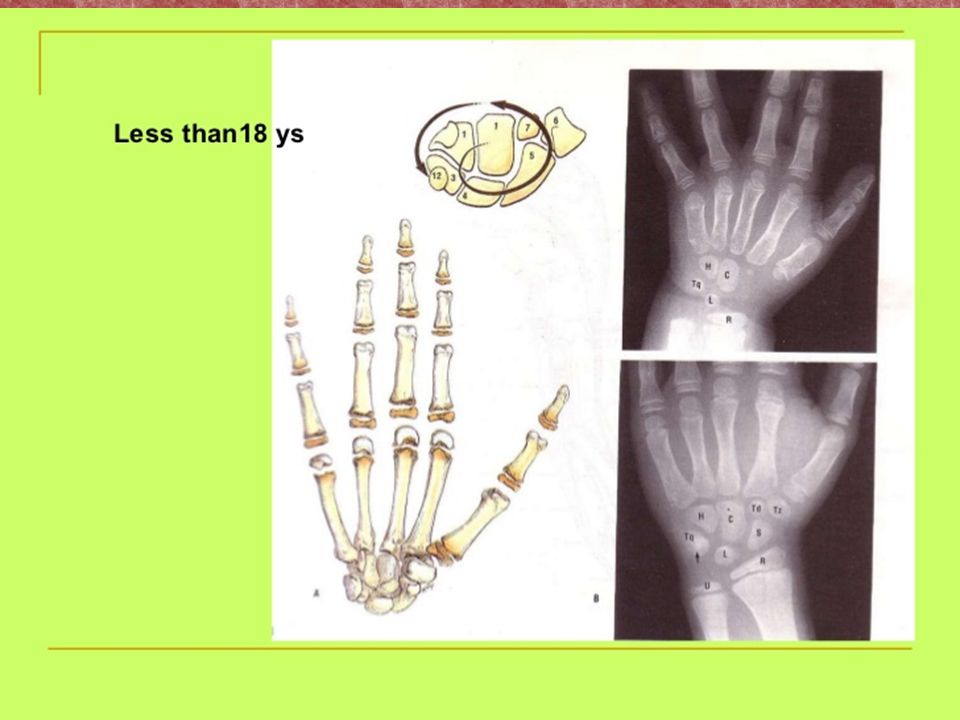
E- Mandible:- The angle of the mandible is obtuse in infants and old ages and right angle in adults. The alveolar margin is resorbed in senile mandible.

42
Mandible can now be easily examined, photographed, and X-rayed.

45
Medicolegal importance
Identification of individual in accidental or homicidal cases Differential identification in mass disaster Estimation of age Determination of sex & blood group Identification of criminal through bite marks Detection of poison

46
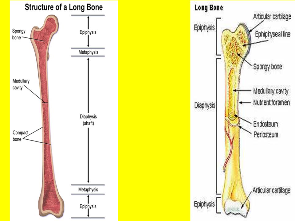
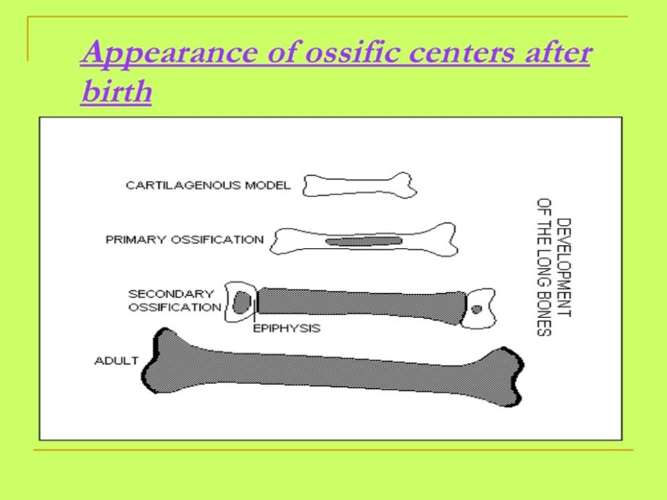
II- Ossific centers These are centers of bone growth that appears as a dark area inside a translucent cartilage. Ossification centers :- Useful only in fetus and babies. May be determined radiologically or by cutting into ossification centers. Most important center in medico-legal work is the distal center of the femur. This is present at birth and indicates a full term baby.

47
Ossific centers that appear after birth :-
1 year at the head of the humerus , femur and tibia. 2 years at the lower end radius. 3 years patella. 4 years upper fibula ,greater trochanter of femur 5 years at lower fibula. 6 years at head of radius , lower ulna.

55
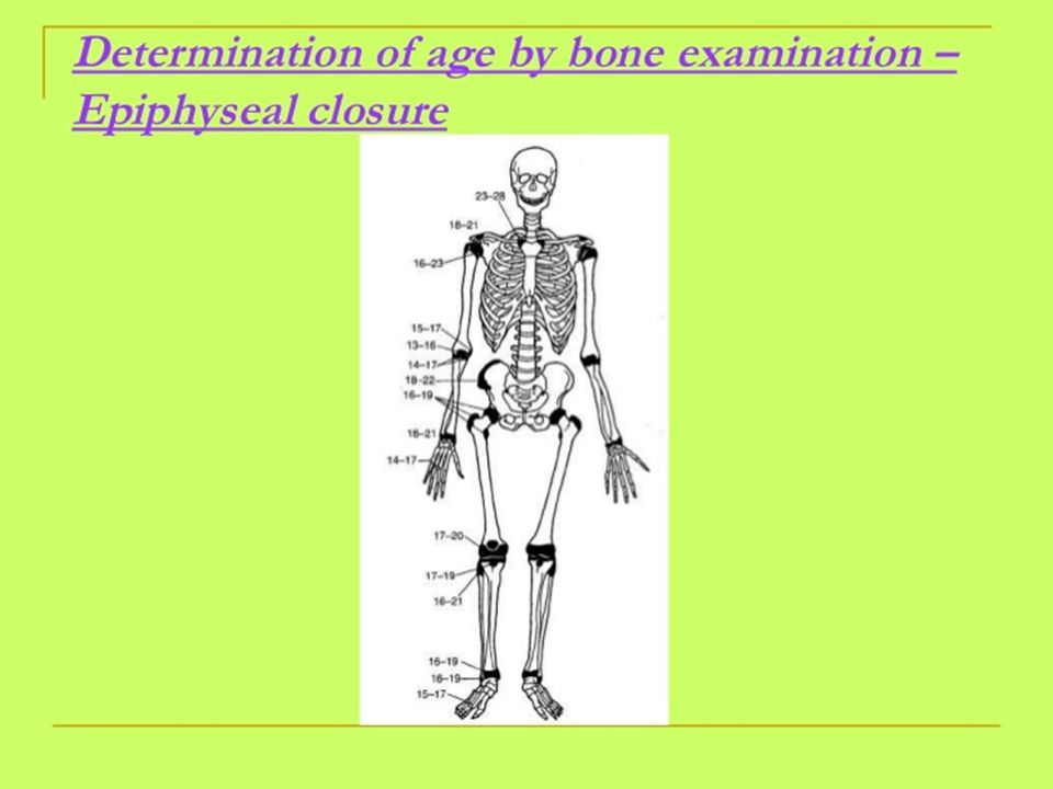
III- Union of epiphysis
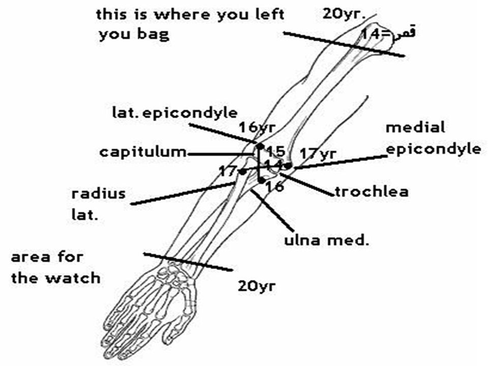
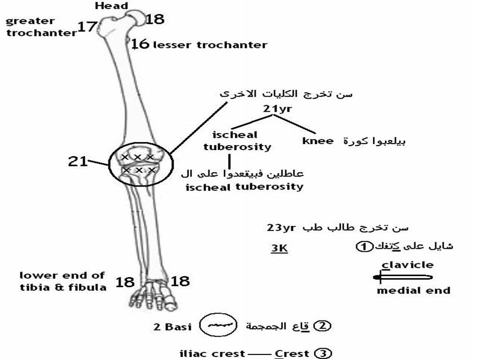
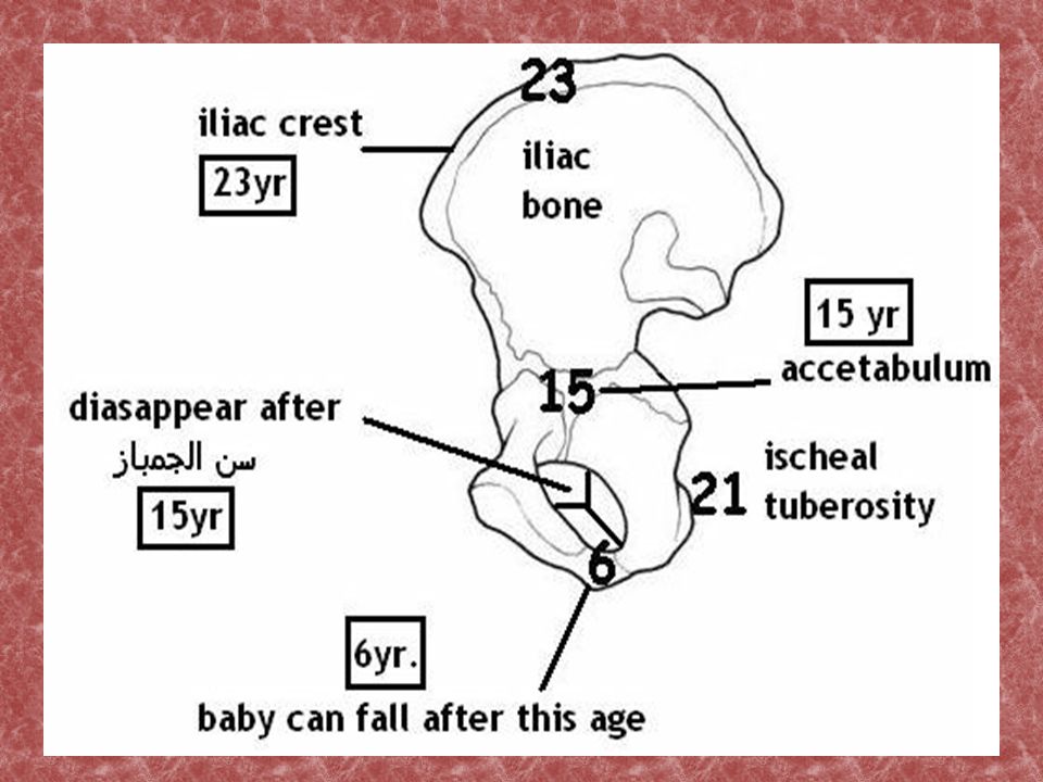
AGE The pubic ramus of ischial bone is united with the ischial ramus of pubic bone. 6 years The trochlea is united with the capitulum of the humerus. 14 years Both trochlea and capitulum united with the shaft of the humerus. Ileum ,ischium and pubis are united to form the acetabulum. 15 years Lateral epicondyle united with the shaft of the humerus. Upper end of the ulna united with shaft. Lesser trochanter of the femur with the shaft. 16 year Medial epicondyle united with the shaft of the humerus. Upper end of radius with the shaft. Greater trochanter of the femur with the shaft. 17 years

56
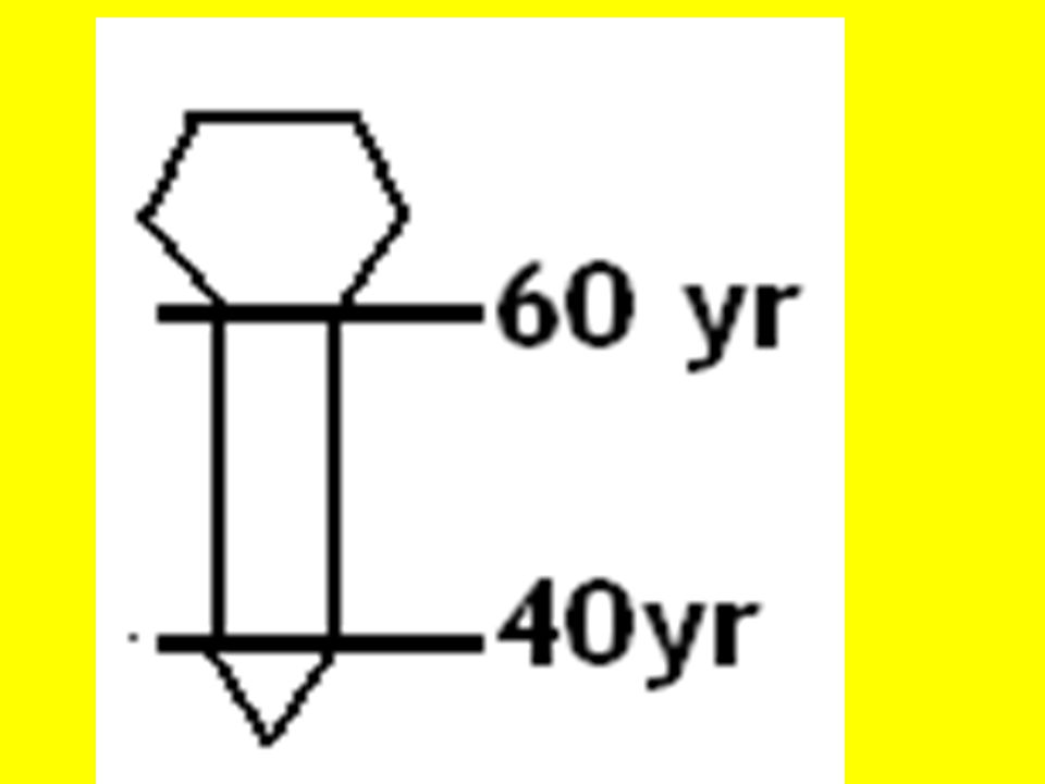
EPIPHYSIS AGE Distal end of metacarpals and the proximal ends of proximal phalanges are united with their shafts. Lower end of tibia and fibula with their shafts. 18 years Lower end of radius and ulna with their shafts. 20-21 years Lower end of femur with shaft. Upper end of radius and ulna with their shafts. Ischial tuberosity with ischium. 21years Iliac crest with ileum. Sternal end of clavicle with shaft. Basiocciput with basishenoid at base of the skull. 23year Closure of sagittal suture (begins from its inner aspect) 30years
 30years.")
57
EPIPHYSIS AGE Xiphoid process with body of sternum. Closure of coronal suture. 40years Greater cornnu of hyaloid bone with its body. Closure of lambdoid suture. 50years Manubrium sterni with body of sternum . 60years Closure of all skull sutures except temporoparietal. 70 years

62
Time pass since death Are they ancient or modern bones?

63
Thank you

Similar presentations


 Traumas (injuries)>")


. Forensic Anthropology: – study of human skeletal remains in a legal setting, most often.>")
