Download presentation
Presentation is loading. Please wait.

1
Copyright Notice! This PowerPoint slide set is copyrighted by Ross Koning and is thereby preserved for all to use from plantphys.info for as long as that website is available. Images lacking photo credits are mine and, as long as you are engaged in non-profit educational missions, you have my permission to use my images and slides in your teaching. However, please notice that some of the images in these slides have an associated URL photo credit to provide you with the location of their original source within internet cyberspace. Those images may have separate copyright protection. If you are seeking permission for use of those images, you need to consult the original sources for such permission; they are NOT mine to give you permission.

2
Biology: What is Life? life study of Properties of Life
Cellular Structure: the unit of life, one or many Metabolism: photosynthesis, respiration, fermentation, digestion, gas exchange, secretion, excretion, circulation--processing materials and energy Growth: cell enlargement, cell number Movement: intracellular, movement, locomotion Reproduction: avoid extinction at death Behavior: short term response to stimuli Evolution: long term adaptation

3
Cell Structure Prokaryotic before nucleus Eukaryotic true nucleus

4
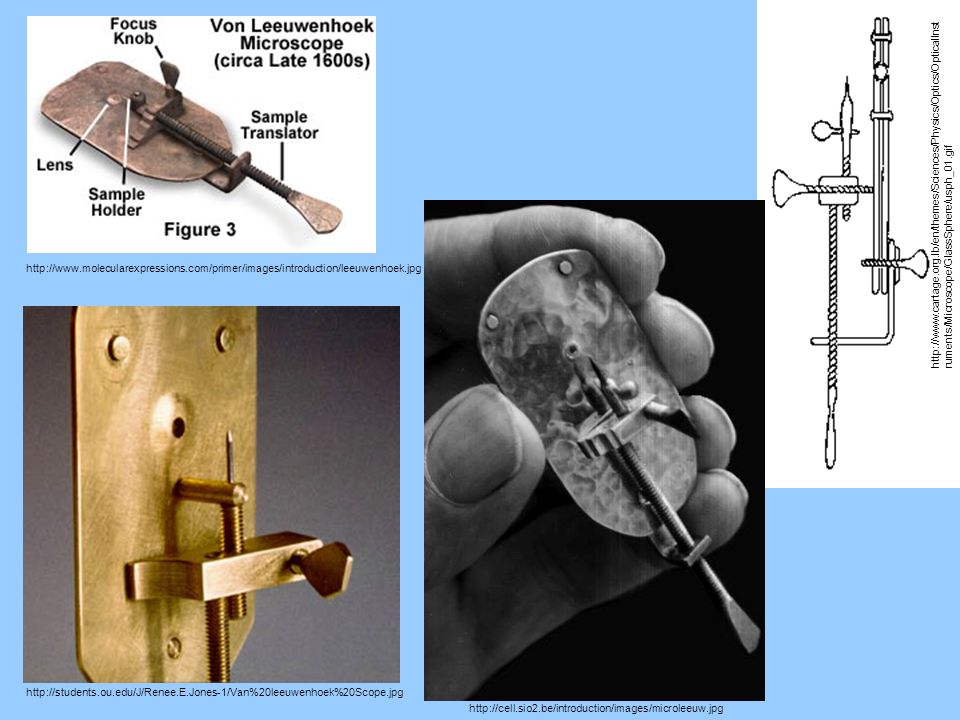
Antonie van Leeuwenhoek 1632-1723
Developed microscopes for observing living organisms 1674 discovered live protist cells 1677 discovered spermatozoa 1682 discovered striated muscle fibers

5
http://www. molecularexpressions
6
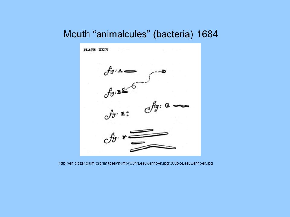
Mouth “animalcules” (bacteria) 1684
7
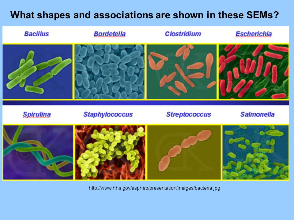
Prokaryotic Cell Shapes
Coccus - cocci Bacillus - bacillus Spirillum - spirilli Vibrio - vibrios

8
Cells are attached to each other by intercellular glue or a secreted sheath made of mucilaginous polysaccharides Cell Associations Coccus Diplococcus The sheath can provide antibiotic resistance too! Streptococcus - filamentous Staphylococcus - colonial ? Streptobacillus

9
What shapes and associations are shown in these SEMs?
10
What are the shapes of these disease bacteria?
Vibrio cholerae Helicobacter pylori Are they motile? If so, by what mechanism?

11
Comparing Cell Sizes Mycoplasma 0.3-0.8 µm E. coli 1x2 µm
Cyanobacteria 10 µm diam Plant Cell 30x75 µm Obviously eukaryotic Nucleus present Mitochondrion Bacterium Chloroplast Cyanobacterium Endosymbiosis: Eukaryotes are Chimeras!

12
Cell Theory 1839 Theodor Schwann Prussian Zoologist 1810-1882
Theodor Schwann Prussian Zoologist Matthias Schleiden German Botanist 1. All living organisms consist of one or more cells. 2. Some organisms are unicellular, so cells are the fundamental unit of life. 3. New cells come from pre-existing cells by cell division. We can now add: 4. Cells must show all the properties of life. 5. All cells are basically similar in chemical and structural composition.

13
Cell Structure: Boundary
Mycoplasma cell membrane bilayer only… glycolipid, sulfolipid transport proteins cytosol regulates input/output ETS for PSN, Resp Gram Positive Gram Negative cell wall-murein peptidoglycan muramic acid - peptide prevents dye release prevents bursting turgor pressure penicillin sensitive additional membrane bilayer glyco- sulfo-lipids releases dye

14
Cell Structure: Cytosol
Mycoplasma Water and enzymes for fermentation, glycolysis, Kreb’s cycle, Calvin cycle, naked circular DNA for transcription, 70S ribosomes for translation cell membrane bilayer glycolipid, sulfolipid transport proteins cytosol regulates input/output ETS for PSN, Resp Gram Positive Gram Negative cell wall-murein peptidoglycan muramic acid - peptide prevents dye release prevents bursting turgor pressure penicillin sensitive additional membrane bilayer glyco- sulfo-lipids releases dye

15
Is this example Gram+ or Gram−?
This cartoon is not labeled, so it merely acts as a key, to orient the viewer to the enlarged portion of the TEM image. Cytoplasm The cytosol area (labeled cytoplasm)shows the nucleoid (DNA) area at the top. The cell membrane shows that it is a bilayer. The cell wall shows that it is multilayered. Plasma membrane Cell wall Figure 7-2 Page 121 Is this example Gram+ or Gram−?
shows the nucleoid (DNA) area at the top. The cell membrane shows that it is a bilayer. The cell wall shows that it is multilayered. Plasma. membrane. Cell wall. Figure 7-2 Page 121. Is this example Gram+ or Gram−")
16
Light microscopy would be even less detailed!
This is a cartoon image created by an artist to emphasize certain structures. Cytosol This is the transmission electron microscopy image that inspired the cartoon. Light microscopy would be even less detailed! Figure 7-1 Page 107

17
The bacterium is prokaryotic (before-nucleus).
These are an SEM (above) and TEM (below). The DNA double helix is highly twisted to form the coils you are seeing here. The area inside the cell including the naked, circular DNA molecule (lacking DNA- binding proteins) is called the nucleoid; it is not a nucleus! The bacterium is prokaryotic (before-nucleus). The functions of the nucleoid are transcription (making mRNA), and replication (making a copy of DNA prior to cell division). Figure 7-2 Page 108
 and TEM (below). The DNA double helix is highly twisted to form the coils you are seeing here. The area inside the cell including the naked, circular DNA molecule (lacking DNA- binding proteins) is called the nucleoid; it is not a nucleus! The bacterium is prokaryotic (before-nucleus). The functions of the nucleoid are transcription (making mRNA), and replication (making a copy of DNA prior to cell division). Figure 7-2 Page 108.")
18
Figure 7-4 Page 121 (3rd edition: not in current edition!)
Ribosomes are 70S in “size” in prokaryotes, mitochondria, and plastids. Those found in the eukaryotic cytosol are 80S in “size.” The function of the ribosome in both kinds of cells is translation; the synthesis of protein from the information in mRNA. Ribosome Large subunit of ribosome Small subunit of ribosome Figure 7-4 Page 121 (3rd edition: not in current edition!)
")
19
Cell Structure: Nucleoid
Nucleoid - genome one circular DNA molecule no histone protein association attached to cell membrane mRNA transcription by RNA polymerase 70S Ribosome rRNA + protein + ribozymes translation of mRNA into protein

20
Prokaryotic Growth Cells are generally very small
Cells may double in volume, but only before binary fission Growth is mostly in terms of cell number or colony size, etc. The doubling time in cell numbers may be 20 minutes in ideal conditions Bacteria could quickly take over the earth if conditions could remain ideal They are very competitive, but often shed by-products that inhibit their own survival, so ideal conditions are usually not sustainable. They are ultimate survivors billion years!

21
Cell Structure: Nucleoid
Nucleoid - genome one circular DNA molecule no histone protein association attached to cell membrane DNA replication by DNA polymerase separation of chromosomes cytokinesis by furrowing Process called binary fission NOT mitosis! Genome and copy are identical Genome is haploid There is no synapsis There is no recombination

22
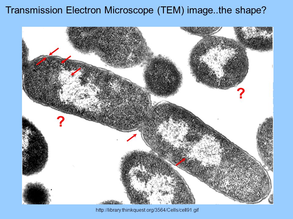
? ? Transmission Electron Microscope (TEM) image..the shape?
23
Cyanobacterial Vegetative Cell:
Photosynthesis Cyanobacterial Vegetative Cell: Respiration cell wall mesosome ETS reactions cell membrane cyanophycean starch cyanophycin gas vacuole lipid droplet polyphosphate granule thylakoids light (ETS) reactions 70S ribosome nucleoid cytosol polyhedral body RuBisCO Calvin cycle sugar synthesis glycolysis Kreb’s cycle light chlorophyll H2O O2 CO2 + + CH2O O2 + CH2O CO2 + H2O + energy
 reactions. 70S ribosome. nucleoid. cytosol. polyhedral body. RuBisCO. Calvin cycle. sugar synthesis. glycolysis. Kreb’s cycle. light. chlorophyll. H2O. O2. CO2 + + CH2O. O2 + CH2O. CO2. + H2O + energy.")
24
Sulfolobus acidocaldarius
TEM or SEM? Of Archaeon Extremophile Sulfur metabolism pH 1 to 6 75°C Optimum Strict aerobe Partial monolayer (C40) membranes Multiple DNA Circles Introns in DNA DNA binding proteins rRNA similarity RNA synthase similarity Operon style regulation 70S ribosomes Shape?
 membranes. Multiple DNA Circles. Introns in DNA. DNA binding proteins. rRNA similarity. RNA synthase similarity. Operon style regulation. 70S ribosomes. Shape")
25
Comparing Cell Sizes Mycoplasma 0.3-0.8 µm E. coli 1x2 µm
Cyanobacteria 10 µm diam Plant Cell 30x75 µm Obviously eukaryotic Nucleus present Mitochondrion Bacterium Chloroplast Cyanobacterium Endosymbiosis: Eukaryotes are Chimeras!

Similar presentations


 to carry out all of life’s processes.>")


