Download presentation
Presentation is loading. Please wait.

1
Approach and Hemodynamic Evaluation of Shocks
Mazen Kherallah, MD, FCCP King Faisal Specialist Hospital & Research Center

2
Shock Definition

3
Question #0 Which of the following is necessary in the definition of shock? (a) A drop in the systolic blood pressure of less than 90 mm Hg (b) A drop in the mean arterial pressure of less than 60 mm Hg (c) A drop in the SBP of 40 mm Hg or 20% from baseline (d) An elevated lactic acid of ≥ 4 mmoL/L (e) Any of the above
 A drop in the mean arterial pressure of less than 60 mm Hg. (c) A drop in the SBP of 40 mm Hg or 20% from baseline. (d) An elevated lactic acid of ≥ 4 mmoL/L. (e) Any of the above.")
4
Question #1 Which of the following is necessary in the definition of shock? (a) Hypotension (b) Tissue hypoxia (c) Use of pressors (d) Multiple organ dysfunction
 Use of pressors. (d) Multiple organ dysfunction.")
5
Shock Profound and widespread reduction in the effective delivery of oxygen leading to first to reversible, and then if prolonged, to irreversible cellular hypoxia and organ dysfunction” Kumar and Parrillo Leads to Multiple Organ Dysfunction Syndrome (MODS) Not defined as hypotension – inadequate tissue oxygenation
 Not defined as hypotension – inadequate tissue oxygenation.")
6
Pathophysiology

7
Oxygen extraction ratio
Pathophysiology DO2 Oxygen delivery VO2 Oxidative metabolism depends on constant supply of oxygen from blood as oxygen not stored in tissues. Extraction increases until maximal – then DO2 at critical level Oxygen uptake Oxygen extraction ratio

8
Physiologic Oxygen Supply Dependency
Oxygen Consumption Lactic Acidosis Critical Delivery Threshold Oxygen Delivery Mizock BA. Crit Care Med. 1992;20:80-93. 8

9
sepsis sepsis sepsis Sepsis

10
Pathologic Oxygen Supply Dependency
Physiologic Oxygen Consumption Oxygen Delivery Mizock BA. Crit Care Med. 1992;20:80-93. 10

11
Pathophysiology Most forms of shock Septic shock
Low cardiac output state Supply-dependency of systemic VO2 Septic shock Systemic DO2 and VO2 are both supranormal, but an oxygen deficit remains Peripheral oxygen extraction may be deranged and VO2 is pathologically supply-dependent Possible microvascular maldistribution of perfusion

12
Question #2 Which is not an important determinant of oxygen delivery?
(a) Hemoglobin level (b) Cardiac output (c) pO2 (d) SaO2
 Hemoglobin level. (b) Cardiac output. (c) pO2. (d) SaO2.")
13
CO x [(1.3 x Hgb x SaO2) + (0.003 x PaO2)]
Oxygen Delivery (DO2) Cardiac Output x Oxygen Content CO x [(1.3 x Hgb x SaO2) + (0.003 x PaO2)] Hemoglobin concentration SaO2 Cardiac output PaO2 (minimal)
![CO x [(1.3 x Hgb x SaO2) + (0.003 x PaO2)]](http://slideplayer.com/slide/3900131/13/images/13/CO+x+%5B%281.3+x+Hgb+x+SaO2%29+%2B+%280.003+x+PaO2%29%5D.jpg "Oxygen Delivery (DO2) Cardiac Output x Oxygen Content. CO x [(1.3 x Hgb x SaO2) + (0.003 x PaO2)] Hemoglobin concentration. SaO2. Cardiac output. PaO2 (minimal)")
14
Cardiac Output Afterload Preload Contractility
Stroke volume is determined by preload, afterload and contractility. Preload can be estimated from the JVP, CVP or pulmonary artery occlusion pressure but afterload and contractility are difficult to assess clinically. Contractility

15
Preload Determined by end-diastolic volume
Pressure and volume related by compliance of the ventricle We use EDP for EDV. Compliance decreased with myocardial ischemia and hypertrophy

16
Left ventricular end-diastolic pressure versus left ventricular end-diastolic volume

17
Cardiac Output Sterling relationship Cardiac output Volume loading
Now we can address the issue of how much fluid we should give. In most patients an increase in fluid loading will result in an increase in cardiac output. However many doctors are reluctant to give fluid, particularly to a patient with a raised JVP, because they are afraid of causing pulmonary oedema Volume loading 17

18
Clinical Adaptation of the Sterling Myocardial Function Curves

19
Global Hemodynamic Relationships
Stroke volume is determined by preload, afterload and contractility. Preload can be estimated from the JVP, CVP or pulmonary artery occlusion pressure but afterload and contractility are difficult to assess clinically. Afterload Preload Contractility 19

20
Question #3 Which can cause low preload? (a) High PEEP
(b) Tension pneumothorax (c) Third spacing (d) Positive pressure ventilation (e) All of the above
 Tension pneumothorax. (c) Third spacing. (d) Positive pressure ventilation. (e) All of the above.")
21
Pathophysiology Inadequate/ineffective DO2 leads to anaerobic metabolism Large/prolonged oxygen deficit causes decrease of high-energy phosphates stores Membrane depolarization, intracellular edema, loss of membrane integrity and ultimately cell death Anaerobic metabolism – lactate production, metabolic acidosis High energy phosphates – ATP, creatine phosphate – causes derangements in key biochemical processes

22
Effects of Shock at Cellular Level

24
Common Histopathology Associated with Tissue Hypoperfusion and Shock?

25
Mitochondrial Dysfunction in Cell Injury
Increased cytosolic Ca2+, oxidative stress, lipid peroxidation Robbins & Cotran Pathologic Basis of Disease: 2005

26
Histopathology of Tissue Hypoperfusion Associated with Shock
Coagulative necrosis Contraction bands Edema Neutrophil infiltrate Myocardium Diffuse alveolar damage Exudate, atelectasis Edema Hyaline membrane Lung Robbins & Cotran Pathologic Basis of Disease: 2005

27
Histopathology of Tissue Hypoperfusion Associated with Shock
Mucosal infarction Hemorrhagic mucosa Epithelium absent Small Intestine Liver Centrilobular hemorrhagic necrosis Nutmeg appearance Robbins & Cotran Pathologic Basis of Disease: 2005

28
Histopathology of Tissue Hypoperfusion Associated with Shock
Bland infarct Punctate hemorrhages Brain Brain Eosinophilia and shrinkage of neurons Neutrophil infiltration Robbins & Cotran Pathologic Basis of Disease: 2005

29
Histopathology of Tissue Hypoperfusion Associated with Shock
Kidney Tubular cells, necrotic Detached from basement membrane Swollen, vacuolated Pancreas Fat necrosis Parenchymal necrosis Robbins & Cotran Pathologic Basis of Disease: 2005

30
Incidence of Ischemic Histopathology in Patients Dying with Shock
Hypovolemic n = 102 (%) Septic n = 93 (%) Cardiogenic n = 197 (%) Heart 37 17 100 Lung 55 65 10 Kidney 25 18 11 Liver 46 30 56 Intestine 9 26 16 Pancreas 7 6 3 Brain 4 McGovern VJ, Pathol Annu 1984;19:15
 Septic. n = 93 (%) Cardiogenic. n = 197 (%) Heart Lung Kidney Liver Intestine Pancreas Brain. 4. McGovern VJ, Pathol Annu 1984;19:15.")
31
Shock Syndromes

32
Shock Cardiogenic shock - a major component of the the mortality associated with cardiovascular disease (the #1 cause of U.S. deaths) Hypovolemic shock - the major contributor to early mortality from trauma (the #1 cause of death in those < 45 years of age) Septic shock - the most common cause of death in American ICUs (the 13th leading cause of death overall in US)
 Septic shock - the most common cause of death in American ICUs (the 13th leading cause of death overall in US)")
33
Cardiac Performance Left ventricular Peripheral Preload resistance
size Peripheral resistance Stroke volume Arterial pressure Myocardial fiber shortening Contractility Cardiac output Heart rate Afterload

34
Compensatory Mechanisms

35
Case 1 34 year old involved in a motor vehicle accident arrived to emergency room with blood pressure of 70/30 and heart rate of 140/min

36
Question #5 Which is typical of hypovolemic shock? (a) High SVR
(b) High cardiac output (c) High oxygen delivery (d) Normal wedge pressure
 High cardiac output. (c) High oxygen delivery. (d) Normal wedge pressure.")
37
Compensatory Mechanism
Hypovolemic Shock Preload Left ventricular size Peripheral resistance Stroke volume Arterial pressure Myocardial fiber shortening Contractility Cardiac output Heart rate Afterload Compensatory Mechanism Adrenaline 37

38
Hypovolemic Shock Hemodynamics
CO SVR PWP EDV Hypovolemic Cardiogenic Obstructive afterload preload Distributive pre-resusc post-resusc

39
Hypovolemic Shock Hemorrhagic Fluid depletion (nonhemorrhagic)
Trauma Gastrointestinal Retroperitoneal Fluid depletion (nonhemorrhagic) External fluid loss - Dehydration - Vomiting - Diarrhea - Polyuria Interstitial fluid redistribution - Thermal injury - Trauma - Anaphylaxis Increased vascular capacitance (venodilatation) Sepsis Anaphylaxis Toxins/drugs Kumar and Parrillo, 2001
 External fluid loss. - Dehydration. - Vomiting. - Diarrhea. - Polyuria. Interstitial fluid redistribution. - Thermal injury. - Trauma. - Anaphylaxis. Increased vascular capacitance (venodilatation) Sepsis. Anaphylaxis. Toxins/drugs. Kumar and Parrillo,")
40
Case 2 54 year old with acute onset chest pain arrived to emergency room with blood pressure of 70/30 and heart rate of 140/min

41
Question #5 Which is typical of cardiogenic shock? (a) Low SVR
(b) High cardiac output (c) Low oxygen delivery (d) Low wedge pressure
 High cardiac output. (c) Low oxygen delivery. (d) Low wedge pressure.")
42
Compensatory Mechanism
Cardiogenic Shock Preload Left ventricular size Peripheral resistance Stroke volume Arterial pressure Myocardial fiber shortening Contractility Cardiac output Heart rate Afterload Compensatory Mechanism Adrenaline 42

43
Cardiogenic Shock Hemodynamics
CO SVR PWP EDV Hypovolemic Cardiogenic Obstructive afterload preload Distributive pre-resusc post-resusc 43

44
Cardiogenic Shock Myopathic Right ventricle
Myocardial infarction (hibernating myocardium) Left ventricle Right ventricle Myocardial contusion (trauma) Myocarditis Cardiomyopathy Post-ischemic myocardial stunning Septic myocardial depression Pharmacologic Anthracycline cardiotoxicity Calcium channel blockers Mechanical Valvular failure (stenotic or regurgitant) Hypertropic cardiomyopathy Ventricular septal defect Arrhythmic Bradycardia Tachycardia Kumar and Parrillo, 2001
 Left ventricle. Right ventricle. Myocardial contusion (trauma) Myocarditis. Cardiomyopathy. Post-ischemic myocardial stunning. Septic myocardial depression. Pharmacologic. Anthracycline cardiotoxicity. Calcium channel blockers. Mechanical. Valvular failure (stenotic or regurgitant) Hypertropic cardiomyopathy. Ventricular septal defect. Arrhythmic. Bradycardia. Tachycardia. Kumar and Parrillo,")
45
Case 3 67 year old with fever, chills, SOB, and ugly looking abdominal wound arrived to emergency room with blood pressure of 70/30 and heart rate of 140/min

46
Question #6 Which is not typical of sepsis? (a) Low SVR
(b) High cardiac output (c) Low oxygen delivery (d) Low wedge pressure
 High cardiac output. (c) Low oxygen delivery. (d) Low wedge pressure.")
47
Compensatory Mechanism
Distributive Shock Preload Left ventricular size Peripheral resistance Stroke volume Arterial pressure Myocardial fiber shortening Contractility Cardiac output Heart rate Afterload Compensatory Mechanism Adrenaline 47

48
Distributive Hemodynamics
CO SVR PWP EDV Hypovolemic Cardiogenic Obstructive afterload preload Distributive pre-resusc post-resusc 48

49
Distributive Shock Septic (bacterial, fungal, viral, rickettsial)
Toxic shock syndrome Anaphylactic, anaphylactoid Neurogenic (spinal shock) Endocrinologic Adrenal crisis Thyroid storm Toxic (e.g., nitroprusside, bretylium) Kumar and Parrillo, 2001
 Endocrinologic. Adrenal crisis. Thyroid storm. Toxic (e.g., nitroprusside, bretylium) Kumar and Parrillo,")
50
Compensatory Mechanism
Obstructive Shock Preload Left ventricular size Peripheral resistance Stroke volume Arterial pressure Myocardial fiber shortening Contractility Cardiac output Heart rate Afterload Compensatory Mechanism Adrenaline 50

51
Obstructive Shock Hemodynamics
CO SVR PWP EDV Hypovolemic Cardiogenic Obstructive afterload preload Distributive pre-resusc post-resusc 51

52
Extracardiac Obstructive Shock
Impaired diastolic filling (decreased ventricular preload) Direct venous obstruction (vena cava) - Intrathoracic obstructive tumors Increased intrathoracic pressure - Tension pneumothorax - Mechanical ventilation (with excessive pressure or volume depletion) - Asthma Decreased cardiac compliance - Constrictive pericarditis - Cardiac tamponade Impaired systolic contraction (increased ventricular afterload) Right ventricle - Pulmonary embolus (massive) - Acute pulmonary hypertension Left ventricle - Saddle embolus - Aortic dissection Kumar and Parrillo, 2001
 Direct venous obstruction (vena cava) - Intrathoracic obstructive tumors. Increased intrathoracic pressure. - Tension pneumothorax. - Mechanical ventilation (with excessive pressure or volume depletion) - Asthma. Decreased cardiac compliance. - Constrictive pericarditis. - Cardiac tamponade. Impaired systolic contraction (increased ventricular afterload) Right ventricle. - Pulmonary embolus (massive) - Acute pulmonary hypertension. Left ventricle. - Saddle embolus. - Aortic dissection. Kumar and Parrillo,")
53
Angio-oedema

55
Clinical Features

56
Symptoms of Shock Hx of Trauma / other illness Vomiting & Diarrhoea
General Symptoms Specific Symptoms Anxiety /Nervousness Dizziness Weakness Faintness Nausea & Vomiting Thirst Confusion Decreased UO Hx of Trauma / other illness Vomiting & Diarrhoea Chest Pain Fevers / Rigors SOB

57
Hypotensive/Oliguric
Signs of Shock Pale Cold & Clammy Sweating Cyanosis Tachycardia Tachypnoea Confused / Aggiatated Unconscious Hypotensive/Oliguric Stridor / SOB

58
Capillary Refill

59
Skin Mottling 59

60
Skin Mottling Features of tissue hypoperfusion include decreased conscious state, which may only manifest itself as confusion, cold peripheries, mottled skin, oliguria, metabolic acidosis and hyperlactataemia 60

61
Diagnosis and Evaluation
Primary diagnosis - tachycardia, tachypnea, oliguria, encephalopathy (confusion), peripheral hypoperfusion (mottled, poor capillary refill vs. hyperemic and warm), hypotension Differential DX: JVP - hypovolemic vs. cardiogenic Left S3, S4, new murmurs – cardiogenic Right heart failure - PE, tamponade Pulsus paradoxus, Kussmaul’s sign – tamponade Fever, rigors, infection focus - septic Poor skin turgor and dry mucous membranes: hypovolemic
, peripheral hypoperfusion (mottled, poor capillary refill vs. hyperemic and warm), hypotension. Differential DX: JVP - hypovolemic vs. cardiogenic. Left S3, S4, new murmurs – cardiogenic. Right heart failure - PE, tamponade. Pulsus paradoxus, Kussmaul’s sign – tamponade. Fever, rigors, infection focus - septic. Poor skin turgor and dry mucous membranes: hypovolemic.")
62
Tachycardia Greater than 160 in infant
Greater than 140 in preschool age child Greater than 120 in school age child Greater than 100 in an adult

63
Unable to produce Tachycardia
Limited cardiac response to catecholamine stimulation: elderly Autonomic dysfunction: DM Concurrent use of beta-adrenergic blocking agents The presence of a pacemaker

64
Severity of Hemorrhage
Comparison of Adult vs Child

65
Classification of Hemorrhagic Shock

68
Shock Evaluation and Monitoring

69
A Clinical Approach to Shock Diagnosis and Management
Initial Therapeutic Steps Admit to intensive care unit (ICU) Venous access (1 or 2 large-bore catheters) Central venous catheter Arterial catheter EKG monitoring Pulse oximetry Urine output monitoring Hemodynamic support (MAP < 60 mmHg) Fluid challenge Vasopressors for severe shock unresponsive to fluids
 Venous access (1 or 2 large-bore catheters) Central venous catheter. Arterial catheter. EKG monitoring. Pulse oximetry. Urine output monitoring. Hemodynamic support (MAP < 60 mmHg) Fluid challenge. Vasopressors for severe shock unresponsive to fluids.")
70
Diagnosis and Evaluation
Laboratory Hgb, WBC, platelets PT/PTT Electrolytes, arterial blood gases BUN, Cr Ca, Mg Serum lactate ECG

71
A Clinical Approach to Shock Diagnosis and Management
Initial Diagnostic Steps CXR Abdominal views* CT scan abdomen or chest* Echocardiogram* Pulmonary perfusion scan* * When indicated

72
A Clinical Approach to Shock Diagnosis and Management
Diagnosis Remains Undefined or Hemodynamic Status Requires Repeated Fluid Challenges or Vasopressors Pulmonary Artery Catheterization Cardiac output Oxygen delivery MVO2 DO and VO Filling pressures Echocardiography Pericardial fluid Cardiac function Valve or shunt abnormalities

73
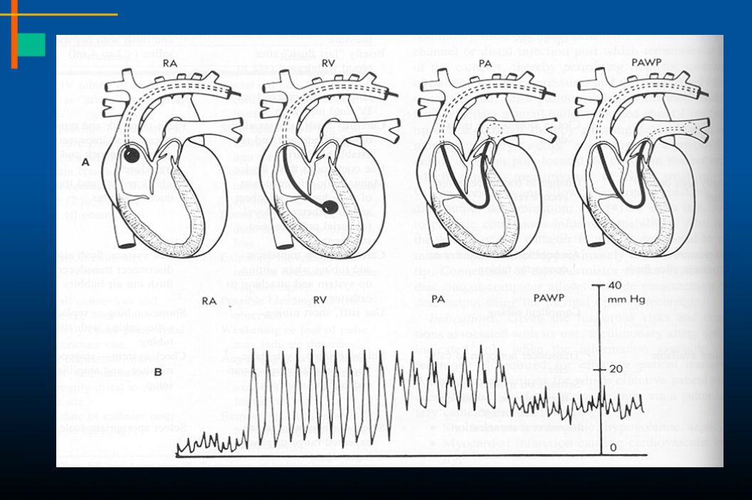
Hemodynamic Monitoring Measurement of PCWP

75
Components of the Atrial Waves

76
The difference between CVP and PCWP waves

77
The mean of the A wave approximates ventricular end-diastolic pressure

78
Reading CVP A V c

79
Reading PCWP A V A V

80
Differences in CVP and PCWP EKG correlation

81
Reading the mean of an A wave
22+10/2=16

82
Question #7 Which is true about the “wedge”? (a) Measures LVEDV
(b) Falsely elevated by PEEP (c) Increased in pulmonary HTN (d) Accurately measured in mitral stenosis
 Falsely elevated by PEEP. (c) Increased in pulmonary HTN. (d) Accurately measured in mitral stenosis.")
83
Wedge Pressure Correlates well with LA and LVEDP if normal anatomy
Reliable measure of preload (volume) only with normal/stable ventricular compliance Falsely elevated by PEEP (and auto-PEEP)
 only with normal/stable ventricular compliance. Falsely elevated by PEEP (and auto-PEEP)")
84
Shock Management

85
Management Treatment of underlying cause Volume Vasopressors

86
A Clinical Approach to Shock Diagnosis and Management
Identify source of blood or fluid loss in hypovolemic shock Intra-aortic balloon pump (IABP), cardiac angiography, and revascularization for LV infarction Echocardiography, cardiac cath and corrective surgery for mechanical abnormality Pericardiocentesis surgical drainage for pericardial tamponade Thrombolytic therapy, embolectomy for pulmonary embolism Source control and early broad antibiotics for septic shock
, cardiac angiography, and revascularization for LV infarction. Echocardiography, cardiac cath and corrective surgery for mechanical abnormality. Pericardiocentesis surgical drainage for pericardial tamponade. Thrombolytic therapy, embolectomy for pulmonary embolism. Source control and early broad antibiotics for septic shock.")
87
Perfusion Goals in Patients with Septic Shock
HEMODYNAMCS MAP > 60 mm Hg PAOP = mmHg Cardiac Index > 2.2 L/min/m2 ORGAN PERFUSION CNS - improved sensorium Skin - warm, well perfused Renal - UOP > 0.5 cc/kg/hr Decreasing lactate (<2.2 mM/L) Improved renal, liver fucntion O2 DELIVERY ADEQUACY Arterial Hgb SpO2 > 92% Hgb concentration > 9 gm/dL SVO2 > 65% Blood Lactate Conc < 2 mM/L 47
 Improved renal, liver fucntion. O2 DELIVERY ADEQUACY. Arterial Hgb SpO2 > 92% Hgb concentration > 9 gm/dL. SVO2 > 65% Blood Lactate Conc < 2 mM/L. 47.")
88
Volume Therapy Lactated Ringer’s solution Normal saline Hetastarch
Crystalloids Lactated Ringer’s solution Normal saline Colloids Hetastarch Albumin Packed red blood cells Infuse to physiologic endpoints

89
Early Goal Directed Therapy
89 Rivers E, Nguyen B, Havstad S, et al 2001;345:

91
Early Goal-Directed Therapy Results: 28 Day Mortality
60 49.2% 50 Vascular Collapse 21% vs 10% p=0.02 P = 0.01* 40 33.3% 30 Mortality % MODS 22% vs 16% P=0.27 20 Cause of in-hospital death: --Sudden Cardiovascular collapse Standard Tx= 25/119 (21%) EGDT 12/117 (10.3%) --MODS Standard Tx 26/119(21.8%) EGDT 19/117 (16.2%) P. 1374 10 Standard Therapy N=133 EGDT N=130 *Key difference was in sudden CV collapse, not MODS NEJM 2001;345: 91
 EGDT 12/117 (10.3%) --MODS. Standard Tx 26/119(21.8%) EGDT 19/117 (16.2%) P Standard Therapy. N=133. EGDT. N=130. *Key difference was in sudden CV collapse, not MODS. NEJM 2001;345:")
92
In-hospital mortality (all patients)
The Importance of Early Goal-Directed Therapy for Sepsis-induced Hypoperfusion 92 In-hospital mortality (all patients) 10 20 30 40 50 60 Standard therapy EGDT 28-day mortality 60-day mortality NNT to prevent 1 event (death) = 6 - 8 Mortality (%) Rivers E, Nguyen B, Havstad S, et al. 2001;345:
 Standard therapy. EGDT. 28-day mortality. 60-day mortality. NNT to prevent 1 event (death) = Mortality (%) Rivers E, Nguyen B, Havstad S, et al. 2001;345:")
93
Therapies Across The Sepsis Continuum
Infection SIRS Sepsis Severe Sepsis Septic Shock CVP > 8-12 mm Hg MAP > 65 mm Hg Urine Output > 0.5 ml/kg/hr ScvO2 > 70% SaO2 > 93% Hct > 30% Early Goal Directed Therapy * Early Goal-Directed Therapy (EGDT): involves adjustments of cardiac preload, afterload, and contractility to balance O2 delivery with O2 demand: Fluids, Blood, and Inotropes Rivers E, Nguyen B, Havstad S, et al. Early goal-directed therapy in the treatment of severe sepsis and septic shock. NEJM 2001;345:1368. 93
: involves adjustments of cardiac preload, afterload, and contractility to balance O2 delivery with O2 demand: Fluids, Blood, and Inotropes. Rivers E, Nguyen B, Havstad S, et al. Early goal-directed therapy in the treatment of severe sepsis and septic shock. NEJM 2001;345:")
94
Type of Fluids

95
Therapy: Resuscitation Fluids
Crystalloid vs. colloid Optimal PWP vs mm Hg 20-40 mL/kg fluid challenge in hypovolemic or septic shock with Re-challenges of mL/kg mL challenges in cardiogenic

96
Vasoactive Agent Receptor Activity
Agent a1 a2 b b2 Dopa Dobutamine Dopamine ++/ ? Epinephrine Norepinephrine / Phenylephrine ++/ ? 34

97
Vasopressors/Inotrops

101
Dopamine Norepinephrine

102
Intra-Aortic Balloon Pump

103
Take Home Points Shock is defined by inadequate tissue oxygenation, not hypotension Oxygen delivery depends primarily on CO, Hgb and SaO2 (not pO2) Volume expand with crystalloids and blood, if indicated; then add vasoactive drugs to improve vital organ perfusion Early treatment of shock is critical

104
Thank You

Similar presentations