Download presentation
Presentation is loading. Please wait.

1
Chest Radiography Interpretation
M Chadi Alraies, MD Chief Medical Resident Case Western Reserve University SVCH M C Alraies

2
Reading CXR’s Have a structured method! Be consistent with that method
Don’t take short cuts LOOK AT ALL YOUR PATIENTS XRAYS YOURSELF (and with your resident of course!) PRACTICE…PRACTICE… PRACTICE
 PRACTICE…PRACTICE… PRACTICE.")
3
What is a Chest Radiograph?
SHADOW

4
Identification! Start at the beginning Are old films available?
Correct patient Correct date and time Correct examination Are old films available? DO THIS EVERYTIME – It buys you time and is vitally important.

5
Approach to the CXR: Technical Aspects
Projection – PA or AP Position – Upright or Supine (Supine folks are sick) Inspiratory effort 9-10 posterior ribs Penetration thoracic intervertebral disc space just visible Positioning/rotation medial clavicle heads equidistant to spinous process
 Inspiratory effort posterior ribs. Penetration. thoracic intervertebral disc space just visible. Positioning/rotation. medial clavicle heads equidistant to spinous process.")
6
Projection

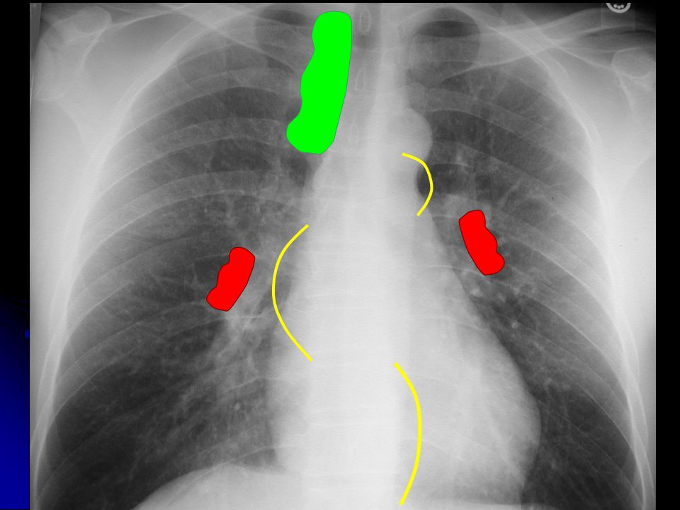
7
Portable (AP or Antero-posterior)
FILM

8
PA (Postero-anterior)
FILM

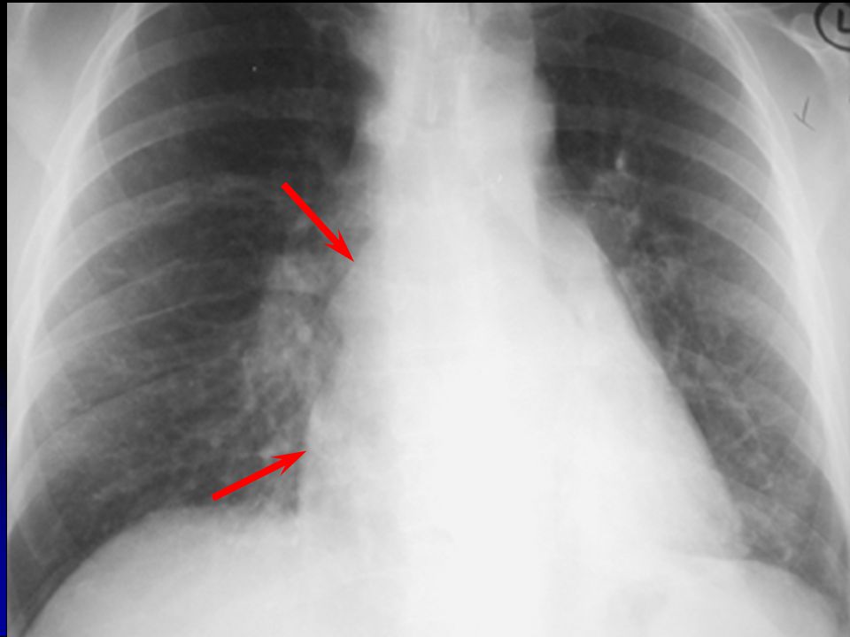
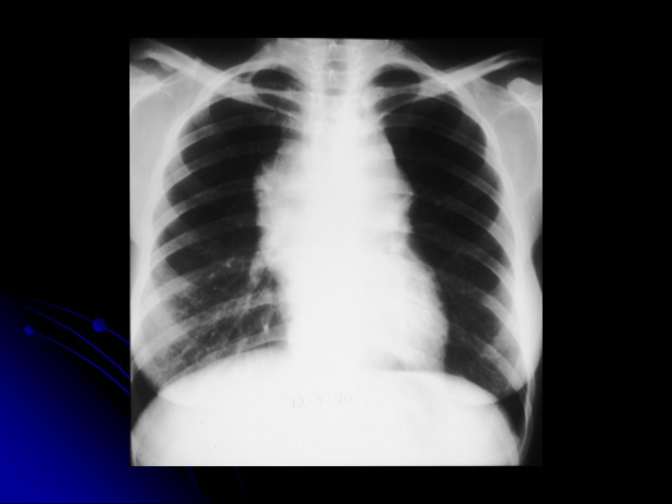
9
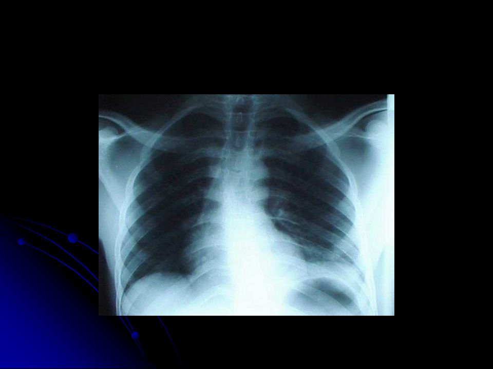
Projection PA AP

10
Low Lung Volumes

11
Over Exposure Proper Exposure

12
9

14
Mental Break

15
Anatomy RUL RML

18
RUL (Right Upper Lung)
")
19
RML (Right Middle Lung)
")
20
RLL (Right Lower Lung)
")
21
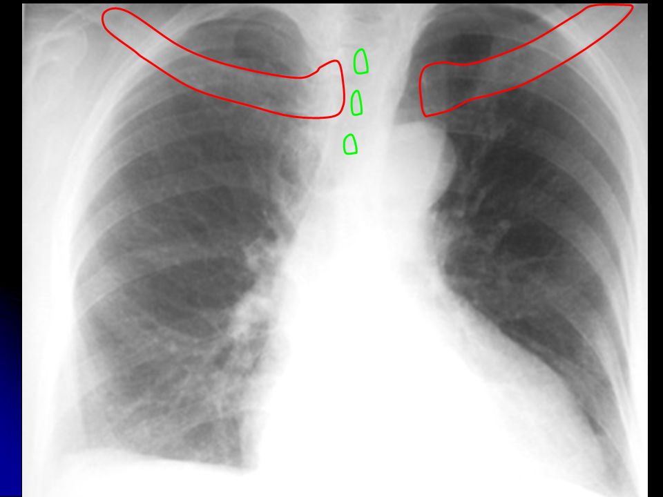
Right Sided Fissures

22
LUL (Left Upper Lung)
")
23
LLL (Left Lower Lung)
")
24
Left Side Fissure LUL LLL

25
What to Evaluate Lungs Pleural surfaces Cardiomediastinal contours
Bones and soft tissues Abdomen

26
Where to Look Apices Retrocardiac areas (left and right)
Below diaphragm

27
Apical TB

28
Left Retrocardiac Opacity

29
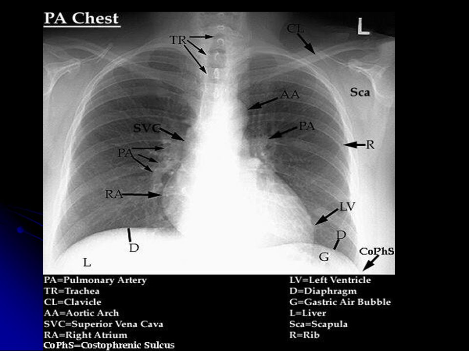
Normal Anatomy: Frontal CXR
Heart Aorta Pulmonary arteries Airways Diaphragm/costophrenic sulci

31
Normal Anatomy: Lateral
Heart Aorta Pulmonary arteries Airways Spine

32
Maximum x-ray Blackest Transmission (least dense tissue) Maximum x–ray
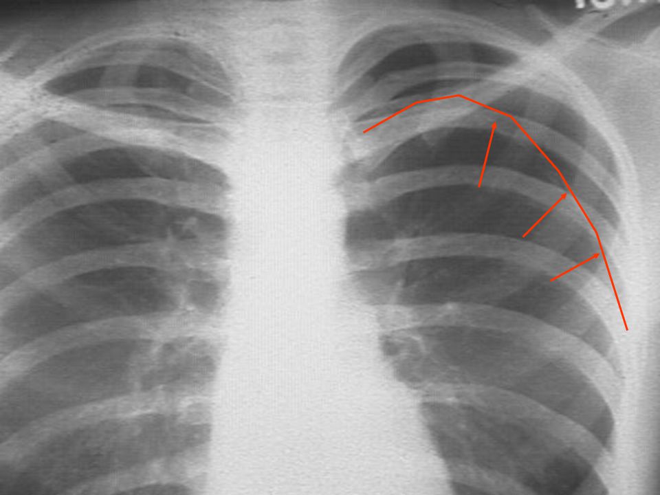
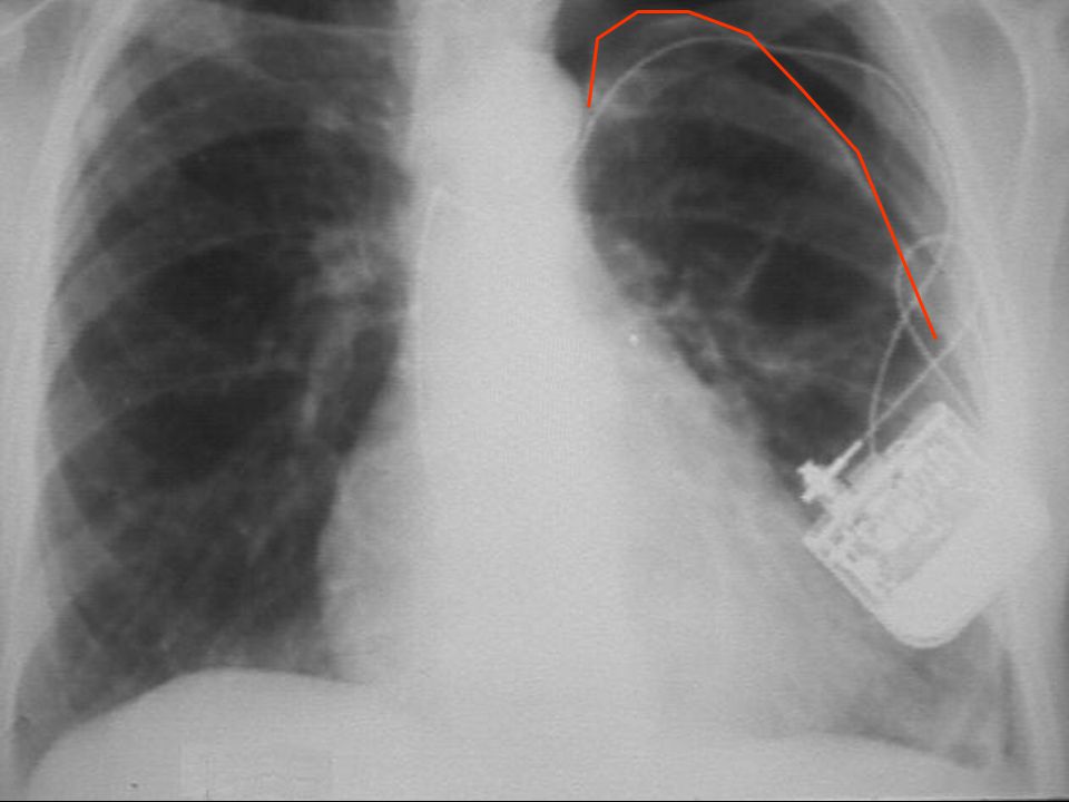
Absorption (densest tissue) Blackest air fat soft tissue calcium bone x-ray contrast metal Whitest
 Blackest. air. fat. soft tissue. calcium. bone. x-ray contrast. metal. Whitest.")
33
Chest Radiography: Basic Principles
A structure is rendered visible on a radiograph by the juxtaposition of two different densities

34
Silhouette Sign Loss of the expected interface normally created by juxtaposition of two structures of different density No boundary can be seen between two structures of similar density

35
Right Lower Lobe Pneumonia

36
Differential X-Ray Absorption
The absence of a normal interface may indicate disease; The presence of an unexpected interface may also indicate disease The presence of interfaces can be used to localize abnormalities

37
Chest Radiographic Patterns of Disease
Air space opacity Interstitial opacity Nodules and masses Lymphadenopathy Cysts and cavities Lung volumes Pleural diseases

38
LUL Pneumonia

39
Air Space Opacity Components:
air bronchogram: air-filled bronchus surrounded by airless lung confluent opacity extending to pleural surfaces segmental distribution

40
Air Space Opacity: DDX Blood (hemorrhage) Pus (pneumonia)
Water (edema) hydrostatic or non-cardiogenic Cells (tumor) Protein/fat: alveolar proteinosis and lipoid pneumonia
 hydrostatic or non-cardiogenic. Cells (tumor) Protein/fat: alveolar proteinosis and lipoid pneumonia.")
41
Interstitial Opacity: Small Nodules

42
Interstitial Opacity:
Lines

43
Interstitial Opacity: Lines & Reticulation

44
Interstitial Opacity Hallmarks: small, well-defined nodules lines
interlobular septal thickening fibrosis reticulation

45
Interstitial Opacity: DDX
Idiopathic interstitial pneumonias Infections (TB, viruses) Edema Hemorrhage Non–infectious inflammatory lesions sarcoidosis Tumor
 Edema. Hemorrhage. Non–infectious inflammatory lesions. sarcoidosis. Tumor.")
46
Well-Defined Calcification Ill-Defined Mass

47
Nodules and Masses Nodule: any pulmonary lesion represented in a radiograph by a sharply defined, discrete, nearly circular opacity mm in diameter Mass: larger than 3 cm

48
Nodules and Masses Qualifiers: single or multiple size
border definition presence or absence of calcification location

49
Right Paratracheal Lymphadenopathy

50
Right Hilar LAN

51
Right Hilar LAN

52
Left Hilar LAN

54
Subcarinal LAN *

55
AP Window LAN

56
Lymphadenopathy Non-specific presentations: Specific patterns:
mediastinal widening hilar prominence Specific patterns: particular station enlargement

58
Cysts & Cavities Cyst: abnormal pulmonary parenchymal space, not containing lung but filled with air and/or fluid, congenital or acquired, with a wall thickness greater than 1 mm epithelial lining often present

59
Cysts & Cavities Cavity: abnormal pulmonary parenchymal space, not containing lung but filled with air and/or fluid, caused by tissue necrosis, with a definitive wall greater than 1 mm in thickness and comprised of inflammatory and/or neoplastic elements

60
Benign Lung Cyst : PCP Pneumatocele
Uniform wall thickness 1 mm Smooth inner lining

61
Benign Cavities : Cryptococcus max wall thickness 4 mm minimally irregular inner lining

62
Indeterminate Cavities
max wall thickness 5-15 mm mildly irregular inner lining

63
Malignant Cavities: Squamous Cell Ca
max wall thickness 16 mm Irregular inner lining

64
Cysts & Cavities Characterize: wall thickness at thickest portion
inner lining presence/absence of air/fluid level number and location

65
Pleural Effusion

66
Pleural Effusion

67
Pleural Calcification

68
Pleural Disease: Basic Patterns
Effusion angle blunting to massive mobility Thickening distortion, no mobility Mass Air Calcification

69
Thoracic Aorta Aneurysm

70
Chest breast implants

71
Rib fx’s Mediast. OK Pulmonary contusion Subcu air Chest tube NG tube

72
MVC victim

73
Deep Right Mainstem Intubation
Carina Tip of ET tube Deep Right Mainstem Intubation

74
Tip of ET Pneumomediastinum

75
Potential X ray findings
wide mediastinum obliteration of aortic knob Rt mainstem shift up and right NG deviate to right pleural cap Major Vessel Injury

76
Pneumothoraces

78
Expiration reduces lung volume, making a small pneumo easier to see

87
Irregular linear opacities are present in both lungs, especially in the periphery and the bases of the lungs. The heart is slightly enlarged, but this is not related to the pulmonary abnormalities in this case.

90
Hodgkin’s Disease

91
Ao SVC Mediastinal Hematoma

92
Tracheal deviation to Rt. ET tube First rib fx Obliterated aortic knob
NG shift to Rt. Chest tube

93
Lt. Internal Carotid Artery
Rt. Subclavian Art. ET Lt. Subclavian Artery NG Aortic Rupture

94
Tension Pneumothorax on CT
Mediastinum Rt. Lt. Ao

95
Hemothoraces

96
Hemothorax Supine Upright

97
Hemopneumothorax

99
Indistinct diaphragm

100
Elevated, irregular hemidiaphragm

101
Indistinct, elevated diaphragm
Clavicle fx Suspicious Close-up Rib fxs Indistinct, elevated diaphragm Chest tube

102
Crushed right chest

103
After ventilated with PEEP

Similar presentations


 –Partial.>")


