Download presentation
Presentation is loading. Please wait.

1
X-Ray Rounds Plain Chest Radiographs
Garry W. K. Ho, M.D. VCU / Fairfax Family Practice July 13, 2005

3
} } } The 12-Step Program Pre-read Quality Control Findings 1: Name
2: Date 3: Old films 4: What type of view(s) 5: Penetration 6: Inspiration 7: Rotation 8: Angulation 9: Soft tissues / bony structures 10: Mediastinum 11: Diaphragms 12: Lung Fields Pre-read } Quality Control } Findings
 5: Penetration. 6: Inspiration. 7: Rotation. 8: Angulation. 9: Soft tissues / bony structures. 10: Mediastinum. 11: Diaphragms. 12: Lung Fields. Pre-read. } Quality Control. } Findings.")
4
Pre-Reading 1. Check the name 2. Check the date
3. Obtain old films if available 4. Which view(s) do you have? PA / AP, lateral, decubitus, AP lordotic
 do you have PA / AP, lateral, decubitus, AP lordotic.")
5
Quality Control 5. Penetration Should see ribs through the heart
Barely see the spine through the heart Should see pulmonary vessels nearly to the edges of the lungs

6
Overpenetrated Film Lung fields darker than normal—may obscure subtle pathologies See spine well beyond the diaphragms Inadequate lung detail

7
Underpenetrated Film Hemidiaphragms are obscured
Pulmonary markings more prominent than they actually are

8
Quality Control 6. Inspiration
Should be able to count 9-10 posterior ribs Heart shadow should not be hidden by the diaphragm 1 2 3 4 5 6 7 8 9 10

9
About 8 posterior ribs are showing 9-10 posterior ribs are showing
Poor inspiration can crowd lung markings producing pseudo-airspace disease 9-10 posterior ribs are showing 9 With better inspiration, the “disease process” at the lung bases has cleared

10
Quality Control 7. Rotation
Medial ends of bilateral clavicles are equidistant from the midline or vertebral bodies

12
If spinous process appears closer to the right clavicle (red arrow), the patient is rotated toward their own left side If spinous process appears closer to the left clavicle (red arrow), the patient is rotated toward their own right side
, the patient is rotated toward their own right side.")
13
Quality Control 8. Angulation Clavicle should lay over 3rd rib 1 2 3

14
Same patient, not lordotic
Pitfall Due to Angulation Apical lordotic Same patient, not lordotic A film which is apical lordotic (beam is angled up toward head) will have an unusually shaped heart and the usually sharp border of the left hemidiaphragm will be absent
 will have an unusually shaped heart and the usually sharp border of the left hemidiaphragm will be absent.")
15
Findings 9. Soft tissue and bony structures Check for Symmetry
Deformities Fractures Masses Calcifications Lytic lesions

16
Findings 10. Mediastinum Check for Cardiomegaly
Mediastinal and Hilar contours for increase densities or deformities

17
Findings 11. Diaphragms Check sharpness of borders
Right is normally higher than left Check for free air, gastric bubble, pleural effusions

19
Findings 12. The Lung Fields!
To help you determine abnormalities and their location… Use silhouettes of other thoracic structures Use fissures

20
Lung Fields: Using Structures / Silhouettes
Silhouette / Structure Contact with Lung Upper right heart border/ascending aorta Anterior segment of RUL Right heart border RML (medial) Upper left heart border Anterior segment of LUL Left heart border Lingula (anterior) Aortic knob Apical portion of LUL (posterior) Anterior hemidiaphragms Lower lobes (anterior)
 Upper left heart border. Anterior segment of LUL. Left heart border. Lingula (anterior) Aortic knob. Apical portion of LUL (posterior) Anterior hemidiaphragms. Lower lobes (anterior)")
21
Lung Fields: Using Structures / Silhouettes
Upper right heart border / ascending aorta (anterior RUL) Aortic knob (Apical portion of LUL ) Upper left heart border (anterior LUL) Right heart border (medial RML) Left heart border (lingula; anterior) Anterior hemidiaphragms (anterior lower lobes)
 Aortic knob. (Apical portion of LUL ) Upper left heart border. (anterior LUL) Right heart border. (medial RML) Left heart border. (lingula; anterior) Anterior hemidiaphragms. (anterior. lower lobes)")
22
Lung Fields: Fissures The fissures can also help you to determine the boundaries of pathology Major Oblique Fissure Separates the LUL from the LLL Right Major Fissure Separates the RUL/RML from the RLL Right Minor Fissure Separates the RUL from the RML

23
Remember… be systematic!
Now for the Cases… Remember… be systematic!

24
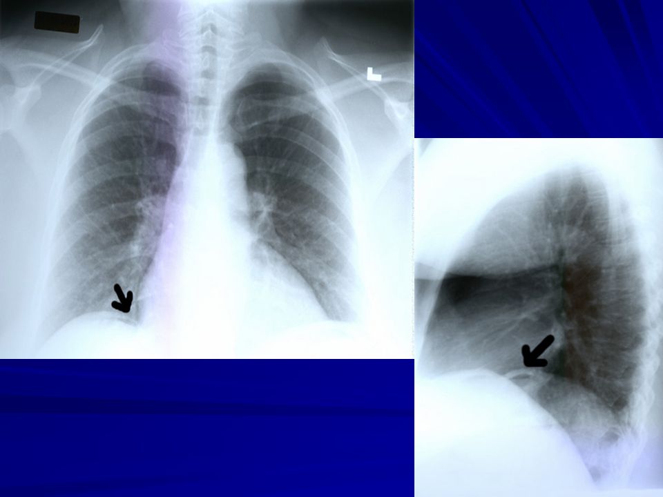
PA view: RML consolidation and loss of right heart silhouette
PA view of a patient with right middle lobe pneumonia, showing consolidation of the right middle lobe and loss of the right heart silhouette. Lateral view of the same patient. The right middle lobe appears wedge-shaped on this view. PA view: RML consolidation and loss of right heart silhouette Lateral View: RML wedge shaped consolidation RML pneumonia

25
RUL infiltrate / consolidation, bordered by minor fissure inferiorly
Underpenetrated; right upper lobe pneumonia (bordered inferiorly by the minor fissure) and a more patchy left lower lobe pneumonia. RUL infiltrate / consolidation, bordered by minor fissure inferiorly Patchy LLL infiltrate that obscures the left hemidiaphragm; right and left heart borders obscured RUL and LLL pneumonia
 and a more patchy left lower lobe pneumonia. RUL infiltrate / consolidation, bordered by minor fissure inferiorly. Patchy LLL infiltrate that obscures the left hemidiaphragm; right and left heart borders obscured. RUL and LLL pneumonia.")
26
Some nonspecific obscuring of L heart border in an mildly underpenetrated film; ?? Breast implants?? mostly normal Underpenetrated; possible nonspecific obscuring of left heart border; mostly normal

27
Patient with multiple bilateral pulmonary abscesses, due to tuberculosis. Note the air-fluid levels within several of these cavitary lesions. Multiple bilateral cavitary lesions with air-fluid levels c/w pulmonary abscesses Tuberculosis

28
28 y/o female with sudden onset SOB while jogging this morning
This 28 y.o. female was jogging this morning when she experienced sudden onset of shortness of breath. Diagnosis: Left Spontaneous Pneumothorax Trivia Question: How much air is required to see a pneumothorax on a chest radiograph? Answer: About 500 ml Not-so-trivia Question: Besides pleural blebs featured in this case, what are some other causes of a pneumothorax? Answer: Bullae, emphysema and interstitial lung disease can also cause spontaneous pneumothoraces. Traumatic and iatrogenic causes include penetrating wounds, line placements, lung biopsies and mechanical ventilators. Well demarcated paucity of pulmonary vascular markings in right apex Left spontaneous pneumothorax

29
RML consolidation that appears wedge shaped on lateral view
RML pneumonia

30
RLL infiltrate / consolidation
Plain film of pneumonia infiltrating the right lower lobe RLL infiltrate / consolidation RLL pneumonia

31
Increased vascular markings; otherwise normal
Increased pulm vasc markings, otherwise normal Increased vascular markings; otherwise normal

32
Concern for aortic injury
Patient BIBA to ER s/p airplane crash. Widened mediastinum; concern for aortic injury (s/p airplane crash) Widened mediastinum Concern for aortic injury
 Widened mediastinum. Concern for aortic injury.")
33
Explain the prominence of the right atrium on this AP radiograph
The patient was rotated to their right (left shoulder forward) when the film was taken Explain the prominence of the right atrium on this AP radiograph Patient rotated to their right (left shoulder forward)
 when the film was taken. Explain the prominence of the right atrium on this AP radiograph. Patient rotated to their right (left shoulder forward)")
34
Dilatation of the main pulmonary artery with decreased peripheral vascular markings
There is dilatation of the main pulmonary artery with decreased peripheral vascular markings. ?? Pulmonary embolism ??

35
Obscuring of the right and left heart borders; infiltrate at the bases
Bilateral aspiration pna Management: Antibiotics First Line Clindamycin mg IV q8 hours Alternative Cefoxitin 2 grams IV q8 hours Ticarcillin-Clavulanate (Timentin) 3.1 grams IV q6h Piperacillin-Tazobactam (Zosyn) g IV q6 hours Obscuring of the right and left heart borders; infiltrate at the bases Bilateral aspiration pneumonia
 3.1 grams IV q6h. Piperacillin-Tazobactam (Zosyn) g IV q6 hours. Obscuring of the right and left heart borders; infiltrate at the bases. Bilateral aspiration pneumonia.")
36
Diffuse bilateral fluffy interstitial infiltrates
Diffuse bilateral Interstitial Infiltrates (80-95%) : Pneumocystis carinii Pneumonia Management: Antibiotics First Line treatment Bactrim PO or IV (15 mg/kg of trimethoprim/day) Adverse reactions occur in 40-60% within 3 weeks Other agents (Bactrim intolerance) Pentamidine 4 mg/kg/day IV or IM Atovaquone 750 mg PO bid Dapsone and Trimethoprim Indicated for mild-moderate Pneumocystis Fewer dose limiting adverse reactions Primaquine and Clindamycin Indicated for intolerance for other regimens Diffuse bilateral fluffy interstitial infiltrates Pneumocystis carinii pneumonia
 : Pneumocystis carinii Pneumonia. Management: Antibiotics. First Line treatment. Bactrim PO or IV (15 mg/kg of trimethoprim/day) Adverse reactions occur in 40-60% within 3 weeks. Other agents (Bactrim intolerance) Pentamidine 4 mg/kg/day IV or IM. Atovaquone 750 mg PO bid. Dapsone and Trimethoprim. Indicated for mild-moderate Pneumocystis. Fewer dose limiting adverse reactions. Primaquine and Clindamycin. Indicated for intolerance for other regimens. Diffuse bilateral fluffy interstitial infiltrates. Pneumocystis carinii pneumonia.")
37
LUL pna LUL pneumonia

38
Severe pulmonary TB Severe pulmonary TB

39
Later diagnosed as lung cancer
The chest x-ray shows a shadow in the left lung, which was later diagnosed as lung cancer Left lung opacity Later diagnosed as lung cancer

40
Cardiomegaly and pulmonary congestion with fluid in horizontal fissure.
Cardiomegaly, increased pulmonary vascular markings, fluid in the horizontal fissure CHF

41
Kerley B Lines What do the arrows indicate?
Short (1 -2 cm) white lines at the lung bases, perpendicular to the pleural surface representing distended interlobular septa
 white lines at the lung bases, perpendicular to the pleural surface representing distended interlobular septa.")
42
} } } The 12-Step Program Pre-read Quality Control Findings 1: Name
2: Date 3: Old films 4: What type of view(s) 5: Penetration 6: Inspiration 7: Rotation 8: Angulation 9: Soft tissues / bony structures 10: Mediastinum 11: Diaphragms 12: Lung Fields Pre-read } The point is to read radiographs SYSTEMATICALLY. FYI: residents must review all X-rays and EKGs with a faculty member Quality Control } Findings
 5: Penetration. 6: Inspiration. 7: Rotation. 8: Angulation. 9: Soft tissues / bony structures. 10: Mediastinum. 11: Diaphragms. 12: Lung Fields. Pre-read. } The point is to read radiographs SYSTEMATICALLY. FYI: residents must review all X-rays and EKGs with a faculty member. Quality Control. } Findings.")
43
The End Questions?

Similar presentations


 –Partial.>")


