Download presentation
Presentation is loading. Please wait.

1
Introduction to Orthopaedics
Stephen P. England, MD MPD Department of Orthopaedic Surgery Park Nicollett Clinic

2
Introduction to Orthopaedics

3
Test Yourself List the bones of the body. (More pts more bones!)
Bone forming cells are called ______. Local stress stimulates bone formation. T or F? The knee is a/an _______joint.

4
What do you know from the slides?
Which is the hand of the elderly adult? How old do you think the individual is on slide A? A B

5
Bone Structure: Orthopaedic Implications
Periosteum Diaphysis Epiphysis Endosteum Epiphyseal plates; bone growth, injury

6
What is the significance of the epiphyseal plate?

7
Bone Formation and Maintenance
Types Bone = cells, protein matrix, mineral deposits Types of bone cells Function of each type bone cell Protein matrix: 98% collagen, 2% other Mineral salts: insoluble Ca/Phos = hydroxyapitite + Process of ossification

8
Factors Influencing Bone Growth and Formation
Parathyroid What effect of low Ca? Calcitonin Effect on Ca? Source? Thyroxin Estrogen Glucocorticoids What effect on bones with long term use of glucocorticoids? Vit C & D

9
Types of Joints: Identification
Amphiarthrosis Synarthrosis Diarthrosis

10
Diarthroidal Joint

11
Significance of Diarthrotic Joint
Joint Capsule surrounded by ligaments Synovial Membrane: secretes synovial fluid; lines tendon and muscle sheaths Bursea: painful, but protective!

12
Othropaedic Terminology

13
Descriptive Orthopaedic Terms
Valgus: part of body distal to joint directed away from midline Varus: Part of body distal to joint directed toward midline Hallus Genu varus Genu valgus pes varus metatarus valgus metatarus varus

14
Which foot has a valgus deformity?
Hallus valgus How do you describe this foot deformity?

15
Stressors of the Musculoskeletal System
Trauma Infection Altered Metabolism

16
For the person with a musculoskeletal condition:
Peripheral neurovascular dysfunction Pain (acute, chronic) Impaired skin integrity Infection, high risk for Disuse syndrome Activity intolerance Trauma. high risk for Knowledge deficit Impaired adjustment Fear, anxiety List effects on person List “most “ frequent orthopaedic diagnosis
 Impaired skin integrity. Infection, high risk for. Disuse syndrome. Activity intolerance. Trauma. high risk for. Knowledge deficit. Impaired adjustment. Fear, anxiety. List effects on person. List most frequent orthopaedic diagnosis.")
17
How has orthopedic injury affected this PERSON?

18
Components of Assessment
Chief Complaint Why seeking care Acute and chronic problem History taking; its significance Pain characteristics location character what effects Associated conditions Pain Complications!

19
Principles of Assessment
Test your skills Changes with age Nurtitional status Skin integrity Rashes Color changes, esp with cold; arterial vs. venous Character of joints Bruises, swelling Normal first Bilateral comparision Inspect then gentle palpation shape, size , contour signs inflammation, ecchymosis muscle condition deformity

20
Assessment of the Knee Fluid in the Knee
Bulge sign: medial aspect knee, displace fluid upward, tap lateral patellar margin and note fluid return Ballottment:force fluid into joint space; displace patella

21
Ballottment:force fluid into joint space; displace patella

22
Knee Stability Anterior cruciate ligament: limits anterior motion
Posterior cruciate ligament: limits posterior motion Lateral collateral ligament: limits adduction Medial collateral ligament: limits abduction Meniscal injury: McMurray’s sign

23
Knee Support and Stability Anterior and posterior cruciate ligaments connect the inner surfaces of the head of the femur with the head of the tibia. They cross each other, anterior ligament extend from the inside of the lateral condyle of the femur to the medial side of the tibial head, and posterior ligament extend from the inside of the medial condyle of the femur to the lateral side of the tibial head.

24
Anterior Drawer test McMurray’s sign

25
Diagnostic Tests CT Scan Bone Scan MRI Dual-Photon Absorptiometry
Arthrography Arthrocenthesis Arthroscopy

26
Diagnostic Tests Arthrocenthesis Arthrography
Aspiration synovial fluid; reduce pain; dx; treatment Analysis joint fluid: usual clear, high viscosity, scant fluid Teaching: no restrictions; consent form; slight pain Post-op: RICE Arthrography Radiographic exam, use air or contrast medium:; 90-95% accuracy Teaching Complications: infection, allergy Post-op: Rest joint 6-12 hrs, use ice

27
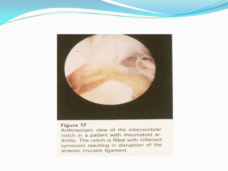
Arthroscopy Therapeutic /diagnostic
Visual recording; surgical removal of meniscus, foreign bodies, etc Rare complications; depends on procedure, operative length, use of tourniquet Teaching Post-op care

30
Orthopaedic Interventions!
Traction Casts External Fixators Pin, plates and screws CPM Crutch-walking

31
Assistive Devices Traction Crutch-walking
Definition Uses Types Counter traction is provided by: a. body weight b. pulleys c. traction weight d. splints Crutch-walking Two-point Three-point Four-point Swing-through swing-to Safety in crutch-walking Cane

33
CPM Purpose Guidelines for Use Teaching

34
Bone Stimulators Indications Electronegativity Bone Remodeling
Internal Percutaneous External

36
External Bone Stimulator

37
Autologous Blood Transfusions
Indications for Ortho Cell Savers Criteria for Use

38
Cell Savers Autologous Blood

39
Surgical /Medical Interventions
Tissue Allografts Abductor Pillow, Carter Pillow Hot Ice Machines that Aren’t! Bone Paste!

40
Tissue allografts, synthetic grafts

41
Pins, plates, screws ORIF (open reduction, internal fixation)
")
42
Casts Purposes Casting Material Plaster Fiberglass

43
Application of Cast Principles Skin Assessment Skin Protection
Heat Generated Time to Dry

44
Cast Types Sugar Tong/Splint Spica Type Body Cast Care Cast Syndrome
Hip spica Gauntlet Cast-Brace Body Cast Care Cast Syndrome Hip Spica Turning Cast Drying

45
External Fixators How They Work Principles of Care The Iliazarov

46
External Fixator

47
Conclusion

Similar presentations


>")


, F.R.C.S.(C )>")


 Medial condyle (8 left) Intercondylar fossa (7 left)>")

