Download presentation
Presentation is loading. Please wait.

1
Cells as Units of Life Chapter 3

2
Cell Theory Cells represent the basic structural and functional unit of life. Important unifying concept in biology. All organisms are composed of one or more cells. All tissues & organs are composed of cells. There is no life without cells!

3
Cell Theory Cell theory states that all living organisms are composed of cells. Cells come from preexisting cells.

4
Prokaryotic vs. Eukaryotic Cells
All cells: Have DNA Use the same genetic code Synthesize proteins Use ATP in similar ways This implies common ancestry.

5
Prokaryotic vs. Eukaryotic Cells
Prokaryotic cells – no nucleus or other membrane-bound organelles. Kingdom Archaebacteria Kingdom Eubacteria Eukaryotic cells – do have nucleus and membrane-bound organelles. Kingdom Protista Kingdom Fungi Kingdom Plantae Kingdom Animalia

6
Components of Eukaryotic Cells
The plasma membrane surrounds the cell. The nucleus is the largest organelle. Double layered nuclear envelope. Cell Model

7
Components of Eukaryotic Cells
Cytoplasm refers to the cellular material between the cell membrane and nuclear envelope. Organelles such as the mitochondria, Golgi complex, centrioles, and endoplasmic reticulum are found in the cytoplasm.

8
Plasma Membrane Plasma membrane structure is described using the fluid mosaic model.

9
Plasma Membrane Two layers of phospholipid molecules oriented with hydrophilic heads toward the outside and hydrophobic tails inside. Fluid-like – flexible

10
Plasma Membrane The nonpolar nature of the hydrophobic ends in the interior of the membrane prohibit polar substances from crossing the membrane. Glycoproteins embedded in the membrane function in the transport of molecules across the membrane.

11
Nucleus The nuclear envelope contains pores to allow molecules to move between nucleus & cytoplasm. Chromosomes are contained in the nucleus. Chromatin refers to loosely condensed DNA & proteins.

12
Nucleus Nucleoli are specialized parts of certain chromosomes that carry multiple copies of the DNA used to synthesize ribosomal RNA. This rRNA combines with protein to from the two subunits of ribosomes. Ribosomes leave the nucleus through pores in the nuclear envelope.

13
Endoplasmic Reticulum
The nuclear envelope joins with a cytoplasmic membranous system – the endoplasmic reticulum (ER). Rough endoplasmic reticulum (RER) is covered with ribosomes. Smooth (SER) is not.
. Rough endoplasmic reticulum (RER) is covered with ribosomes. Smooth (SER) is not.")
14
Endoplasmic Reticulum
Ribosomes on the RER synthesize proteins that enter the ER that will either be incorporated into the plasma membrane, exported from the cell, or they may be bound for lysosomes. Lipids and phospholipids are synthesized in the SER.

15
Golgi Complex The Golgi complex is a stack of membranous vesicles where storage, modification, and packaging of protein products occurs.

16
Assembling & Secreting Proteins

17
Lysosomes Lysosomes contain enzymes (proteins) that can breakdown foreign material like bacteria or worn out cellular components. Contents of lysosome would kill cell if membrane ruptured. May pour enzymes into food vacuoles.

18
Mitochondria Mitochondria are the powerhouses of cells – they contain enzymes that carry out the energy-yielding steps of aerobic metabolism. ATP is produced here. Composed of a double membrane – the inner membrane is folded into cristae. Mitochondria are self-replicating, containing their own circular DNA molecule.

19
Cytoskeleton Eukaryotic cells have a cytoskeleton that provides support and often locomotion and movement of organelles. Composed of microfilaments, microtubules, and intermediate filaments.

20
Cytoskeleton Microfilaments are made of the proteins actin and myosin and function in a cell’s ability to contract as seen in muscle cells. Actin microfilaments move molecules and organelles through the cytoplasm.

21
Cytoskeleton Microtubules are larger tubular structures composed of the protein tubulin. Move chromosomes during cell division. Part of the structure of cilia & flagella.

22
Cytoskeleton Microtubules radiate out from the centrosome – the microtubule organizing center. Located near nucleus. Not membrane bound. Centrioles are found in the centrosome. Centrioles composed of 9 triplets of microtubules. Replicate before cell division.

23
Cytoskeleton Intermediate fibers fall in between microfilaments and microtubules in size. There are five biochemically distinct types of intermediate fibers.

24
Cilia & Flagella Cilia & flagella are motile extensions of the cell surface. In many single celled organisms they are a source of locomotion. In multicellular animals they usually sweep material past the fixed cell. Nine pairs of microtubules enclose a central pair. At the base is a basal body - identical to a centriole.

25
Pseudopodia Some single-celled organisms, migrating cells in embryos, and white blood cells show ameboid movement. Cytoplasmic streaming through the action of actin microfilaments extends a pseudopodium outward. Some have specialized pseudopodia with microtubules that are assembled & disassembled to allow movement.

26
Junctions Tight junctions form when cell membranes ofadjacent cells fuse. Function as seals. Adhesion junctions occur under tight junctions. Transmembrane proteins link across a small space and connect to microfilaments.

27
Junctions Desmosomes act as spot welds and increase the strength of the tissue. Hemidesmosomes are found at the base of cells and anchor them to connective tissue. Gap junctions are canals between cells that provide intercellular communication.

28
Microvilli Microvilli are small fingerlike projections that have bundles of actin microfilaments. They serve to increase the surface area of the tissue as in the intestine.

29
Membrane Function Membranes surround the outside of the cell and the organelles inside it. The plasma membrane acts as a selective gatekeeper. A substance may cross the membrane: By diffusion By a mediated transport system By endocytosis

30
Diffusion & Osmosis Diffusion is the movement of molecules from an area of high concentration to an area of low concentration. This tends to equalize the concentration. Down the concentration gradient. Solutes are molecules (e.g. salt) that are found in a solution.
 that are found in a solution.")
31
Diffusion & Osmosis Cell membranes are selectively permeable – water can pass through, but not most solutes. Gases (oxygen & carbon dioxide), urea, lipid soluble solutes can cross the membrane.
, urea, lipid soluble solutes can cross the membrane. v=sdiJtDRJQEc&feature=player_embedded#!")
32
Diffusion & Osmosis Osmosis - if there is a membrane between two solutions with unequal concentration of solutes that can not cross the membrane, water will flow toward the side with less water / more solute until the two sides have equal concentrations.

33
Diffusion & Osmosis

34
Diffusion & Osmosis Animals utilize osmosis to control internal fluid and solute levels. The blood of marine fishes has 1/3 the salt content of the water. They are hypoosmotic to seawater. Freshwater fishes have blood that is saltier than the water. They are hyperosmotic to the water. If the solute concentrations were the same, the two solutions would be isoosmotic.

35
Diffusion Through Channels
Charged substances, like water and dissolved ions, can’t simply diffuse across the cell membrane. They pass through channels created by transmembrane proteins. Some channels always open. Some are gated channels.

36
Diffusion Through Channels
Gated channels require a signal to open or close them. Chemically-gated channels open or close when a signaling molecule binds to a binding site on the transmembrane protein. Voltage-gated channels open or close when the ionic charge across the membrane changes.

37
Carrier Mediated Transport
Sugars & amino acids must be able to enter cells and waste products must be able to leave. These molecules cross the membrane with the help of transporter proteins. Transporter proteins are specific. Facilitated diffusion Active transport

38
Facilitated Diffusion
In facilitated diffusion, the transporter protein binds to the substrate molecule on one side of the plasma membrane then changes shape to release it on the other side. Takes place in the direction of the concentration gradient.

39
Active Transport Active transport requires energy (ATP) to transport molecules in the direction opposite the concentration gradient.

40
Endocytosis Endocytosis is the ingestion of material by cells.
Phagocytosis – cell eating – method of feeding by single- celled organisms. Pinocytosis – small molecules or ions are enclosed in vesicles called caveolae. Receptor-mediated endocytosis – method of bringing large molecules into a cell with the help of the protein clathrin.

41
Endocytosis and Exocytosis

42
Exocytosis Exocytosis - membranes of a vesicle inside the cell can fuse with the plasma membrane to discharge the contents of the vesicle outside the cell. Transcytosis – a substance may be picked up on one side of the cell, transported completely across the cell and discharged on the other side.

43
Mitosis and Cell Division
Mitosis is the process of nuclear cell division in nonreproductive, or somatic, cells. A fertilized egg, or zygote, divides by mitosis to produce a multicellular organism. Damaged cells are replaced by mitosis.

44
Chromosomes In cells that are not dividing, the DNA is loosely organized so that individual chromosomes can’t be distinguished – it is now referred to as chromatin. Before division, chromatin becomes more compact and chromosomes can be recognized.

45
Chromosomes All nonreproductive cells in a species have the same number of chromosomes. 46 in humans Half of these chromosomes come from each parent. Result is two sets of chromosomes. Diploid Chromosome 1 from Mom and chromosome 1 from Dad are called homologous chromosomes.

46
The Cell Cycle Cells come from preexisting cells through the process of cell division. Cell division – mitosis and cytokinesis – occupy a very small portion of the cell cycle.

47
The Cell Cycle Interphase includes: Mitosis Cytokinesis
G1 – growth phase where RNA and functional proteins are synthesized. S – DNA replication. G2 – growth phase where structural proteins are made. Mitosis Cytokinesis

48
Chromosome Structure During S phase, each of the 2 homologues replicates, resulting in identical copies called sister chromatids. Chromatids remain connected at a linkage site called the centromere.

49
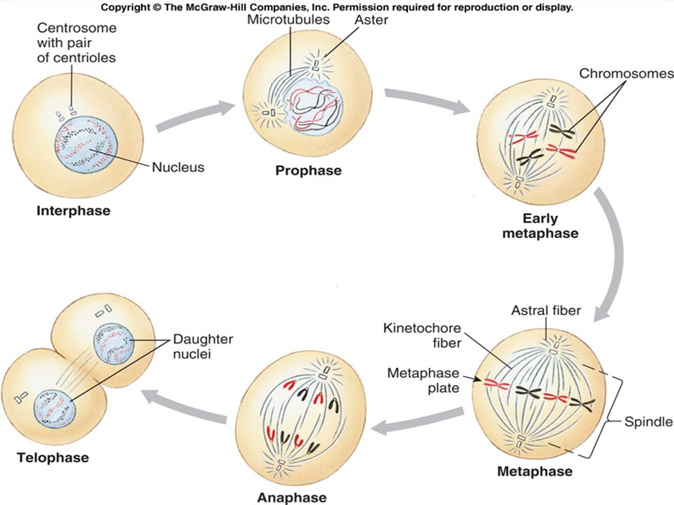
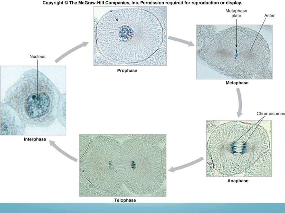
Cell Division There are two phases of cell division:
Mitosis – nuclear cell division Prophase Metaphase Anaphase Telophase Cytokinesis – division of the cytoplasm Multiple nuclear divisions not accompanied by cytokinesis result in a multinucleate cell.

51
Cell Division Prophase – Chromosomes condense enough to be seen with a light microscope. Spindle forms between the 2 centrioles. Spindle fibers attach to kinetochores.

52
Cell Division Metaphase – Alignment of the chromosomes along center of cell (metaphase plate). Fibers attached to kinetochores on both sides of each chromosome.

53
Cell Division Anaphase – Separation of the sister chromatids.
Centromere splits apart – sister chromatids move toward opposite poles. Disassembly of the tubulin subunits shortens the microtubules.

54
Cell Division Telophase – re- formation of the nuclei once the chromosomes are at opposite poles. Chromosomes unwind.

55
Cell Division Cytokinesis – division of the cytoplasm.
Two complete, diploid cells that are identical to the original cell.

56
Cytokinesis During cytokinesis in animal cells, the cell pinches in two. A cleavage furrow produced by microfilaments deepens until the cell splits.

58
Mitosis

Similar presentations


 of cell membrane 2. Structure (and function) of organelles 3. Interconnections between cells to maintain structural.>")


 Relationship.>")