Download presentation
Presentation is loading. Please wait.

1
Etiology and Risk Factor of Cancer
Noorwati Sutandyo Dharmais Cancer Center / Division of Hematology-Medical Oncology Dept Internal Medicine University of Indonesia School of Medicine International Class May 2013

2
CANCER Incidence of cancer is high and going higher every year
Globocan: 12.7 million new cancer cases 2008 Nearly double to 21.4 million cases by 2030 Cancer causes 1 in 8 deaths worldwide becoming a global pandemic Cancer is killing more people in the developing world than HIV/AIDS, tuberculosis, and malaria combined

3
Jakarta Minimum Cancer Incidence (Coverage 70% )
79 Hospitals. 2 Private Clinics, 90 Pathology Laboratories, 44 Municipals Primary health Care (as a coordinator of 301 Primary Health Care in District area). 3
. 3.")
4
4

5
Cancer Definition Cancer is abnormal growth of cells.
Cancer cells will growth fast even with limited of space and nutrition. Heterogenous: more than 100 different types of cancer Some are familial : 5-10% (herediter), and others are sporadic : 90 % (non-herediter)
, and others are sporadic : 90 % (non-herediter)")
6
Tumor = Cancerr??? CYSTS Contain: Liquid/Semisolid LUMP BENIGN Contain: Solid/mass TUMOR MALIGNANT CANCER REMEBER: Every tumor must be considered malignant, until proven malignant/benign

7
Cancer Characteristic

8
Cancer is a gen disease 1.Body consist of billion cells
2.Inside the cells there is nucleolus 3.Inside the nucleolus there are Chromosom 4.Chomosom consist of genes 5.Gene are forms of DNA

9
DNA-Deoxyribonucleic Acid
Is blue print that contain instruction of orgnism “creation” define human characteristic: skin color, eye color, etc. DNA consist of nucleotide Nucleotide: sugar+ phosphat + base 4 types of base: thymine (T), adenine (A), guanine (G), and cytosine (C) The fastest computer in the world (2006): IBM Blue Gene/L supercomputer
, adenine (A), guanine (G), and cytosine (C) The fastest computer in the world (2006): IBM Blue Gene/L supercomputer.")
10
Everyday There are 20.000 DNA damaged Repaired by DNA repair gene
Small part of gene ,can’t be repaired This is the starting point of cancer

11
Cancer Occur due to somatic mutation accumulation in genes that have role in cancer, such as: Oncogenes Tumor supressor genes Mismatch repair genes Carcinogenesis process of the tumor or neoplasm development Carsinogen The cause of cancer

12
Carsinogenesis Defect of Genetic Control Tumor suppressor
Growth promoting oncogenes DNA repair gene Defect Mutation Amplification Mutation Inaktivation Apoptosis Defect Defect of Genetic Control

13
Onkogenes Type of proto-onkogene mutation: point, translocation, amplification, insertion and deletion Cause cell growth or survival become dominan exceed the activity of other normal genes Somatic mutation of proto-onkogene: Occur in various tumor in human Occur during carcinogenesis process Usually, together with other mutations (tumor supresor genes and DNA repair genes)
")
14
Tumor Supressor Genes Failed in DNA repair
Occur two hits mutations during carcinogenesis all of gene functions are diminished p53 protein: guardian of the genome Regulation of mitosis cycle Detect and repair DNA damage Regulation of apoptosis Mutation can be hereditary

15
DNA Repair Genes 1. Repair DNA damaged
2. If there is Silencing mismatch repair gene cause by somatic DNA methylation

16
Cancer Cell Development
Comes from one normal ‘stem cell’ change to be a tumor cell gradually 1 cell divide to be 2 – 4 – and so on different one and others showing the cell heterogenity

18
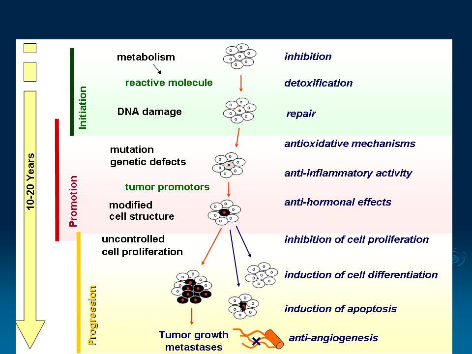
Carsinogenesis Multistep
Inisiation: Permanent change in target cell DNA Promotion: Epigenetic change selectivelly influence cell proliferation that has done inisiation Progression: Cell cancer development that shown autonomy growth, invasive progression, and metastasis.

20
Cancer Etiology Several factors that play a role: Age Genetic Hormone
Immnune system Obesitas Chemical agents (cigarette) Diet Radiation & UV light Virus Stress
 Diet. Radiation & UV light. Virus. Stress.")
21
Age Generally, the risk increases in older person
BUT, cancer can occur to anyone, at any age. Tendency, increasing in younger age

22
Carsinogens Everything causes cancer by causing DNA changes mutagenic Type of carcinogens Chemical agents Virus Radiation Hormone Nutrition

23
CHEMICAL AGENT

24
Chemical Carcinogen Mostly in forms pro-carsinogen need to be metabolise in body to become active ( carcinogen) Convert lipophilic component water soluble metabolite (hydrophilic) can be excreted by urine or bile
 Convert lipophilic component water soluble metabolite (hydrophilic) can be excreted by urine or bile.")
25
Drugs Metabolism Pathway

26
CANCER Chemical Absorption Distribution
Biotransformation (liver, kidney, lung) Excretion (liver, kidney, lung) Activation: Genotoxic & Non-genotoxic mechanism Inactivation Hypermutability Gene instability Loss of proliferation control Resistance to apoptosis Altered signal transduction CANCER Gene Damage
 Excretion. (liver, kidney, lung) Activation: Genotoxic. & Non-genotoxic mechanism. Inactivation. Hypermutability. Gene instability. Loss of proliferation control. Resistance to apoptosis. Altered signal transduction. CANCER. Gene Damage.")
27
Chemical Carcinogens Group Example Cancer
Polycyclic aromatic hydrocarbon (HPA) Cigarette smoke, tobacco, grilled meat, smoked meat/fish Skin, lung, gaster, liver Aromatic amine and azo dyes Textile dyes liver, vesica Nicotine cigarette Lung, oral, respiratory tract Halogenated compound Dioxin: heated plastic, PVC, bleaching agents for paper Kidney, lung, liver Arsenic Soil, water, plant, cosmetic product, seafood, cigarette Skin, lung, liver Nitrosamine Preservatives, coloring agent for meat Nasopharynx, esophagus, gaster, oral Alcohol Beverages containing alcohol Oropharynx, larynx, esophagus, liver, colon, and breast
 Cigarette smoke, tobacco, grilled meat, smoked meat/fish. Skin, lung, gaster, liver. Aromatic amine and azo dyes. Textile dyes. liver, vesica. Nicotine. cigarette. Lung, oral, respiratory tract. Halogenated compound. Dioxin: heated plastic, PVC, bleaching agents for paper. Kidney, lung, liver. Arsenic. Soil, water, plant, cosmetic product, seafood, cigarette. Skin, lung, liver. Nitrosamine. Preservatives, coloring agent for meat. Nasopharynx, esophagus, gaster, oral. Alcohol. Beverages containing alcohol. Oropharynx, larynx, esophagus, liver, colon, and breast.")
28
Chemical carcinogen Cigarette> 4000 chemical agents

29
Chemical Carcinogen Mechanism
Cigarette

30
VIRAL CARCINOGEN

31
Viral Carcinogen Viral oncogenicDNA and RNA
DNA virustumor supressor gene inactivation (p53, Rb) HPV 16, 18, 31cervical cancer EBV Nasopharynx cancer, Burkitt lymphoma, Hodgkin Disease Hepatitis B virushepatoma CMV and herpes kaposi sacoma (AIDS) RNA virus onkogene activation HLTV 1T cell Leukemia, B cell lymphoma Hepatitis C virushepatoma
 HPV 16, 18, 31cervical cancer. EBV Nasopharynx cancer, Burkitt lymphoma, Hodgkin Disease. Hepatitis B virushepatoma. CMV and herpes kaposi sacoma (AIDS) RNA virus onkogene activation. HLTV 1T cell Leukemia, B cell lymphoma. Hepatitis C virushepatoma.")
32
Mekanisme Virus DNA DNA virus contain ds-DNA that can be integrated partially or fully with host chromosom if it lasts longer, it can give rise to mutations

33
Oncogenic DNA virus and its Product
Virus Gene Product Cell Target Adenovirus SV40 Polyomavirus Papillomavirus E1A E1B Rb p53 Large T antigen Large T antigen Middle T antigen Rb, p53 Rb Src, PI3K E7 E6 E5 Rb p53 PDGF receptor

34
HPV 16 Genome

35
Spectrum of Cervical Epithelial Changes due to HPV infection
Normal Cervix HPV Infection/ CIN* 1 CIN 2 / CIN 3 / Cervical Cancer Key Point Integration of HPV into the DNA of the infected host cell is commonly associated with high-risk oncogenic HPV types1 and is linked to the activity of E6 and E7 proteins.2 Background In benign HPV-associated skin lesions, the HPV virus maintains its genome as episomes at low copy numbers (10–200 copies/cell) in the basal cells of the epithelium separate from the host cell DNA. To maintain its viral DNA as an episome, viral E1 and E2 proteins are expressed. Failure to express E1 leads to the integration of the HPV genome into the host cell chromosome.3 Integration of HPV into the DNA of the infected host cell is commonly associated with high-risk oncogenic HPV types1 and is considered an important step in tumor progression.2 In malignant HPV-associated skin lesions, HPV DNA integration into the host cell’s chromosome regularly occurs through a break in the viral genome around the E1/E2 region. Integration-mediated disruption of E2 may trigger uncontrolled expression of E6 and E7, resulting in cellular transformation.2 The E6 protein associates with the tumor suppressor protein p53 and promotes proteolytic destruction of the protein. This leads to malignant transformation and loss of regulated cell growth. The E7 protein associates with the retinoblastoma protein (pRB), which inactivates the cell cycle restriction function of this protein.2 References 1. Gallo G, Bibbo M, Bagella L, et al. Study of viral integration of HPV-16 in young patients with LSIL. J Clin Pathol. 2003;56:532–536. 2. Syrjänen KJ, Syrjänen SM. Molecular biology of papillomaviruses. In: Papillomavirus Infections in Human Pathology. Chichester, United Kingdom: John Wiley & Sons, Inc.; 2000:11–51. 3. Doorbar J. The papillomavirus life cycle. J Clin Virol. 2005;32(suppl):S7–S15. *CIN = cervical intraepithelial neoplasia Adapted from Goodman A, Wilbur DC. N Engl J Med. 2003;349:1555–1564.
 in the basal cells of the epithelium separate from the host cell DNA. To maintain its viral DNA as an episome, viral E1 and E2 proteins are expressed. Failure to express E1 leads to the integration of the HPV genome into the host cell chromosome.3. Integration of HPV into the DNA of the infected host cell is commonly associated with high-risk oncogenic HPV types1 and is considered an important step in tumor progression.2 In malignant HPV-associated skin lesions, HPV DNA integration into the host cell’s chromosome regularly occurs through a break in the viral genome around the E1/E2 region. Integration-mediated disruption of E2 may trigger uncontrolled expression of E6 and E7, resulting in cellular transformation.2. The E6 protein associates with the tumor suppressor protein p53 and promotes proteolytic destruction of the protein. This leads to malignant transformation and loss of regulated cell growth. The E7 protein associates with the retinoblastoma protein (pRB), which inactivates the cell cycle restriction function of this protein.2. References. 1. Gallo G, Bibbo M, Bagella L, et al. Study of viral integration of HPV-16 in young patients with LSIL. J Clin Pathol. 2003;56:532– Syrjänen KJ, Syrjänen SM. Molecular biology of papillomaviruses. In: Papillomavirus Infections in Human Pathology. Chichester, United Kingdom: John Wiley & Sons, Inc.; 2000:11– Doorbar J. The papillomavirus life cycle. J Clin Virol. 2005;32(suppl):S7–S15. *CIN = cervical intraepithelial neoplasia. Adapted from Goodman A, Wilbur DC. N Engl J Med. 2003;349:1555–1564.")
36
Important Function of HPV Protein
Six early Gene (E) regulate mRNA virus synthesis ans virus genome replication E6 & E7 important in cancer process E6 disrupt supressor tumor p53 protein E7 inactivate Retinoblastoma protein E2 suppress E6 & E7 expression Late Gene (L) code protein that involved in viral kapsid assembly L1 major capsid L2 kapsid minor Destruction of p53 will prevent apoptosis of cells with damaged/abnormal DNA and thus will facilitate the accumulation of mutations. Inactivation of Retinoblastoma results in maintaining the maturing cells in a “proliferative mode” such that the DNA replication machinery of the host cell will continue to work.
 regulate mRNA virus synthesis ans virus genome replication. E6 & E7 important in cancer process. E6 disrupt supressor tumor p53 protein. E7 inactivate Retinoblastoma protein. E2 suppress E6 & E7 expression. Late Gene (L) code protein that involved in viral kapsid assembly. L1 major capsid. L2 kapsid minor. Destruction of p53 will prevent apoptosis of cells with damaged/abnormal DNA and thus will facilitate the accumulation of mutations. Inactivation of Retinoblastoma results in maintaining the maturing cells in a proliferative mode such that the DNA replication machinery of the host cell will continue to work.")
37
Cell Division Cycle Cell division cycle keep going with gene mutation
Cycle cell consist of 4 phase: Phase Gap 1 (preparation) Phase Sintesis (DNA synthesis) Phase Gap 2 (Division preparation) Phase Mitosis (one cell divide to be 2 cells) Phase G 0 = resting phase
 Phase Sintesis (DNA synthesis) Phase Gap 2 (Division preparation) Phase Mitosis (one cell divide to be 2 cells) Phase G 0 = resting phase.")
40
RNA Virus Mechanism RNA virus infect cellgenetic material of RNA virus become DNA pro-virus unite with host DNA Genetic material of RNA can bring part of infected host genetic material v-onkogene V-oncogene can be transferred to another cell's genetic material (transduction)
")
41
RNA Virus Mechanism Oncogenic RNA virus create onkogen by: getting
modification Cellular gene deregulation proto-onkogen

42
RADIATION CARCINOGEN

43
Radiation Radiation Non-ionizingUV
Ionizingradio-diagnostic, radio-therapeutic

44
UV Radiation Wave length: 280-320 nm UVB : BCC, SCC
UVC: 1 part per million UVClethal UVA: good penetration but poor absorbtion in DNA. Related with skin cancer kulit (SCC, BCC, and melanoma maligna)especially in white people BCC = basal cell carcinoma SCC=squamous cell carcinoma
especially in white people. BCC = basal cell carcinoma. SCC=squamous cell carcinoma.")
45
UV Radiation Carcinogenesis Mechanism
CANCER NORMAL

46
UV Radiation Carcinogenesis Mechanism
Carsinogenesis occur if: P53 mutationcell-cycle arrest not happen, disrupt DNA repair excessive proliferation Disregulation of pro-apoptosis and anti apoptosis apoptosis not happenimmortal SKIN CANCER

47
Radiation Carcinogen Ionizing radiationradiotherapy, radiodiagnostic
Carsinogenesis mechanism: Direct cell/macro-molecule damage Produce free radical Effect: Enzyme inactivation, protein changes Broken/translocation/point mutation of inisiator Inhibit cellular immunitypromotor

48
HORMONAL CARCINOGENESIS

49
Hormone 1. Is proven as a risk factor of several cancer: breast, endometrium, ovary, prostat, thyroid, testis, bone. 2. Carcinogenesis mechanism of hormone: cause excessive stimulation of hormone to affected organ by its receptor stimulate proliferation

50
Hormone & Breast Cancer
Estrogen-2 (Estradiol) : Has role in cell growth and specific organ function. More than 100 years ago, it’s been known relation of estrogen and breast cancer Estrogen effect is mediated by a protein that called estrogen receptor (ER ).
 : Has role in cell growth and specific organ function. More than 100 years ago, it’s been known relation of estrogen and breast cancer. Estrogen effect is mediated by a protein that called estrogen receptor (ER ).")
51
Estradiol & other steroid hormone stimulate breast cell proliferation facilitate mutation / genetic abnormality expression Henderson BE, Bernstein L, Ross R. Etiology of cancer: hormonal factors. In: DeVita VT, Hellman S, Rosenberg SA. Principles & practice of oncology, fifth edition. Philadelphia,2007.

52
Estrogen Signaling Pathway
Estrogen binds to ER in cytoplasm E-ER complex go to nucleus Binds with Estrogen Response Element (ERE) in DNA It’s called classic pathway/genomic/ nuclear-initiated steroid signaling(NISS)
 in DNA. It’s called classic pathway/genomic/ nuclear-initiated steroid signaling(NISS)")
53
NUTRITION CARCINOGENESIS

54
Nutrition Carcinogen Nutrition:
Can be carsinogenic (negative side) Can be inhibit carsinogenesis, and even can be part of cancer therapy (positive side) Carsinogenic and anticarsinogenic of food role in every carcinogenesis step
 Can be inhibit carsinogenesis, and even can be part of cancer therapy (positive side) Carsinogenic and anticarsinogenic of food role in every carcinogenesis step.")
55
Nutrition Carsinogen Nutrients that affect cancer occurrence can be divided into 2 categories : Micro component Macro componen and total caloric intake Also, can be divided into: Genotoxic agents Non-genotoxic agents

56
Genotoxic Cause DNA damage: Generally in the form of micro components
Point mutations, deletion and insertion, recombination, rearrangements and amplification, aberrant chromosomes Generally in the form of micro components Most common: Heterocyclic amines (HCA) comes from protein, especially overcooked meat.
 comes from protein, especially overcooked meat.")
57
Non-genotoxic Carsinogenesis mechanism has not been clearly understanding Incorporate with genotoxic agents in carsinogenesis process Generally, in the form of macro components, and require higher and longer exposure Example: fat (breatst cancer and colon cancer), Natrium chlorida (ca gaster)
, Natrium chlorida (ca gaster)")
58
Genotoxic and Nongenotoxic Mechanism
DNA Adduct (DNA + Carcinogen) Non-genotoxic Initiation Mutation DNA-repair Normal DNA Abnormal DNA and cell replication Promoter Promotion Apoptosis Obesitas Physical activity Diet Intestinal bacterial colonies Hormone Growth factor Immune system Precancerous lesion Progression Invasive lesion
 Non-genotoxic. Initiation. Mutation. DNA-repair. Normal DNA. Abnormal DNA and cell replication. Promoter. Promotion. Apoptosis. Obesitas. Physical activity. Diet. Intestinal bacterial colonies. Hormone. Growth factor. Immune system. Precancerous lesion. Progression. Invasive lesion.")
59
Nutrition Carsinogen Other micro components
Alphatoxin B1 (AFB1) from mold Aspergillus flavus legumes, corns, soybeans, rice, milk, and cheese Hepatoma Hydrazin and agaritin compound in undercooeked champignon mushroom bone carcinoma, gaster, liver, and lung cancer Glucoside in ferns (Pterigium sp,. Bracken fern) vesica, gasterm breast cancer
 from mold Aspergillus flavus legumes, corns, soybeans, rice, milk, and cheese Hepatoma. Hydrazin and agaritin compound in undercooeked champignon mushroom bone carcinoma, gaster, liver, and lung cancer. Glucoside in ferns (Pterigium sp,. Bracken fern) vesica, gasterm breast cancer.")
60
Nutrition carsinogen Macro Nutrition:
Fat consumption>> positive correlation with breast, colon, prostate cancer incidence Fatty acid influence tumorigenesis through immune system (eicosanoid metabolite) Saturated fat acids in animal and unsaturated fatty acids (Ω -6 PUFA) from corn oil, sunflower seed oil associated with carsinogenesis & tumorigenesis
 Saturated fat acids in animal and unsaturated fatty acids (Ω -6 PUFA) from corn oil, sunflower seed oil associated with carsinogenesis & tumorigenesis.")
61
Summary 1.Cancer is a disease start from the gene, and end with organ metastases (death) and caused by gene mutation 2.Genes that play role in cancer are oncogenes, tumor suppressor genes, mismatch repair genes 3.The etiology of cancer is called carsinogen, consist of chemical agents, virus, radiation, hormone, and nutrition

62
4. Some cancer can be prevented
4. Some cancer can be prevented. Information about risk factor is important. 5. Cancer will be an epidemic disease, every health worker, especially doctors have to know it better.

63
Thank You

Similar presentations


 Ms. Gaynor Honors Genetics.>")


