Download presentation
Presentation is loading. Please wait.

1
Human Reproduction

2
The Male Reproductive System
This consists of: A pair of testes situated in a scrotum The epididymis, vas deferens, ejaculatory duct and urethra The accessory glands: seminal vesicles, prostate glands and Cowper’s glands Penis

3
The Male Reproductive System

4
The Testis Testis are the male sex organs,
They occur in a bag like scrotum which lies outside the abdominal cavity The testes produce sperm, which cannot survive long at body temperature. Sperm leave the body in semen, a fluid produced by the seminal vesicles. Within each testis are found seminiferous tubules

5
The Testis These are lined by germinal epithelium cells
These cells produce the spermatozoa by spermatogenesis Inside the seminiferous tubules, there are specialised cells called sertoli cells

7
nucleus containing DNA
Sex cells – sperm In males, the sex cells are called sperm. tail middle piece head cell membrane nucleus containing DNA Sperm are produced in sex organs called testes.

9
Sperm cell

10
The Testis These cells secrete the male sex hormone called testosterone When sexual maturity (puberty) occurs, testosterone is responsible for the development of male characteristics
 occurs, testosterone is responsible for the development of male characteristics.")
11
Ducts responsible for carrying the spermatozoa from the testis to the penis
Epididymis Vas deferens Ejaculatory ducts Urethra

12
The Testis These cells are rich in glycogen
This glycogen serves as nutrients for the spermatids as they develop into sperm cells Between the seminiferous tubules are intestitial cells called cells of Leydig

13
Tubes Responsible for carrying the spermatozoa
The epididymis is a coiled tube lying outside the testis but within the scrotum It leads from seminiferous tubules, stores sperm temporarily Later passes the sperm into vas deferens

14
Tubes responsible for carrying the spermatozoa- vas deferens
The vas deferens (sperm duct) carries the spermatozoa from the epididymis through the abdomen into the ejaculatory duct
 carries the spermatozoa from the epididymis. through the abdomen into the ejaculatory duct.")
16
Tubes responsible for carrying the spermatozoa- ejaculatory ducts
The two ejaculatory ducts join the urethra just after it leaves the bladder Contraction of the muscular wall of the ducts forces its content (semen) through the urethra
 through the urethra.")
17
Tubes responsible for carrying the spermatozoa - urethra
The urethra is a tube which runs through the penis and opens at the tip It is a common tube for the passage of urine or semen

18
Accessory Glands The tubes transporting the spermatozoa are joined by seminal vesicles: prostate glands and Cowper’s gland

19
Functions of accessory glands
These glands: Secrete a fluid which promotes movement of the spermatozoa Secretes a fluid which provides nutrition to the spermatozoa

20
Functions of the accessory glands
Seminal Vesicles Secretes a fluid that nourishes and enables sperm to move. Prostate gland Secretes an alkaline fluid that neutralizes the acidity. Cowper’s glands Two glands by prostate that secrete a fluid that neutralizes acidity.

22
The Penis The penis is the external reproductive organ of the male
Made up of spongy tissue Becomes filled with blood causing the penis to become erect Then inserted into the female organ

24
The female reproductive system

25
An overview This system consists of: A pair of ovaries
A fallopian tube or oviduct The uterus or womb The vagina or birth canal The vulva or external opening

27
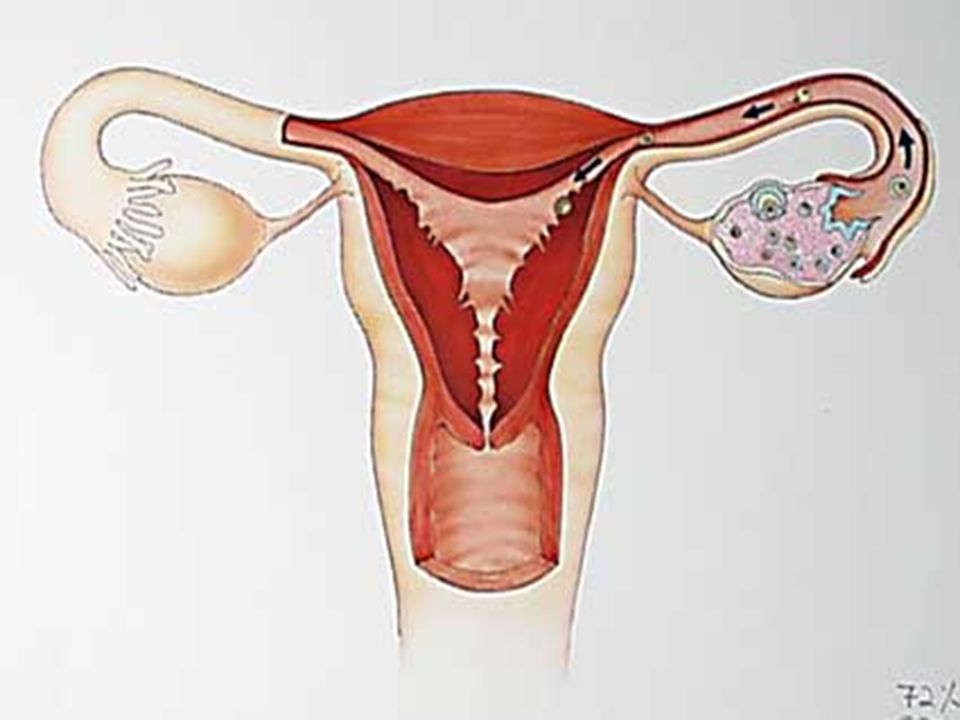
Front View of female reproductive system

28
The Ovaries The ovaries are the female sex organs
Occur in the lower part of the abdominal cavity and are held in place by ligaments Each ovary is made up of a covering of germinal epithelium with a large number of follicles within it

29
The Ovaries The germinal epithelium produces the follicles
Oogenesis takes place within the follicles to produce the ova Follicles secrete the female hormones oestrogen and progesterone

32
The fallopian tubes (oviducts)
The fallopian tubes convey ova from the ovaries to the uterus Upper part is expanded into ciliated funnels Partially enclosed ovaries

33
The Uterus The neck of the uterus, called the cervix, extends into the vagina The uterus serves for the attachment of the embryo Fertilisation takes place

34
The Uterus Two fallopian tubes open into the uterus which is pear shaped, hollow organ with muscular walls The ling of the uterus is called endometrium, is richly supplied with blood vessels

Similar presentations