Download presentation
Presentation is loading. Please wait.

1
Excretory Systems semester-2 MBBS
Dr. Kumar K.V (14/2/2011)
")
2
Relationship of the Kidneys to Vertebra and Ribs

3
External Anatomy of Kidney
Paired kidney-bean-shaped organ 4-5 in long, 2-3 in wide, 1 in thick Found just above the waist between the peritoneum & posterior wall of abdomen retroperitoneal along with adrenal glands & ureters Protected by 11th & 12th ribs with right kidney lower

4
Kidneys: Gross and Sectional Anatomy
Retroperitoneal Anterior surface covered with peritoneum Posterior surface against posterior abdominal wall Superior pole: T-12 Inferior pole: L-3 Right kidney ~ 2cm lower than left Adrenal gland on superior pole

5
Kidney Location Lateral to vertebral column high on body wall, under floating ribs, in retro-peritoneal position (posterior to the parietal peritoneum) The right kidney is slightly inferior to the left kidney in order to accommodate the liver Surrounded by the renal capsule with a fat pad 12 x 6 x 3 cm Bean shaped Hilus – indentation Retroperitoneal - between the body wall and peritoneum
 The right kidney is slightly inferior to the left kidney in order to accommodate the liver. Surrounded by the renal capsule with a fat pad. 12 x 6 x 3 cm. Bean shaped. Hilus – indentation. Retroperitoneal - between the body wall and peritoneum.")
6
Kidney External Anatomy
Average size – 12cm x 6cm x 3 cm Weights 150 grams or 5 oz Surrounded by three membranes (deep to superficial) Renal capsule – fibrous barrier for kidneys. Adipose capsule – fatty tissue designed for protection / stability. Renal fascia – dense fibrous CTP anchors kidneys/ adrenals/ membrane 1 and 2 to surroundings.
 Renal capsule – fibrous barrier for kidneys. Adipose capsule – fatty tissue designed for protection / stability. Renal fascia – dense fibrous CTP anchors kidneys/ adrenals/ membrane 1 and 2 to surroundings.")
7
Kidney - general information
Lie against posterior abdominal wall at level of T12-L3. Right kidney is lower than left kidney due to the shape of the liver. Lateral surface of kidney is convex while medial is concave. Concave side has a cleft – Renal Hilus Inside hilus is Renal sinus Where kidneys receive renal vessels and nerves.

8
Kidneys: Gross and Sectional Anatomy
Surrounding tissues, from deep to superficial: Fibrous capsule (renal capsule) Dense irregular CT Covers outer surface Perinephric fat (adipose capsule) Also called perirenal fat Completely surrounds kidney Cushioning and insulation Renal fascia Anchors kidney to posterior wall and peritoneum Paranephric fat Between renal fascia and peritoneum
 Dense irregular CT. Covers outer surface. Perinephric fat (adipose capsule) Also called perirenal fat. Completely surrounds kidney. Cushioning and insulation. Renal fascia. Anchors kidney to posterior wall and peritoneum. Paranephric fat. Between renal fascia and peritoneum.")
10
External Anatomy of Kidney
Protective coverings of kidney renal capsule---you will see in lab over sheep kidney adipose surrounds that layers of thin fascia hold everything against back body wall peritoneum over all Blood vessels & ureter enter hilus of kidney Renal capsule = transparent membrane maintains organ shape Adipose capsule that helps protect from trauma Renal fascia = dense, irregular connective tissue that holds against back body wall

11
Kidney- External Anatomy
Lateral surface- convex Medial is concave- Renal Hilum Opening to Kidney Renal Sinus Space within hilus Kidneys receive blood vessels and nerves.

12
The Position of the Kidneys
Figure 26.2a, b

14
Figure 26.3 The Urinary System in Gross Dissection

15
Kidney Internal Anatomy I
Renal arteries and veins Bring blood in and out of kidney Renal cortex Outer layer of Kidney Renal medulla Inner layer of Kidney Nephron

16
Human Kidney

17
Kidneys: Gross and Sectional Anatomy
Minor calyx: Funnel shaped Receives renal papilla 8 to 15 per kidney, one per pyramid Major calyx Fusion of minor calyces 2 to 3 per kidney Major calyces merge to form renal pelvis Renal Lobe Pyramid plus some cortical tissue 8 to 15 per kidney

18
a. Right renal vein enters IVC directly
Detailed Anatomy, cont … 2. Renal Veins a. Right renal vein enters IVC directly b. Left renal vein passes anterior to aorta, posterior to SMA, then into IVC c. Venous pattern complex

19
Variations in Renal Veins
Note the duplication in the Left Renal Vein Branches surround aorta

20
Origin of Renal, Gonadal Arteries
Note the right gonadal artery arising from the aorta, then branching to form the inferior capsular artery. R and L Renal Arteries Gonadal Arteries

21
d. R. renal artery courses from aorta posterior to IVC into hilus
Detailed Anatomy, cont … d. R. renal artery courses from aorta posterior to IVC into hilus e. Left renal artery course is from aorta directly to hilus f. May see 2 or 3 pairs of renal arteries g. Gonadal arteries: 1. may arise from renal artery usually arise from aorta

22
a. arise from abdominal aorta
Detailed Anatomy, cont … C. Blood supply 1. Renal arteries: a. arise from abdominal aorta b. Divide into 2 or 3 branches before entering kidney c. If 3 branches, may form: 1. “vascular fork” 2. may constrict ureter

24
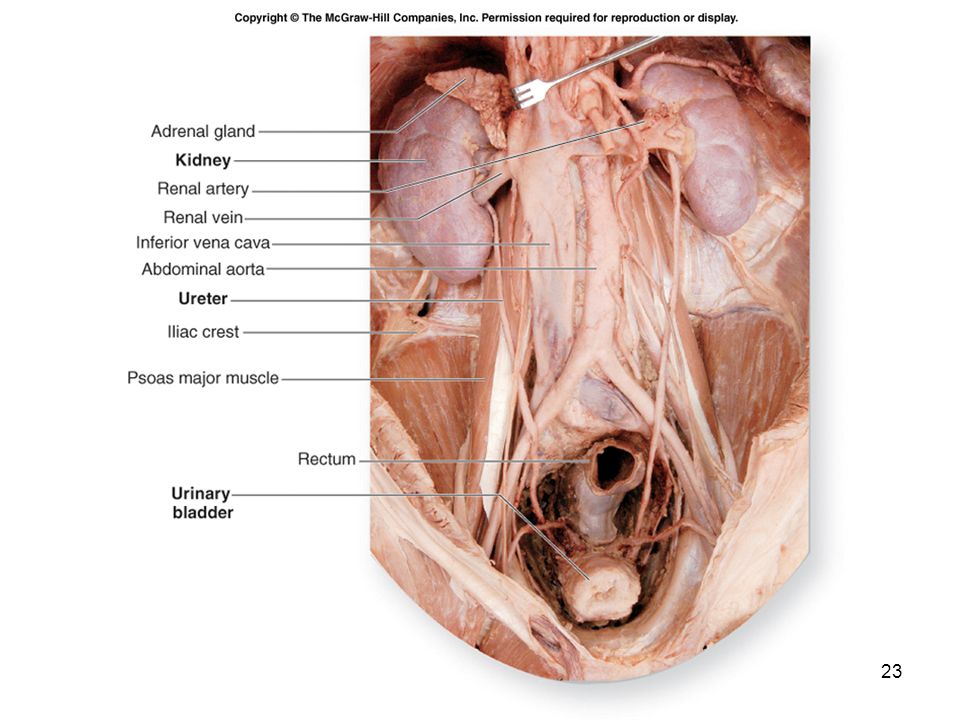
Right Kidney: Relationships
Note: Liver Descending duodenum Psoas muscle Quadratus lumborum muscle IVC

25
Left Kidney: Relationships
Note: Tail of pancreas Splenic flexure of colon Aorta Psoas and quadratus lumborum muscles

28
Anatomy of Urinary Bladder
Hollow, distensible muscular organ with capacity of mL Trigone is smooth flat area bordered by 2 ureteral openings and one urethral opening

29
Location of Urinary Bladder
Posterior to pubic symphysis In females is anterior to vagina & inferior to uterus In males lies anterior to rectum

30
Urinary Tract – Urinary Bladder
The urinary bladder: expandable, muscular container serves as a reservoir for urine positioned immediately superior and posterior to the pubic symphysis. in females the urinary bladder is in contact with the uterus posterosuperiorly and with the vagina posteroinferiorly. in males it is in contact with the rectum posterosuperiorly and is immediately superior to the prostate gland. is a retroperitoneal organ. when empty exhibits an upside-down pyramidal shape. Filling with urine distends it superiorly until it assumes an oval shape.

31
Urinary Bladder Located in pelvic cavity, posterior to pubic symphysis
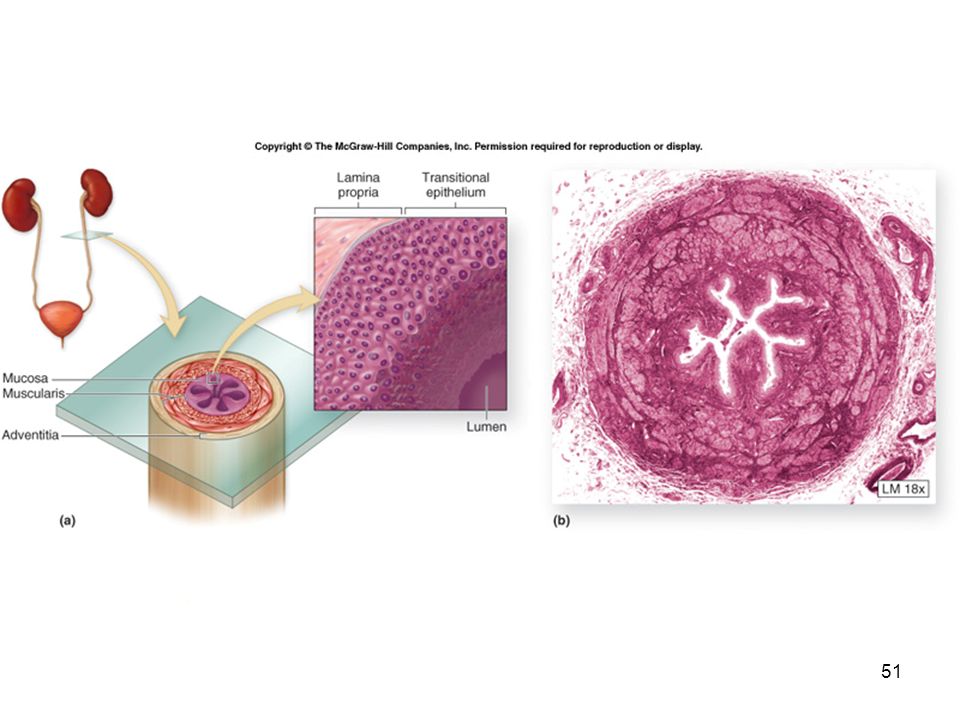
3 layers parietal peritoneum, superiorly; fibrous adventitia rest muscularis: detrusor muscle, 3 layers of smooth muscle mucosa: transitional epithelium trigone: openings of ureters and urethra, triangular rugae: relaxed bladder wrinkled, highly distensible capacity: moderately full ml, max ml

34
Urinary Tract – Urinary Bladder
Trigone posteroinferior triangular area of the urinary bladder wall formed by imaginary lines connect the two posterior ureteral openings and the anterior urethral opening. The trigone remains immovable as the urinary bladder fills and evacuates. It functions as a funnel directs urine into the urethra as the bladder wall contracts four tunics mucosa submucosa Muscularis: called the detrusor muscle adventitia. Internal urethral sphincter (smooth muscle)
")
35
Urinary Bladder Smooth, collapsible, muscular sac
Temporarily stores urine Figure 15.6 Slide 15.21a Copyright © 2003 Pearson Education, Inc. publishing as Benjamin Cummings

36
Urinary Bladder Trigone – three openings Two from the ureters
One to the urethra Figure 15.6 Slide 15.21b Copyright © 2003 Pearson Education, Inc. publishing as Benjamin Cummings

37
Urethra Release of urine is controlled by:
Internal urethral sphincter (involuntary) External urethral sphincter (voluntary) Copyright © 2003 Pearson Education, Inc. publishing as Benjamin Cummings
 External urethral sphincter (voluntary) Copyright © 2003 Pearson Education, Inc. publishing as Benjamin Cummings.")
38
Anatomy of Ureters 10 to 12 in long Varies in diameter from 1-10 mm
Extends from renal pelvis to bladder Retroperitoneal Enters posterior wall of bladder Physiological valve only bladder wall compresses arterial opening as it expands during filling flow results from peristalsis, gravity & hydrostatic pressure

39
Urinary Tract : Ureters
long, fibromuscular tubes conduct urine from the kidneys to the urinary bladder. average 25 centimeters in length retroperitoneal. ureters originate at the renal pelvis extend inferiorly to enter the posterolateral wall of the base of the urinary bladder. wall is composed of three concentric tunics. mucosa muscularis adventitia.

40
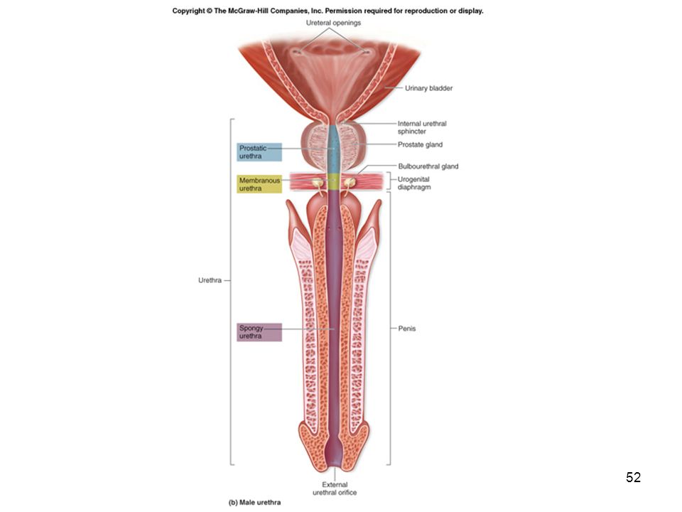
Urethra, Male Male – cm 1. prostatic urethra – from base of bladder through prostate gland 2. membranous urethra – between prostate gland & base of penis 3. penile (spongy) urethra – traverses penis to orifice
 urethra – traverses penis to orifice.")
41
Male Bladder and Urethra
18 cm long Internal urethral sphincter External urethral sphincter 3 regions prostatic urethra receives semen membranous urethra passes through pelvic cavity penile urethra

42
Urethra Fibromuscular tube conducts urine to the exterior of the body.
exits the urinary bladder through the urethral opening at anteroinferior surface conducts urine to the exterior of the body. Tunica mucosa: is a protective mucous membrane houses clusters of mucin-producing cells called urethral glands. Tunica muscularis: primarily smooth muscle fibers help propel urine to the outside of the body. Two urethral sphincters: Internal urethral sphincter restrict the release of urine until the pressure within the urinary bladder is high enough External urethral sphincter and voluntary activities needed to release the urine are activated.

43
Anatomy of the Urethra Females Males
length of 1.5 in., orifice between clitoris & vagina histology transitional changing to nonkeratinized stratified squamous epithelium, lamina propria with elastic fibers & circular smooth muscle Males tube passes through prostate, UG diaphragm & penis 3 regions of urethra prostatic urethra, membranous urethra & spongy urethra circular smooth muscle forms internal urethral sphincter & UG diaphragm forms external urethral sphincter

44
Urethra, Female External urethral sphincter
– voluntary at pelvic floor 3-5 cm – from base of bladder to vestibule UTIs (esp. E.coli)
")
45
Urethra: Gender Differences
Length Females – 3–4 cm (1 inch) Males – 20 cm (8 inches) Location Females – anterior to the vagina Males – through the prostate and penis Slide 15.24a Copyright © 2003 Pearson Education, Inc. publishing as Benjamin Cummings
 Males – 20 cm (8 inches) Location. Females – anterior to the vagina. Males – through the prostate and penis. Slide 15.24a. Copyright © 2003 Pearson Education, Inc. publishing as Benjamin Cummings.")
46
Female Urethra 3 to 4 cm long External urethral orifice
between vaginal orifice and clitoris Internal urethral sphinctermuscle, involuntary control

47
Male vs. Female Fig 23.17

48
Urethra The internal urethral sphincter
involuntary (smooth muscle) superior sphincter surrounding the neck of the bladder, where the urethra originates. a circular thickening of the detrusor muscle controlled by the autonomic nervous system The external urethral sphincter inferior to the internal urethral sphincter formed by skeletal muscle fibers of the urogenital diaphragm. a voluntary sphincter controlled by the somatic nervous system this is the muscle children learn to control when they become “toilet-trained”
 superior sphincter surrounding the neck of the bladder, where the urethra originates. a circular thickening of the detrusor muscle. controlled by the autonomic nervous system. The external urethral sphincter. inferior to the internal urethral sphincter. formed by skeletal muscle fibers of the urogenital diaphragm. a voluntary sphincter. controlled by the somatic nervous system. this is the muscle children learn to control when they become toilet-trained")
49
Female Urethra Has a single function:
to transport urine from the urinary bladder to the vestibule, an external space immediately internal to the labia minora 3 to 5 centimeters long, and opens to the outside of the body at the external urethral orifice located in the female perineum.

50
Male Urethra Urinary and reproductive functions:
passageway for both urine and semen Approximately 18 to 20 centimeters long. Partitioned into three segments: prostatic urethra is approximately 3 to 4 centimeters long and is the most dilatable portion of the urethra extends through the prostate gland, immediately inferior to the male bladder, where multiple small prostatic ducts enter it membranous urethra is the shortest and least dilatable portion extends from the inferior surface of the prostate gland through the urogenital diaphragm spongy urethra is the longest part (15 centimeters) encased within a cylinder of erectile tissue in the penis called the corpus spongiosum extends to the external urethral orifice
 encased within a cylinder of erectile tissue in the penis called the corpus spongiosum. extends to the external urethral orifice.")
53
Disorders of Urinary System
Renal calculi Urinary tract infections Glomerular disease Renal failure Polycystic kidney disease

Similar presentations


 2 Ureters (Passage tubes ?) Bladder (Storage) Urethra (excretion)>")


