Download presentation
Presentation is loading. Please wait.

23
Bone Formation and Growth

63
PG. 106 TABLE 6-3

68
ankylosis

69
Kyphosis – known as hunchback. It is an abnormal humped curvature In the thoracic region of the spine. Most common in adolescents

70
Osgood Schlatter Disease A painful, incomplete separation of the epiphysis of the tibial tubercle from the bone shaft.

72
Rheumatoid Arthritis – involves connective throughout the entire body, but Mainly involves the synovium in the joint.

73
Pathologic Bone Healing

84
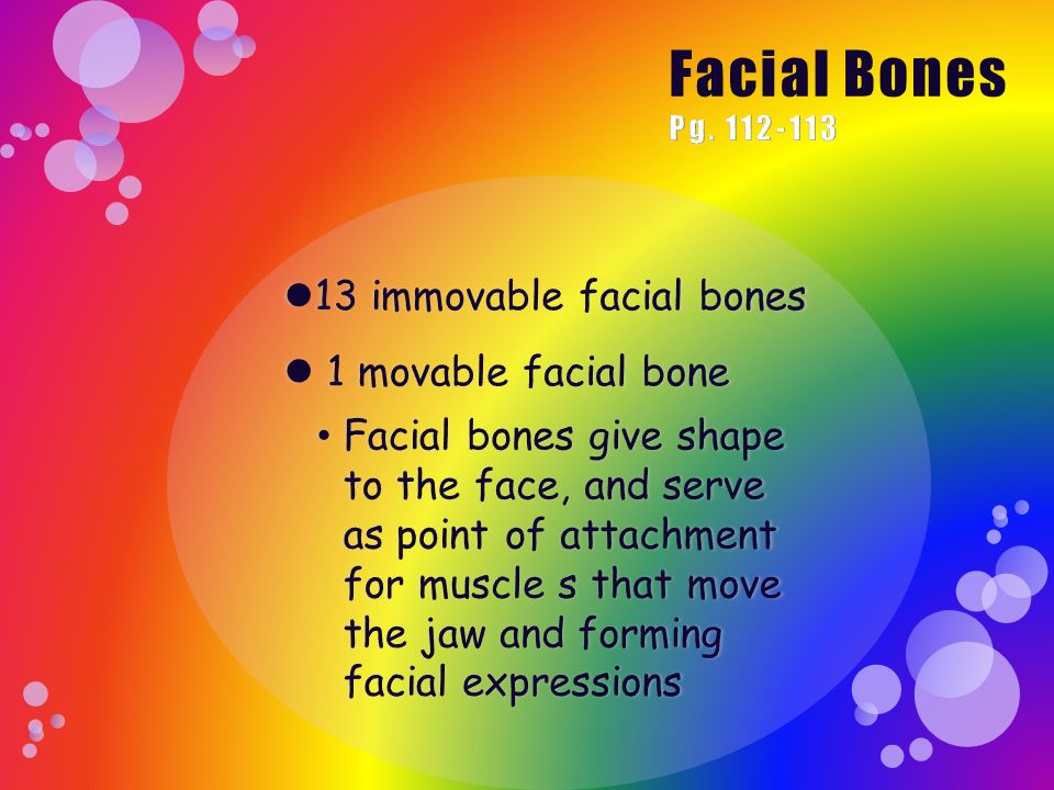
The Facial Bones Include: Nasal Bones (2) Vomer Inferior Nasal Conchae (2) Lacrimal Bones (2) Palatine Bones (2) Zygomatic (2) Maxillary Bones (2) Mandible
 Vomer Inferior Nasal Conchae (2) Lacrimal Bones (2) Palatine Bones (2) Zygomatic (2) Maxillary Bones (2) Mandible")
86
Development of The Infantile Skull The bones of the skull have not matured and completed their growth. Fibrous connective tissue is located between the bones and these areas are called fontanels, commonly referred to as soft spots. The Four Fontanels are: Posterior fontanel Anterior fontanel Mastoid fontanel Sphenoid fontanel

87
Thoracic Cage The thoracic cage, also called the rib cage, consist of the ribs and the sternum. The function of the rib cage is to enclose and protect the lungs and heart. The ribs are also flexible so as to allow the lungs to expand during respiration.

88
There are 12 pairs of ribs. The first 7 pairs are referred to as the true ribs and they join the sternum via the costal cartilage. The next 3 pairs are called the false ribs, since they are not attached to the sternum but are attached to the seventh rib by costal cartilage. The last 2 pairs are called floating ribs, since they are not attached to the sternum or other ribs.

90
Vertebral Column The vertebral column begins close to the foramen magnum of the skull and extends posteriorly to the pelvis. Functions of the column include: 1.Support head and trunk 2.Being flexible to allow the person to bend forward, backward, and to the side, and at the waist. 3.Forming the passageway called the vertebral canal or vertebral foramen to allow the spinal cord to pass through.

91
The column consists of bone called the vertebrae, which are separated by a fibrocartilage called intervertebral disks and are connected to each other by ligaments. The vertebral column has four natural curvatures: 1.Cervical curvature: Located in the neck and curves in a convex anterior direction. 2.Thoracic curvature: Located in the thoracic region; concave anterior curvature. 3.Lumbar curvature: Located in the lower back; convex anterior curvature. 4.Pelvic curvature: Located in the pelvic region; concave anterior curvature.

93
There are three types of vertebrae : 1.Cervical Vertebrae: 7 located in the neck, They are the smallest of the vertebrae. The transverse processes contain transverse foramina, which are openings for arteries to pass through to the brain. Atlas: First cervical vertebra, Supports the head Axis: Second cervical vertebra 2.Thoracic Vertebrae: 12 thoracic vertebrae, have long spinous process that ends in a point, slightly curving downward. Facets are located laterally on the body and articulate with the rib. Starting with the third thoracic vertebrae and traveling downward, each of the vertebrae increases in size in order to aid in weight bearing of the body. 3. Lumbar Vertebrae: 5 lumbar vertebrae, they perform the majority of the weight bearing function as compared to the other vertebrae of the body’ thus they have large bodies, thicker, shorter spinous processes, and the transverse processes project posteriorly at an increased angle.

95
Sacrum o Consist of five vertebrae that are fused into a triangular shape. o Forms the posterior portion of the pelvic cavity. Sacroiliac joints mark where the sacrum joins the Ilium. o Anterior edge of the first sacral vertebra is called the sacral promontory, which is an important anatomic landmark used in determining the size of the pelvis in a female.

96
Coccyx Better known as the tailbone. Most inferior portion of the vertebral column. Composed of four fused vertebrae.

Similar presentations


 are the smallest, lightest vertebrae C3-C7 are distinguished with an oval body, short spinous processes, and.>")
 and intervertebral disks Function: (1) supports head,>")


