Download presentation
Presentation is loading. Please wait.

1
Ass. Prof. Faculty of Medicine
Essentials of Human Anatomy The Skeletal System 2 The Axial Skeletal System Chapter 5 Dr Fadel Naim Ass. Prof. Faculty of Medicine IUG 1

2
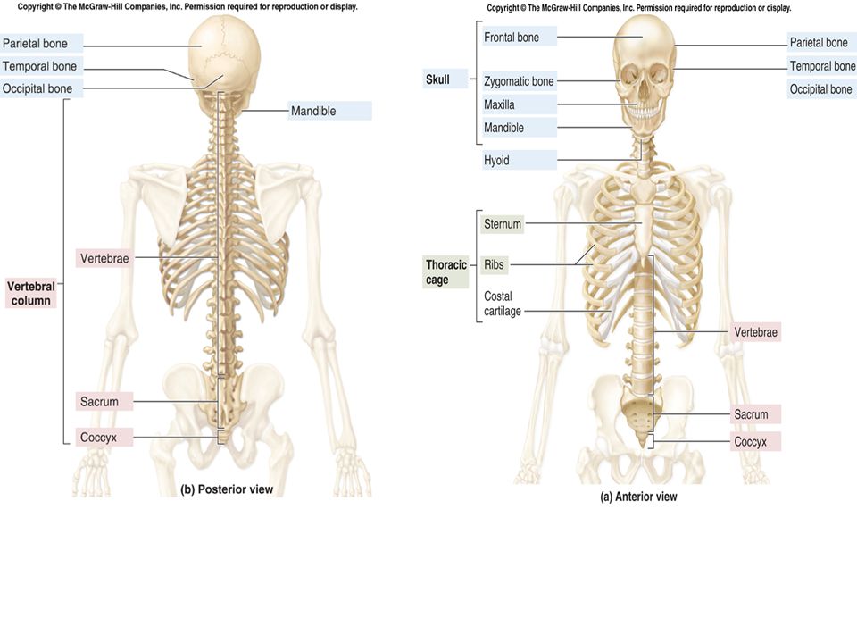
Skeletal Organization
Axial Skeleton head neck trunk Appendicular Skeleton upper limbs lower limbs pectoral girdle pelvic girdle

4
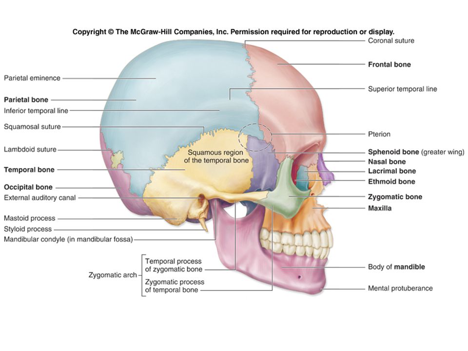
The Skull Cranial bones form the rounded cranium, which completely surrounds and encloses the brain. Facial bones form the bones of the face. They also protect the entrances to the digestive and respiratory systems as well as provide attachment sites for facial muscles

5
Cranium Frontal (1)
")
6
Cranium Parietal (2)
")
7
Cranium Occipital (1)
")
8
Cranium Temporal (2)
")
9
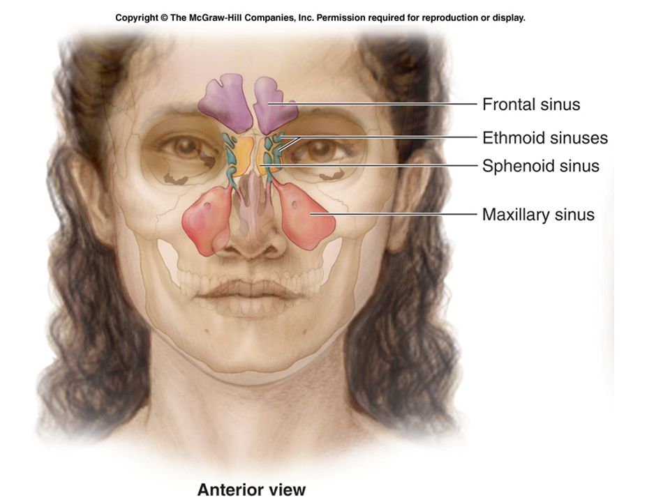
Cavities of The Skull The largest cavity is the cranial cavity, which encloses, cushions, and supports the brain. The skull also has several smaller cavities, including the orbits (eye sockets), the oral cavity (mouth), the nasal cavity, and the paranasal sinuses.
, the oral cavity (mouth), the nasal cavity, and the paranasal sinuses.")
10
Cranium Sphenoid (1)
")
11
Cranium Ethmoid (1)
")
12
Facial Skeleton Maxillary (2)
")
13
Facial Skeleton Palatine (2)
")
14
Facial Skeleton Zygomatic (2)
")
15
Facial Skeleton Lacrimal (2) Nasal (2)
 Nasal (2)")
16
Facial Skeleton Vomer (1)
")
17
Facial Skeleton Inferior Nasal Conchae (2)
")
18
Facial Skeleton Mandible (1)
")
19
Sinuses Have a mucous lining that helps to humidify and warm inhaled air. Cause these skull bones to be lighter. Provide resonance to the voice.

21
Hyoid Bone

22
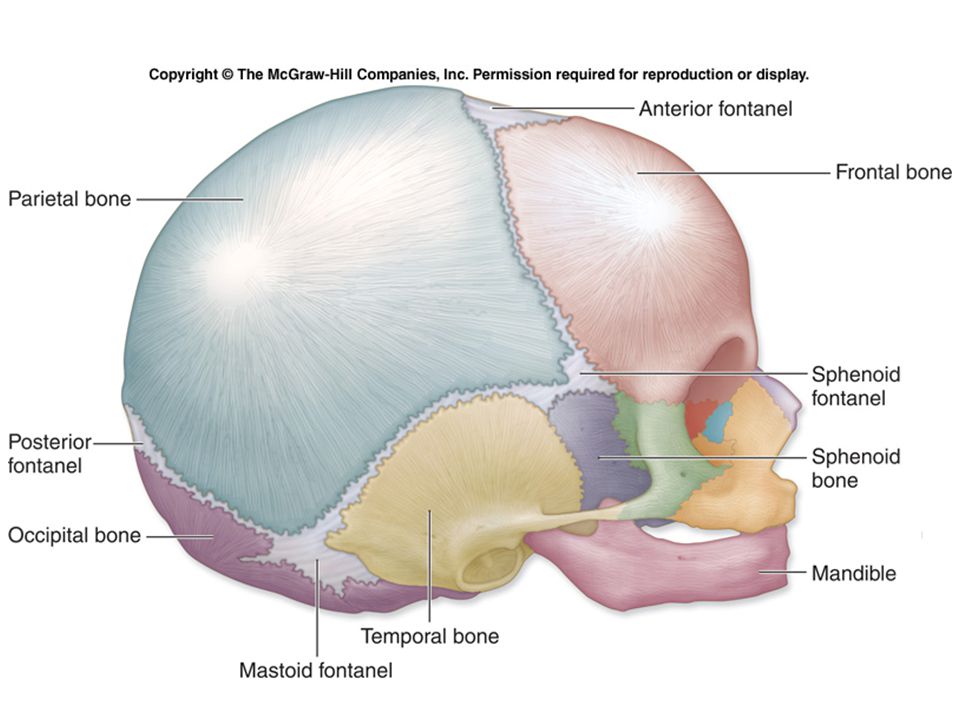
Fontanels The regions between the cranial bones are thickened, fibrous membrane remnants that are not yet ossified. Sometimes referred to as the “soft spots” on a baby’s head. They close by 15 months of age. When a baby travels through the birth canal, the cranial bones overlap at these fontanels, in order to ease the baby’s passage. Newborns frequently have a “cone-shaped” head due to this temporary deformation. Fontanels – fibrous membranes

24
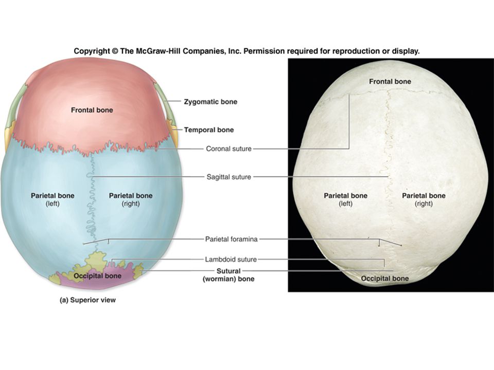
Sutures of the Skull Sutures are immovable fibrous joints that form the boundaries between the cranial bones. Dense regular connective tissue seals cranial bones firmly together at a suture. Allow the cranium to grow and expand during childhood. In adulthood, when cranial growth has stopped, the sutures fuse and are obliterated.

27
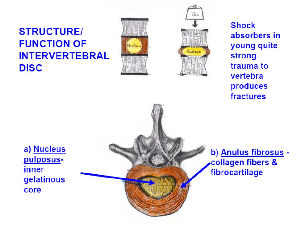
Functions Of Vertebral Column
1) Support weight Transmits weight to pelvis and lower limbs 2) Houses and protects spinal cord spinal nerves leave cord between vertebrae 3) Permits movements 4) Provides for muscle attachments muscles of back muscles of head Neck upper extremity thorax
 Support weight. Transmits weight to pelvis and lower limbs. 2) Houses and protects spinal cord. spinal nerves leave cord between vertebrae. 3) Permits movements. 4) Provides for muscle attachments. muscles of back. muscles of head. Neck. upper extremity. thorax.")
28
Regions and Normal Curvatures
Formed from 33 bones in the adult Divided into five major regions Cervical vertebrae 7 vertebrae of the neck region Thoracic vertebrae 12 vertebrae of the thoracic region Lumbar vertebrae 5 vertebrae of the lower back Sacrum Inferior to lumbar vertebrae Articulates with coxal bones Coccyx Most inferior region of the vertebral column

29
Regions and Normal Curvatures
Four distinct curvatures give vertebral column an S-shape Cervical and lumbar curvature concave posteriorly Thoracic and sacral curvatures convex posteriorly Curvatures increase the resilience of the spine

30
General Structure of Vertebrae

32
Cervical Vertebrae Atlas – 1st; supports head
Axis – 2nd; dens pivots to turn head transverse foramina bifid spinous processes vertebral prominens – useful landmark

33
Thoracic Vertebrae long spinous processes rib facets

34
Lumbar Vertebrae large bodies thick, short spinous processes

35
Sacrum five fused vertebrae median sacral crest
posterior sacral foramina posterior wall of pelvic cavity sacral promontory

36
Coccyx tailbone four fused vertebrae

37
The Axial Skeleton Throughout Life
Curvatures of the vertebral column Primary curvatures thoracic and sacral curvatures An infant's spine is C-shaped at birth Secondary curvatures cervical and lumbar curvatures Develop when a baby begins to walk Redistributes weight of the upper body over the lower limbs

38
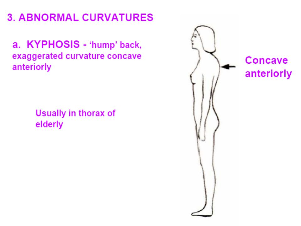
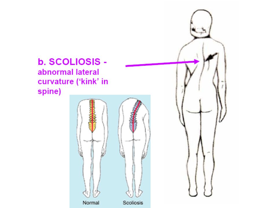
Three Main Spinal Curvature Deformities
Kyphosis is an exaggerated thoracic curvature that is directed posteriorly, producing a “hunchback” look. Lordosis is an exaggerated lumbar curvature, often called “swayback,” that is observed as a protrusion of the abdomen and buttocks. Scoliosis is an abnormal lateral curvature that sometimes results during development when both the vertebral arch and body fail to form, or form incompletely, on one side of a vertebra. scoliosis is the most common spinal curvature deformity.

41
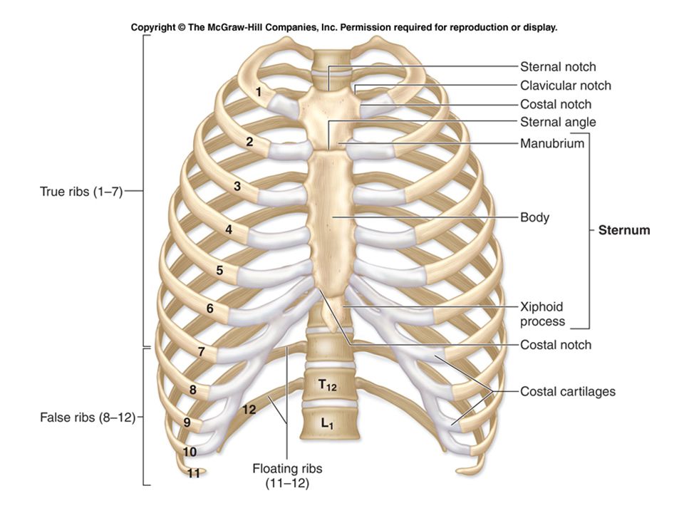
Thoracic Cage Consists of the thoracic vertebrae posteriorly, the ribs laterally, and the sternum anteriorly. Acts as a protective cage around vital organs, such as the heart, lungs, trachea, and esophagus. Provides attachment points for many muscles supporting the pectoral girdles, the chest, the neck, the shoulders, the back, and the muscles involved in respiration.

43
Ribs Both males and females 12 pairs
Ribs 1–7 are called true ribs. At the anterior body wall, the true ribs connect individually to the sternum by separate cartilaginous extensions called costal cartilages. Ribs 8–12 are called false ribs because their costal cartilages do not attach directly to the sternum. The costal cartilages of ribs 8–10 fuse to the costal cartilage of rib 7 and thus indirectly articulate with the sternum. The last two pairs of false ribs (ribs 11 and 12) are called floating ribs because they have no connection with the sternum.
 are called floating ribs because they have no connection with the sternum.")
44
THE END

Similar presentations