Download presentation
Presentation is loading. Please wait.

1
Fig. 18.1(TE Art) Pineal gland Pituitary gland Hypothalamus Thyroid gland Thymus Adrenal glands Pancreas Testes Ovaries Gonads Parathyroid glands
 Pineal gland Pituitary gland Hypothalamus Thyroid gland Thymus Adrenal glands Pancreas Testes Ovaries Gonads Parathyroid glands.")
2
Fig. 18.3(TE Art) Endocrine system Endocrine cells Hormone in bloodstream Nervous system
 Endocrine system Endocrine cells Hormone in bloodstream Nervous system")
3
Fig. 18.1(TE Art) Pineal gland Pituitary gland Hypothalamus Thyroid gland Thymus Adrenal glands Pancreas Testes Ovaries Gonads Parathyroid glands
 Pineal gland Pituitary gland Hypothalamus Thyroid gland Thymus Adrenal glands Pancreas Testes Ovaries Gonads Parathyroid glands.")
4
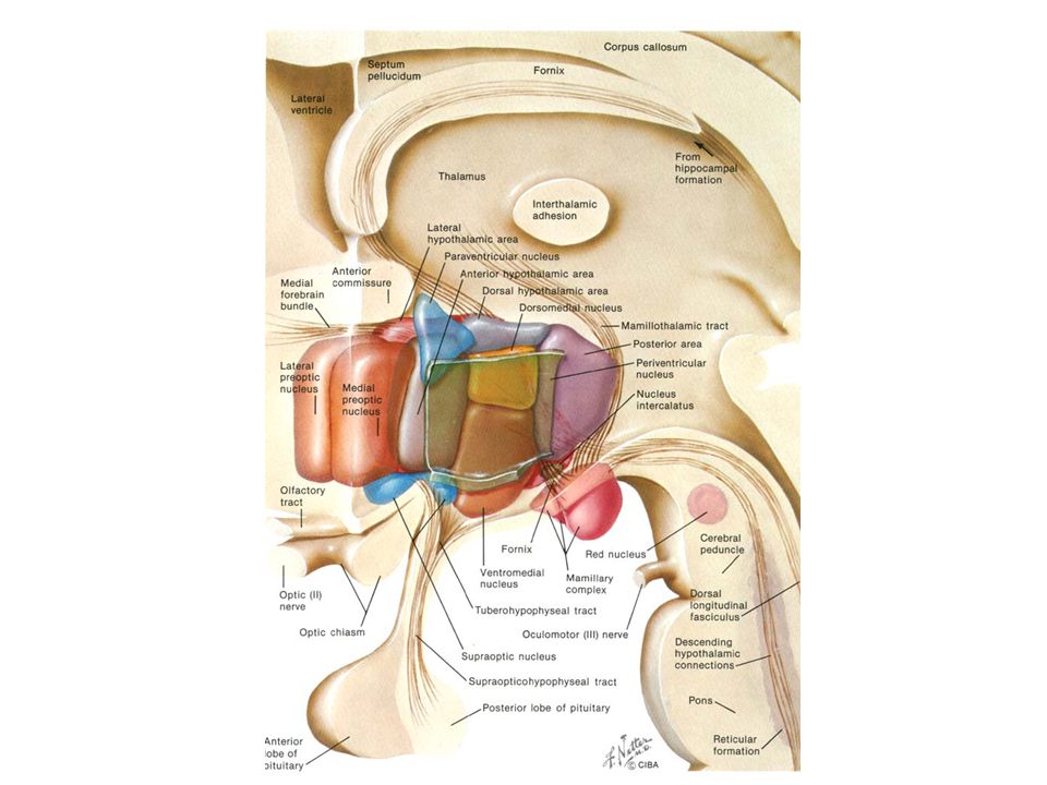
hypothalamus Pituitary gland

6
Hypothalamo- hypophyseal tract Stalk Neurohypophysis Posterior lobe Pars tuberalis Anterior lobe Adenohypophysis Pituitary gland

7
Fig. 18.4a(TE Art) Hypothalamohypophyseal tract Posterior lobe Paraventricular nucleus Supraoptic nucleus Oxytocin ADH Anterior lobe Oxytocin = uterus & mammary glands Antidiuretic hormone = kidneys
 Hypothalamohypophyseal tract Posterior lobe Paraventricular nucleus Supraoptic nucleus Oxytocin ADH Anterior lobe Oxytocin = uterus & mammary glands Antidiuretic hormone = kidneys.")
8
Superior hypophyseal artery Posterior pituitary Anterior pituitary Releasing hormones “go and do something” hormones

9
Fig. 18.6(TE Art) Growth hormone ACTH TSH prolactin Liver Fat, muscle, bone LH FSH TRH GnRH CRH Hypothalamus Adrenal cortex OvaryTestis Thyroid Mammary gland IGF
 Growth hormone ACTH TSH prolactin Liver Fat, muscle, bone LH FSH TRH GnRH CRH Hypothalamus Adrenal cortex OvaryTestis Thyroid Mammary gland IGF.")
10
Fig. 18.10a(TE Art) Adrenal gland Kidney Adrenal cortex Adrenal medulla epinephrine norepinephrine
 Adrenal gland Kidney Adrenal cortex Adrenal medulla epinephrine norepinephrine")
11
Adrenal gland Kidney Adrenal cortex mineralcorticoids glucocorticoids sex steroids Adrenal medulla Aldosterone (mineralcorticoid): kidney = retain Na, excrete K (water retained, BP) Cortisol (glucocorticoid): fat & protein breakdown, glucose synthesis, fatty acid & glucose release into blood, help body adapt to stress, repair damaged tissues Dehydroepiandrosterone (DHEA): weak testosterone = libido, 2 nd sex characteristics
: kidney = retain Na, excrete K (water retained, BP) Cortisol (glucocorticoid): fat & protein breakdown, glucose synthesis, fatty acid & glucose release into blood, help body adapt to stress, repair damaged tissues Dehydroepiandrosterone (DHEA): weak testosterone = libido, 2 nd sex characteristics")
12
Cushing’s Syndrome – adrenal cortex – hypersecretion of cortisol

13
Thyroid Thymus Trachea

14
Thyroid gland Follicular cells: T3 & T4 – increase metabolic rate C cells: calcitonin – inhibits osteoclasts…..

15
Pharynx Thyroid gland Esophagus Parathyroid glands Trachea Detect low calcium Secrete PTH increase Ca absorption inhibits Ca excretion stimulates osteoclasts Posterior view

16
Fig. 18.11a(TE Art) Bile duct Duodenum Head of pancreas Pancreatic ducts Body of pancreas Tail of pancreas
 Bile duct Duodenum Head of pancreas Pancreatic ducts Body of pancreas Tail of pancreas.")
17
Pancreatic islet cell-- insulin cell -- somatostatin cell -- glucagon Insulin: controls glucose transport into cells Diabetes Type I: low or no B-cells, no insulin Diabetes Type II: insulin insensitivity (receptor) hyperglycemia emaciation atherosclerosis (fatty deposits) ketoacidosis (low blood pH) = coma, death
 hyperglycemia emaciation atherosclerosis (fatty deposits) ketoacidosis (low blood pH) = coma, death")
18
Fig. 18.1(TE Art) Pineal gland Pituitary gland Hypothalamus Thyroid gland Thymus Adrenal glands Pancreas Testes Ovaries Gonads Parathyroid glands
 Pineal gland Pituitary gland Hypothalamus Thyroid gland Thymus Adrenal glands Pancreas Testes Ovaries Gonads Parathyroid glands.")
Similar presentations


>")


