Download presentation
Presentation is loading. Please wait.

1
URINARY TRACT ANATOMY Ali Haddad and Matt Newman

2
A 20 year old male is subject to an awkward tackle during a rugby match and landed heavily on his back. 4 hours later the man’s urine was coloured red. What structure(s) are most likely to have been damaged (explain)?
 are most likely to have been damaged (explain) .")
3
Blunt force trauma to kidney Cracked ribs (ribs 11 and 12)
")
4
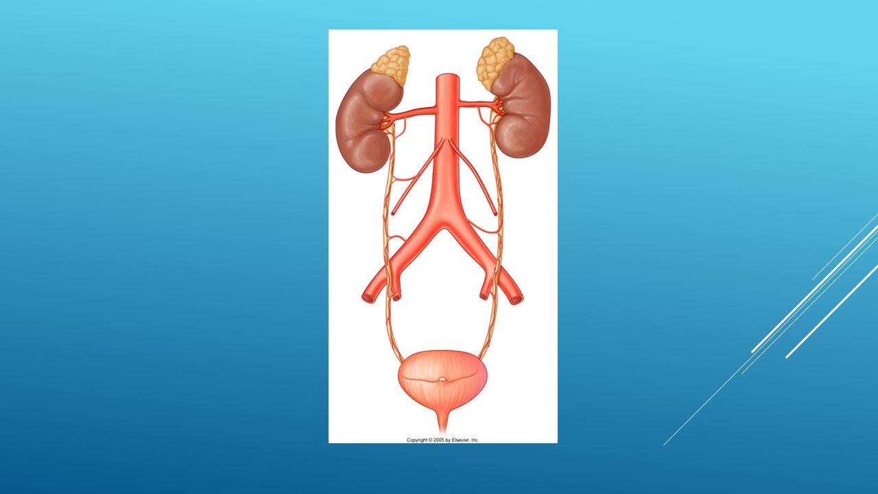
The kidneys are paired retroperitoneal organ located on posterior abdominal wall L3 L1 T10

5
Where could a kidney stone get stuck in the ureter (explain)?
")
6
Renal pelvis L1 Passes over the pelvic brim Vesico-uteric junction

7
Why might the pain from inflammation of the renal pelvis or proximal ureter be made worse by flexing the hip? Where would pain from the stated regions be felt?

9
Psoas major flexes the hip (don’t worry too much about this) but when it moves it irritates the inflamed proximal ureter. Pain referral via the afferent nerves of the renal plexus (T12) Hence the loin pain
 Hence the loin pain.")
10
Where would a patient with ureteric colic from a kidney stone complain of pain?

11
Kidney A renal calculus passing down ureter shows classic shifting loin-groin pain Renal calculus

12
Visceral afferents neurons run to CNS with sympathetic nerves Renal plexus Abdomino-aortic plexus Hypogastric plexus (superior) T12 L1/2 Sensory nerve route
 T12 L1/2 Sensory nerve route")
13
What is the main differential concern for an elderly patient presenting with presumed left sided renal colic?

14
AAA

15
During abdominal surgery the ureter needs to be moved. Which way should it be moved and why?

17
Blood supply from multiple arteries: Renal Gonadal Aortic Internal iliac Vesical/prostatic Displace ureter medially in abdo cavity Displace ureter laterally in pelvic cavity

18
A newborn presents with a clear fluid leaking from the umbilicus. Tests show the fluid is urine. Explain this case.

19
Allantois passes from cloaca to umbilicus Allantoic blood vessels become umbilical arteries & veins Formation of blood cells The bladder develops from the anterior part of the cloaca with the allantois attached Remnants of the allantois can cause clinical problems

20
Why might childbirth cause urinary incontinence? (think nerves, support and sphincters)
")
21
Pubo-vesical ligament (female) Bladder support is derived from multiple structures including the pelvic floor, perineal membrane & ligaments Levator ani in both sexes Somatic = Stop (pudendal nerve) Contracts external urethral sphincter Need to relax to urinate During birth, the muscles can get stretched and weakened. The pudendal nerve can be damaged causing the sphincter to loose it’s tone

24
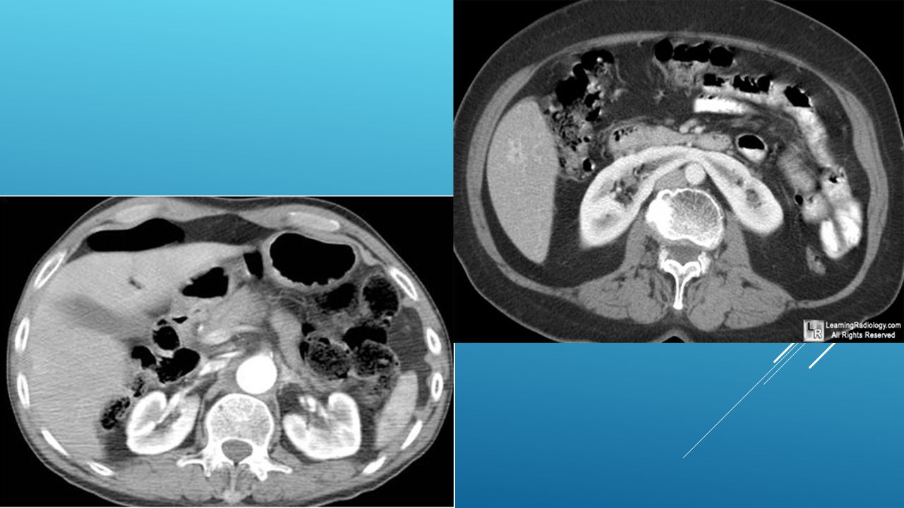
Horse Shoe Kidney

Similar presentations