Download presentation
Presentation is loading. Please wait.

1
Kidneys and Adrenal Glands
Department of Regional Anatomy and Operative Surgery

2
Position, relation Structure Blood supply Lymphatic drainage Innervations

3
Position Retroperitoneal Upper poles T12 vertebra Lower poles
L3 vertebra Right is lower than left

4
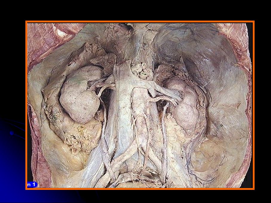
Cadaveric kidneys

5
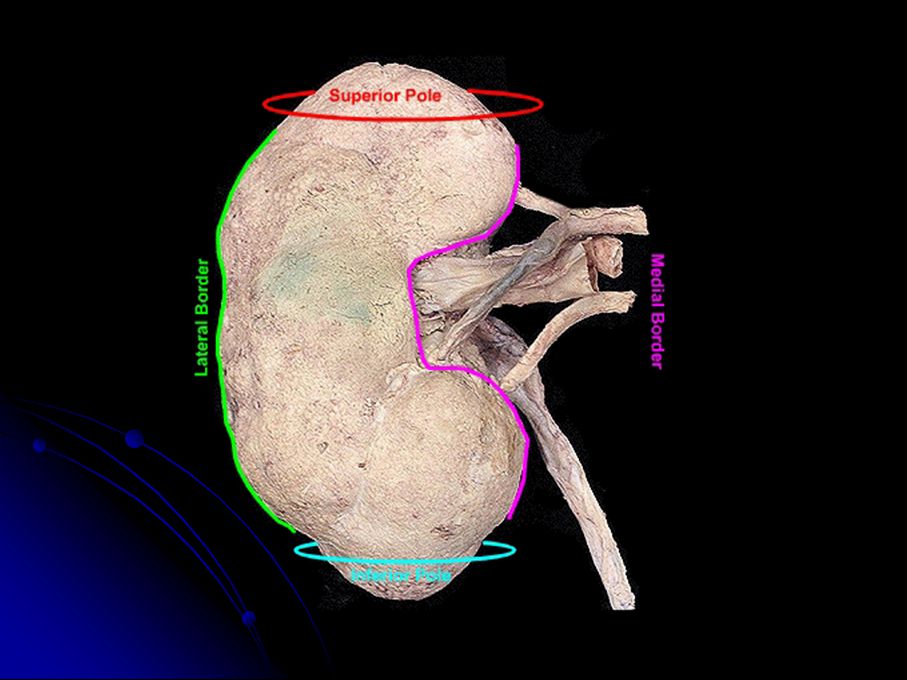
Surface Projection of kidney

6
Renal Angle Tenderness or percussing pain caused by kidney disease is localized here

7
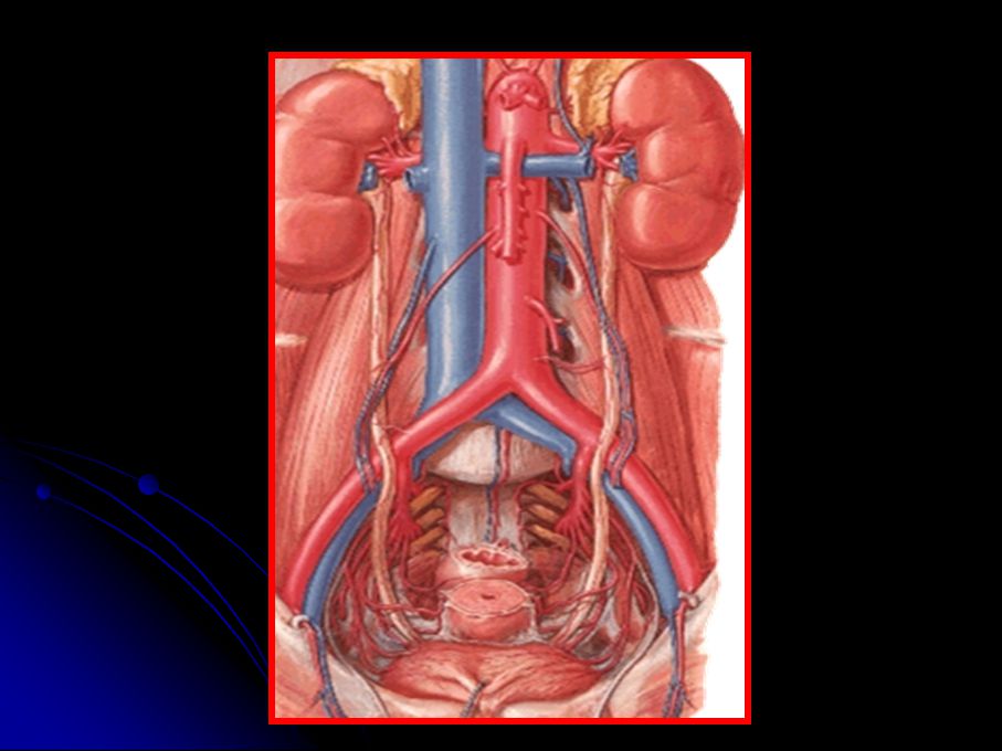
Neighbor of Kidney

9
Anterior Surface of the kidney

11
Posterior Surface

12
Costodiaphragmatic recess of the pleural cavity

13
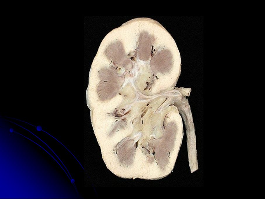
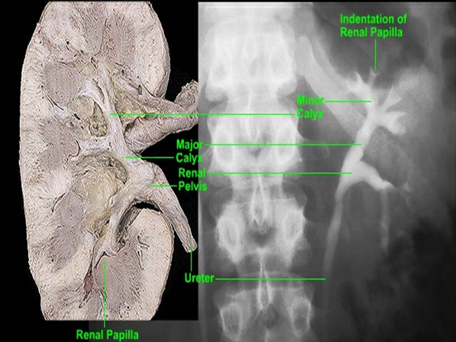
Renal hilum, Renal sinus and Renal pedicle
cortex Renal pyramid Renal column Renal sinus calyx Renal hilum papilla Renal pedicle

16
From anterior to posterior
renal vein renal artery renal pelvis From above downwards the renal artery

17
Hydronephrosis

18
renal artery T11

19
Renal artery

20
Vascular renal segment
Superior (apical) Anterior superior (upper) posterior Anterior inferior (middle) Inferior (lower)
 Anterior superior (upper) posterior. Anterior inferior (middle) Inferior (lower)")
21
variation of renal artery
!

22
Variation of Renal Artery

23
Renal veins

25
Aorta-renal artery-segmental artery-lobar artery-interlobar artery-arcuate artery-interlobulor artery-afferent arteriole-glomerulus (capillaries)-efferent arteriole-peritubular capillaries and vasa recta-interlobular vein-arcuate vein-interlobar vein-renal vein-interior vena cava
-efferent arteriole-peritubular capillaries and vasa recta-interlobular vein-arcuate vein-interlobar vein-renal vein-interior vena cava")
26
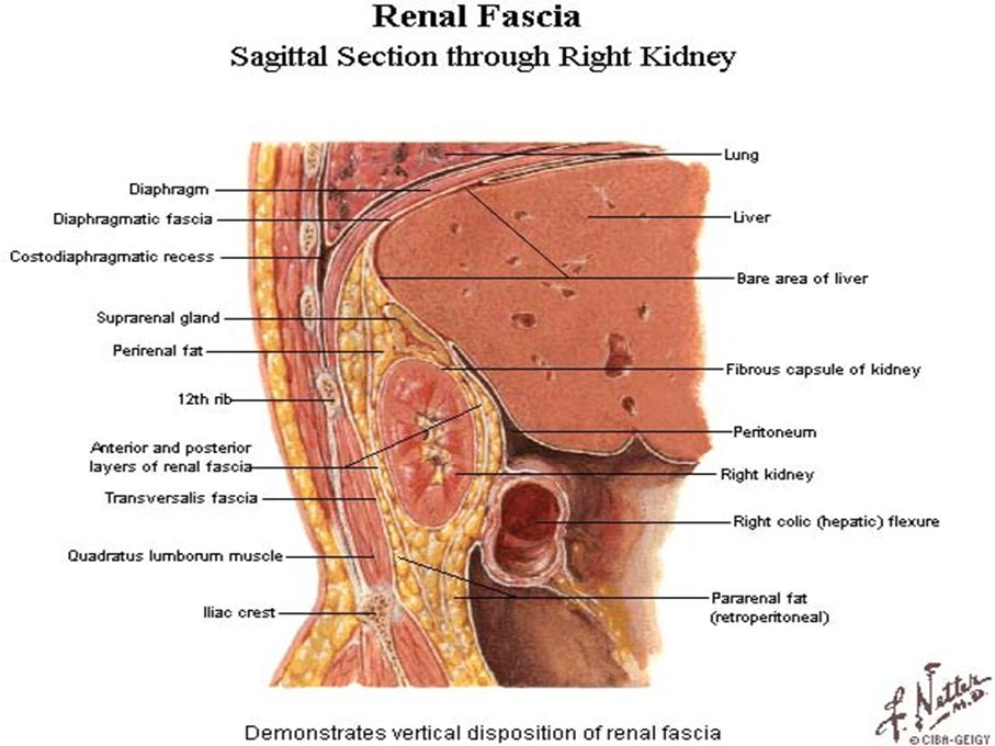
Renal Capsule Renal fascia Adipose capsule Fibrous capsule

27
Renal fascia

29
Fibrous capsule Adipose capsule Perirenal fat Pararenal fat

32
Fibrous Capsule

33
Ureter

34
Ureter is divided into 3 parts:
①abdominal part ②pelvis part ③intramural part

35
28 to 34 cm 3 narrowing sites the pelviureteric junction crossing the pelvic brim traversing the bladder wall

38
Horseshoe kidney

39
Suprarenal Gland

40
Endocrine gland T11 level Right is triangular Left is semi-lunar

41
tail of pancreas & spleen vessel
Relation Left right anterior superior stomach liver inferior tail of pancreas & spleen vessel posterior diaphragm medial abdominal aorta Inferior vena cava

43
Artery of Adrenal Gland

46
Veins of adrenal gland Left suprarenal vein, into the left renal vein
Right suprarenal vein, into the inferior vena cava

49
Anterior transperitoneal approach
incision Posterior retroperitoneal approach Anterior transperitoneal approach

50
Case A A 55-year-old woman was found rolling on her kitchen floor, crying out from agonizing pain in her abdomen. The pain came in waves and extended from the right loin to the groin and to the front of the right thigh. An anteroposterior radiograph of the abdomen revealed a calculus in the right ureter.

51
Question What causes the pain when a ureteral calculus is present?
Why is the pain felt in such an extensive area? Where does one look for the course of the ureter in a radiograph? Where along the ureter is a calculus likely to be held up?

52
Case B An explorer in the Amazon jungle was found alive after having lost contact with the outside world for six months. On physical examination, he was found to be in an emaciated condition. On palpation of the abdomen, a rounded, smooth swelling appeared in the right loin at the end of inspiration. On expiration, the swelling moved upward and could no longer be felt. What anatomical structure could produce such a swelling?

53
Case C An intravenous pyelogram revealed that a patient’s left kidney was in its normal position, but the right kidney was situated in front of the right sacroiliac joint. Can you explain this on embryological grounds?

54
Case D An examination of a patient revealed that she had a horseshoe kidney. What anatomical structure prevents a horseshoe kidney from ascending to a level above the umbilicus?

55
Case E An intravenous pyelogram revealed that the calyces and pelvis of a patient’s right kidney were grossly dilated (a condition known as hydronephrosis). What embryological anomaly may be responsible for this condition?
. What embryological anomaly may be responsible for this condition")
56
Case F Which congenital anomaly of the ureter is likely to present as a case of urinary incontinence?

57
operation procedure of kidney transplant

58
nephrectomy For a nephrectomy, the kidney commonly is exposed in the loin. After an oblique incision midway between the twelfth rib and the iliac crest, the posterior free border of the external oblique is identified, and divided to reveal the peritoneum, which is pushed forward to reveal the renal fascia. The subcostal nerve and vessels are preserved; the renal fascia is opened; and the kidney exposed. Care must be taken not to damage the pleura, since it is separated from the upper pole of the kidney only by the diaphragm. Posterior surgical approach Since the kidneys lie in part above the twelfth rib, a direct posterior approach might pass first into the thorax, perforating the pleural cavity. A posterior approach to the kidney also puts at risk the subcostal and iliohypogastric nerves which pass laterally behind the kidney.

Similar presentations


 Glands Anatomy & Embryology>")


