Download presentation
Presentation is loading. Please wait.

1
Spinal Cord and Spinal Nerve Roots (Sensory and Motor) Fig. 1.4.1
 Fig")
2
CRANIAL NERVES Fig. 1.4.2

3
Autonomic Nervous System Sympathetic and Parasympathetic Divisions are shown. Sympathetic: Promotes energy expenditure, activated by emotion and stress (e.g. increases heart rate, blood pressure) Parasympathetic: Promotes digestion and excretion (e.g., stimulates salivation, stomach and intestinal activity); can be activated by severe stress Fig. 1.4.3
 Parasympathetic: Promotes digestion and excretion (e.g., stimulates salivation, stomach and intestinal activity); can be activated by severe stress Fig")
4
TELENCEPHALON DIENCEPHALON MESENCEPHALON METENCEPHALON MYELENCEPHALON Five Sections of the Adult Brain Fig. 1.4.4

5
TELENCEPHALON DIENCEPHALON MESENCEPHALON METENCEPHALON MYELENCEPHALON MEDULLA Fig. 1.4.5

6
TELENCEPHALON DIENCEPHALON MESENCEPHALON METENCEPHALON MYELENCEPHALON PONS CEREBELLUM Fig. 1.4.6

7
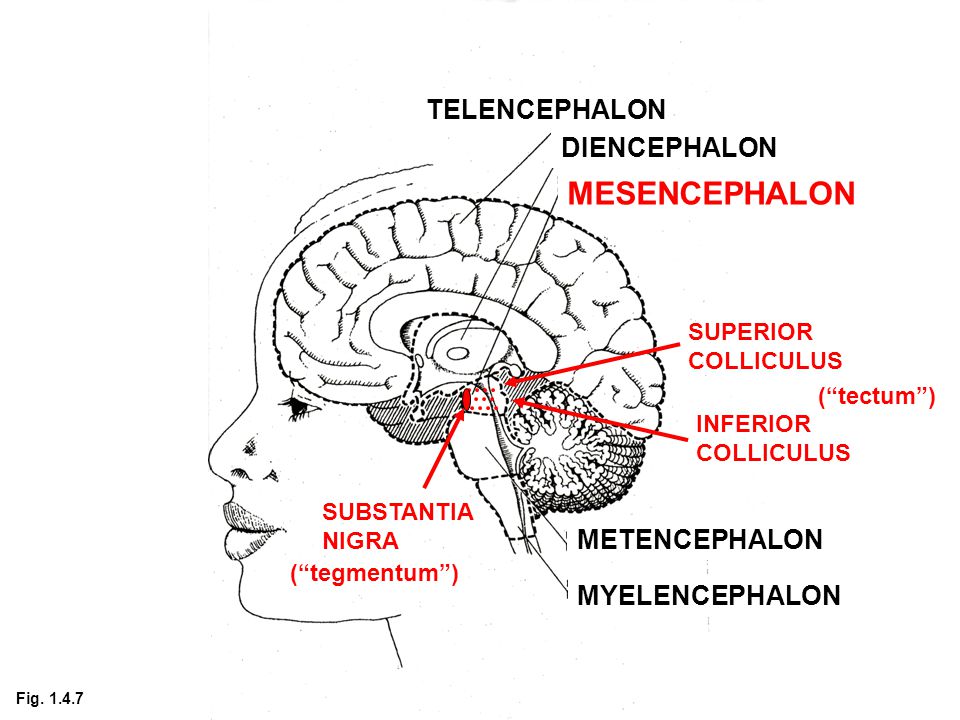
TELENCEPHALON DIENCEPHALON MESENCEPHALON METENCEPHALON MYELENCEPHALON SUPERIOR COLLICULUS INFERIOR COLLICULUS SUBSTANTIA NIGRA (“tectum”) (“tegmentum”) Fig. 1.4.7
8
TELENCEPHALON DIENCEPHALON MESENCEPHALON METENCEPHALON MYELENCEPHALON THALAMUS HYPOTHALAMUS Fig. 1.4.8

9
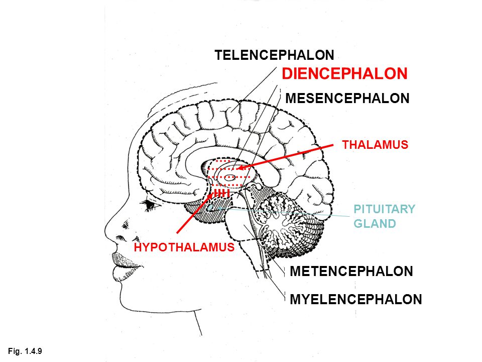
TELENCEPHALON DIENCEPHALON MESENCEPHALON METENCEPHALON MYELENCEPHALON THALAMUS HYPOTHALAMUS PITUITARY GLAND Fig. 1.4.9
10
TELENCEPHALON DIENCEPHALON MESENCEPHALON METENCEPHALON MYELENCEPHALON NEOCORTEX LIMBIC SYSTEM And BASAL GANGLIA (not shown) Fig. 1.4.10

11
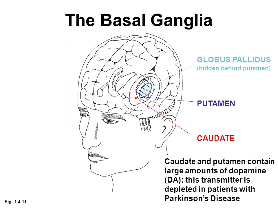
The Basal Ganglia CAUDATE PUTAMEN GLOBUS PALLIDUS (hidden behind putamen) Caudate and putamen contain large amounts of dopamine (DA); this transmitter is depleted in patients with Parkinson’s Disease Fig. 1.4.11
13
NEOCORTEX (showing convolutions) CENTRAL SULCUS PRECENTRAL GYRUS (primary motor; somatotopically organized) POSTCENTRAL GYRUS (primary Somatosensory; Somatotopically organized) Frontal lobe Parietal lobe Fig. 1.4.13

14
NEOCORTEX Fig. 1.4.14

15
FRONTAL LOBE CENTRAL SULCUS PRECENTRAL GYRUS (primary motor) Frontal lobe Supplementary Motor and Premotor Cortex Prefrontal Cortex Fig. 1.4.15

16
PARIETAL LOBE CENTRAL SULCUS POSTCENTRAL GYRUS (primary somatosensory) Parietal lobe Somatosensory Association Cortex Fig. 1.4.16

17
Occipital lobe OCCIPITAL LOBE Primary Visual Cortex Visual Association Cortex Fig. 1.4.17

18
TEMPORAL LOBE Temporal lobe Primary Auditory Cortex Auditory Association Cortex Fig. 1.4.18

19
TELENCEPHALON DIENCEPHALON MESENCEPHALON METENCEPHALON MYELENCEPHALON NEOCORTEX CORPUS CALLOSUM (axons that connect the two hemispheres) Fig. 1.4.19

Similar presentations


>")


