Download presentation
Presentation is loading. Please wait.

2
OCT OF MCULAR DISEASES DEHGHANI.A

3
near infrared light near infrared light cross-sectional images of tissue cross-sectional images of tissue High resolution) 10 µ ( High resolution) 10 µ ( Non contact Non contact
 10 µ ( High resolution) 10 µ ( Non contact Non contact")
4
highest reflections →red and white colours highest reflections →red and white colours Lowest reflections →blue and black Lowest reflections →blue and black

5
1. 2 red bands →RNFL and RPE 2. green/yellow band →Ganglions cell layer 3. blue/black band → Photoreceptor layer

7
Reflectivity Increased : inflammatory infiltrate, fibrosis, exudates and hemorrhage Increased : inflammatory infiltrate, fibrosis, exudates and hemorrhage Decreased reflectivity : retinal edema, hypopigmentation of the RPE Decreased reflectivity : retinal edema, hypopigmentation of the RPE decreased uniformly :abnormalities of the media )small pupil ( decreased uniformly :abnormalities of the media )small pupil (
small pupil ( decreased uniformly :abnormalities of the media )small pupil (")
8
Retinal thickness Retinal thickness Increased : accumulation of intraretinal fluid (diabetic retinopathy, cystoid macular oedema, retinal traction ( Decreased :scarring or atrophy Increased : accumulation of intraretinal fluid (diabetic retinopathy, cystoid macular oedema, retinal traction ( Decreased :scarring or atrophy

9
1. Diabetic Macular Edema 2. AMD 3. CSCR 4. Macular Hole 5. Retinal Vascular Occlusions 6. Retinal Vasculitis 7. Epiretinal Membranes 8. CNV 9.Juxtafoveal Telangiectasia 10.Heredodystrophic Disorders Photic Maculopathy 11. Inflammatory Diseases of Retina-choroid 12.Retinal Angiomatosis Proliferation 13.Trauma 14.Macular Evaluation following Retinal Detachment Surgery 15.Foveal Hemorrhage 16.Intraocular Metastasis

10
DIABETIC MACULAR EDEMA

11
ROLE OF OCT IN DIABETIC MACULAR EDEMA A. Defining the Disease Pattern B. Defining Indications for Pars Plana Vitrectomy C. Longitudinal Tracking of Tissue Alteration following An Intervention

12
Defining the Disease Pattern 1.Sponge-like retinal thickness 2.Cystoid macular edema 3. Subfoveal serous retinal detachment 4. Foveal tractional retinal detachment 5. Taut posterior hyloid membrane 5. Taut posterior hyloid membrane

16
After PRP

20
OCT IN AMD Disease categorization Disease categorization Management issues Management issues Define indications for therapy Define indications for therapy Monitor response to the therapy Monitor response to the therapy

21
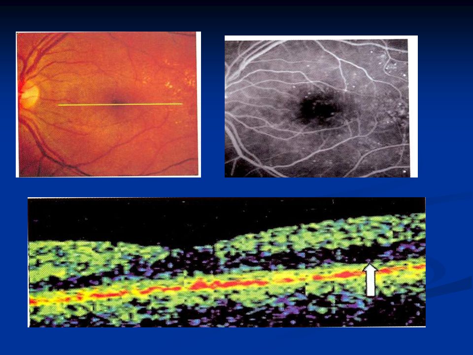
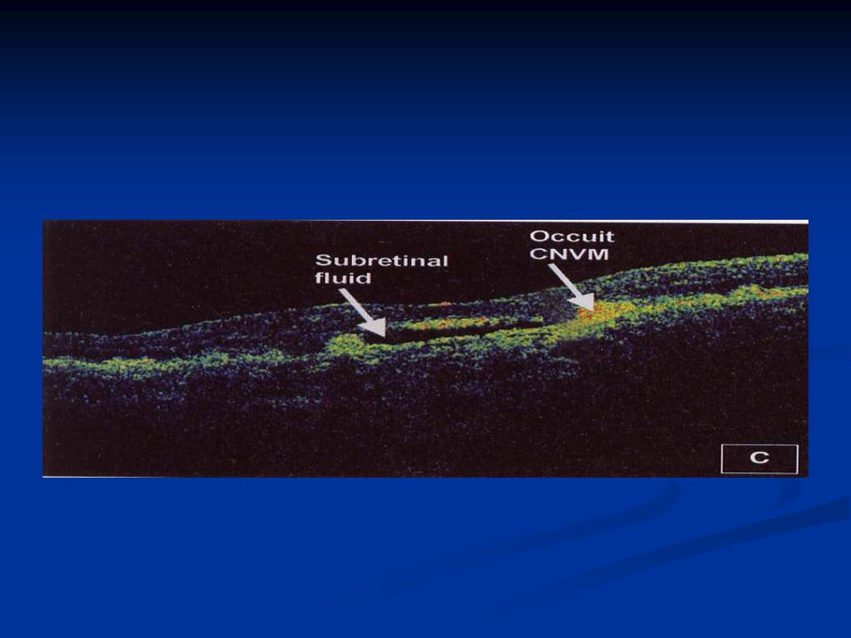
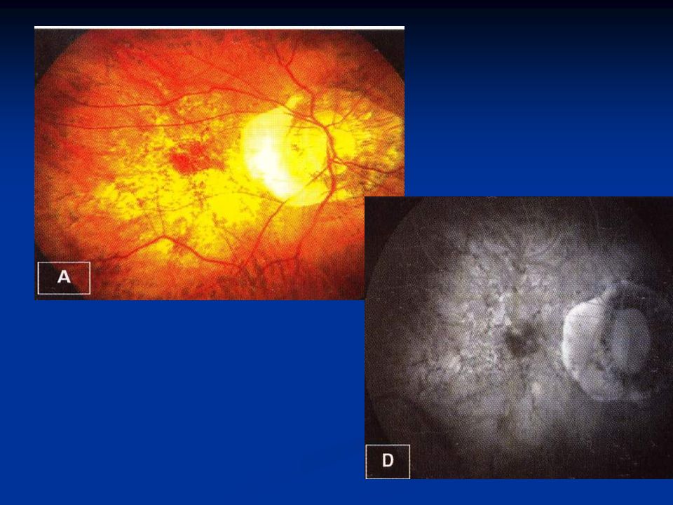
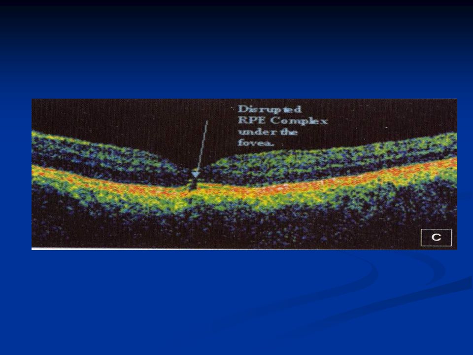
NON-NEOVASCULAR ARMD NON-NEOVASCULAR ARMD -Drusens -Drusens - Geographic Atrophy Neovascular ARMD Neovascular ARMD -classic CNV -classic CNV -occult CNV -occult CNV -serous PED -serous PED -hemorrhagic PED -hemorrhagic PED -fibrovascular PED -fibrovascular PED

22
OCT helps in the management of ARMD in the following ways: OCT helps in the management of ARMD in the following ways: -Disease categorization -Disease categorization -Early occult CNVM -Early occult CNVM -Associated changes -Associated changes -Response to treatment -Response to treatment

36
OCT IN CSCR Typical CSCR Typical CSCR -Serous retinal detachment -Serous retinal detachment -Serous retinal detachment with PED -Serous retinal detachment with PED Atypical CSCR Atypical CSCR -Small PEDs -Small PEDs -Chronic CSCR -Chronic CSCR -CSCR in elderly -CSCR in elderly

37
OCT in diagnosing complications of CSCR OCT in diagnosing complications of CSCR → CNV → CNV → Subretinal fibrin → Subretinal fibrin → RPE rip → RPE rip → Neurosensory atrophy of fovea → Neurosensory atrophy of fovea

43
SOLAR BURN Diagnose subtle changes in the RPE- photoreceptorcomplex Diagnose subtle changes in the RPE- photoreceptorcomplex

47
OCT IN EPIRETINAL MEMBRANE Confirming the diagnosis Confirming the diagnosis Identify the structural alterations Identify the structural alterations Longitudinal tracking of these eye following of vitrectomy Longitudinal tracking of these eye following of vitrectomy

51
OCT IN INTRAOCULAR METASTASIS Localizing the site of metastatic deposits Localizing the site of metastatic deposits Monitoring response to the therapy Monitoring response to the therapy

54
CONCLUSION: 1.Adjunct to thorough clinical examination and standard diagnostic examinations such as FFA and visual fields 2.Powerful diagnostic tool for macular disorders 3.Final diagnostic and therapeutic decision

55
The end

Similar presentations


 Waxman MD PhD>")


 pigment.>")
 1)Noninvasive 2) non-contact imaging 3)Millimeter penetration Aproximately 2-3 mm in tissue with micrometer scale (axial.>")


(C.S.C)>")

 occludes a branch of the central retinal vein Blockage causes bleeding from that branch Concerned about neovascularization.>")
