Download presentation
Presentation is loading. Please wait.

2
Posterior Segment Trauma Dr.Ali Salehi

4
BLUNT TRAUMA Ocular trauma is a significant cause of visual loss. 2.50 millions injuries occur annually in USA 40000 cause serious visual loss

5
BLUNT TRAUMA 75% are monocularly blind Vision is lost because of primary mechanical damage of vital structures and secondary complications such as infectious endophthalmitis and RD due to intraocular fibrosis, proliferation and contracture.

6
Serious Sequelae of Blunt Trauma 1- Angle recession 2- Hyphema 3- Vitreous hemorrhage 4- Retinal tears or RD 5- Subluxated or dislocated lens 6- Commotio Retinae 7- Choroidal rupture 8- Macular hole 9- Avulsed optic nerve 10- scleral rupture

7
Complete Ophthalmologic Examination Is essential because an eye with no anterior damage may have a severe posterior injury A patient without hyphema or iritis may have : A large retinal tear, choroidal rupture, or blowout fracture

8
Vitreous Hemorrhage Result from damage to blood vessels of iris, ciliary body, retina or choroid and retinal tear. As soon as possible 1) Indirect ophthalmoscopy 2) B- scan sonography RD, PVD, most retinal tears can be detected by B scan
 Indirect ophthalmoscopy 2) B- scan sonography RD, PVD, most retinal tears can be detected by B scan.")
9
Vitreous hemorrhages Retinal tear (11.4-44%) Posterior vitreous detachment with retinal vascular tear (3.7-11.7%) Rhegmatogenous retinal detachment (7-10%) Proliferative sickle cell retinopathy (0.2-5.9%) Macroaneurysm (0.6-7.4%) Age-related macular degeneration (0.6-4.3%) Terson syndrome (0.5-1.0%) Trauma (12.0-18.8%) Retinal neovascularization as a result of branch or central retinal vein occlusion (3.5-16%) Proliferative diabetic retinopathy is the most common cause(31.5-54% in the United States)
 Posterior vitreous detachment with retinal vascular tear ( %) Rhegmatogenous retinal detachment (7-10%) Proliferative sickle cell retinopathy ( %) Macroaneurysm ( %) Age-related macular degeneration ( %) Terson syndrome ( %) Trauma ( %) Retinal neovascularization as a result of branch or central retinal vein occlusion (3.5-16%) Proliferative diabetic retinopathy is the most common cause( % in the United States)")
10
Vitreous Hemorrhage It is important to assume that a retinal break is present until proved otherwise.

11
Causes Of Low Vision Due to Vit Hemorrhage Macular hole Choroidal rupture in the macula Traumatic maculopathy RD Berlin,s edema

12
Commotio Retinae The damage to the outer retinal layers caused by shock waves that traverse the eye from the site of impact following blunt trauma Most commonly seen in the posterior pole

13
Mechanisms For The Retinal Opacification 1- Extracellular edema 2- Glial swelling 3- Photoreceptor outer segment disruption With foveal involvement A cherry red spot may appear because the cells involved in the whitening are not present in the fovea

14
Berlin Edema Commotio Retinae in the posterior pole. May decrease visual acuity to as low as 20/200. Prognosis for visual recovery is good. The condition clears in 3-4 weeks.

15
visual recovery is limited by: 1- Associated macular pigment epitheliopathy 2- Choroidal rupture 3- Macular hole formation there is no acute treatment

16
Choroidal Rupture When the eye is compressed along its anterior – posterior axis, the eye wall becomes stretched in horizontal axis because of hydraulic displacement of the vitreous.

17
Choroidal Rupture Burch's membrane, which has little elasticity may tear along with the overlying RPE, and underlying choriocapillaris. Associated adjacent subretinal hemorrhage is common

18
Continue Choroidal ruptures may be single or multiple, commonly in the periphery and may be concentric to the optic disc. Ruptures that extend through the central macular area may cause permanent visual loss. There is no immediate treatment

19
continue Occasionally CNV develops as a late complication in response to the damage to Burch's m. A patient with Choroidal rupture near the macula should be alerted to the risk of CNV and advised to use an Amsler grid for self – testing

20
continue Treatment may be indicated if the CNV does not involve the foveal center. CNV may recur despite treatment May not be as poor as in AMD. PDT may be indicated.

21
Posttraumatic macular hole The fovea is extremely thin so blunt trauma may cause a full – thickness macular hole by mechanisms : 1- contusion necrosis 2- vitreous traction

23
Holes may be noted immediately after 1- Severe Berlin edema 2- After a subretinal hemorrhage caused by a Choroidal rupture 3- Following severe cystoids macular edema 4- After a whiplash separation of the vitreous from the retina

25
MACULAR HOLE Posttraumatic macular holes may be successfully closed with deep vitrectomy and I.L.M peeling and gas injection

26
Retinitis sclopetaria High – speed missile injuries to the orbit A- Large areas of Choroidal and retinal rupture and necrosis B- extensive subretinal and retinal hemorrhage often involving as much as 2 quadrants of the retina

27
sclopetaria As the blood resorbs, the injured area is filled in by extensive scar formation and widespread pigmentary alteration. The macula is almost always involved, leading to significant visual loss. Secondary RD rarely develops.

28
Scleral Rupture Severe blunt trauma can rupture the globe. Most common locations Limbus parallel to and under the insertions of the rectus muscles (thinnest sclera).
..")
29
Important diagnostic signs of ruptured globe: 1- marked decrease in ocular ductions 2- very boggy conj chemosis with hemorrhage 3- deepened AC 4- severe vitreous hemorrhage 5- IOP is usually reduced but may be normal or even elevated

30
Traumatic Breaks Eye trauma can cause retinal breaks or dialysis by contusion or vitreous traction. Fibrocellular proliferation occurring later at the site of an injury may cause vitreoretinal traction and RD

31
Retinal breaks Blunt trauma can cause retinal breaks by direct contusive injury to the globe through 2 mechanisms: 1- coup : adjacent to the point of trauma 2- countercoup : opposite the point of trauma

32
Continue Blunt trauma compresses the eye along its anterior posterior diameter and expands it in the equatorial plane. Rapid compression of the eye results in severe traction at the vitreous base that may cause retinal breaks.

33
Retinal breaks Traumatic breaks are often multiple and commonly found in the inferotemporal and supranasal quadrants. Contusion injury may cause large equatorial breaks, dialysis or a macular hole.

34
Traumatic tears The most common injuries are dialyses, which may be as small as 1 ora bay or extend 90 or more. Dialyses are usually located at the posterior border of the vitreous base but can also be found at the anterior border.

35
Retinal Tears Avulsion of the vitreous base ( Anterior vitreous detachment ) may be associated with dialysis and is pathogonomic of ocular contusion. less common types of breaks due to blunt trauma. 1.horseshoe – shaped tears 2.operculated holes

36
Retinal breaks a - Large U-tear with ‘ subclinical RD ’ - treat b - Large symptomatic U-tear - treat c - Operculated tear bridged by blood vessel - treat d - Asymptomatic operculated tear - do not treat

37
Trauma In Young Eyes young patients have a higher incidence of eye injury than other age groups They rarely develop an acute RRD following blunt trauma because their vitreous has not yet undergoing syneresis, or liquefaction.

38
Continue The vitreous provides an internal tamponade to the retina in spite of retinal tears or dialysis. However with time the vitreous may liquefy over a tear, allowing fluid to pass through the break to detach the retina.

39
Continue The clinical presentation of the retinal detachment is usually delayed due to blunt trauma in young patients as follows: 1- 12% of RD are found immediately 2- 30% are found within 1 month 3- 50% are found within 8 months 4- 80% are found within 24 months

40
Continue Traumatic retinal detachments in young patients may be shallow and often show signs of chronicity including : 1- multiple demarcation lines 2- subretinal deposits 3- intraretinal cysts.

41
Optic Disk Avulsion Multiple hemorrhage around the nerve head and edema of the peripapillary retina.

42
Optic Disk Avulsion

43
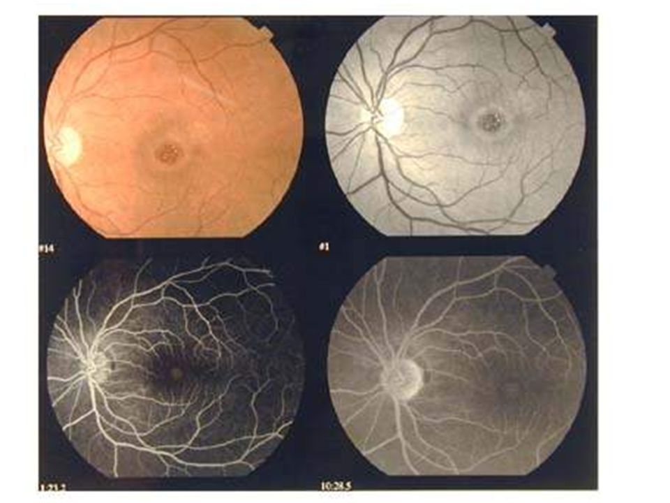
Subretinal hemorrhage

44
Purtscher Retinopathy Following acute compression injuries to the thorax or head visual loss seen due to: Large cotton-wool spots,hemrrhages and retinal edema are found most commonly around of disk.

45
Purtscher Retinopathy

46
THE END

Similar presentations


 Waxman MD PhD>")


 pigment.>")


>")

 2-Perforating Injury 3-Perforating Injury & retained foreign body 4-Chemicals ( acid – alkaline ) & burns 5-Sonar.>")
