Download presentation
Presentation is loading. Please wait.

2
Oct interpretation Ghanbari MD

3
This is what we wanted

8
Qualitative analysis of the OCT scan includes observation of the reflective qualities of the retinal structures.

10
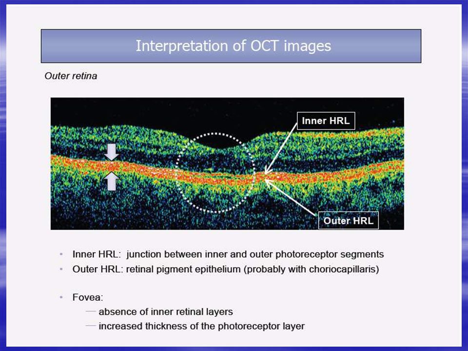
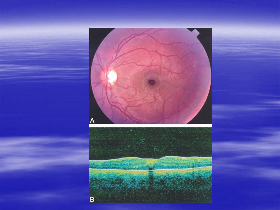
NORMAL MACULA The OCT image closely approximates the histological appearance of the macula and for this reason it bas been referred to as an in vivo optical biopsy.

11
Differences in signal intensity are represented by a false color coding system which is represented by the colors of the visible color spectrum.

12
Highly reflective structures are represented by red. Medium reflections appear yellow or green structures with low reflectivity are blue Black signal designates the absence of a reflective signal.

14
When evaluating an individual radial line scan, the clinician reads the left side of the scan as the beginning of the scan and the right side of the scan as the end.

15
The top of the scan image corresponds to the vitreous cavity. ln a normal patient (Fig. 87-1), this will be optically silent (black), without any significant reflections being noted except for perhaps identification of the posterior hyaloid face (in a patient with complete posterior vitreous detachment) or normal insertion of the posterior hyaloid near the macula in young patients.
, this will be optically silent (black), without any significant reflections being noted except for perhaps identification of the posterior hyaloid face (in a patient with complete posterior vitreous detachment) or normal insertion of the posterior hyaloid near the macula in young patients..")
16
The posterior vitreous face appears as a thin horizontal or oblique line above or inserting in the retina.

17
The Anterior surface of the retina demonstrates high reflectivity (red) and its horizontal expanse demonstrates the normal contour of the macula with the central foveal depression
 and its horizontal expanse demonstrates the normal contour of the macula with the central foveal depression")
18
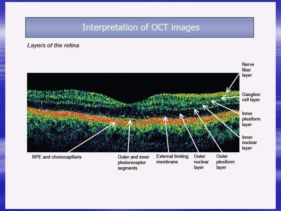
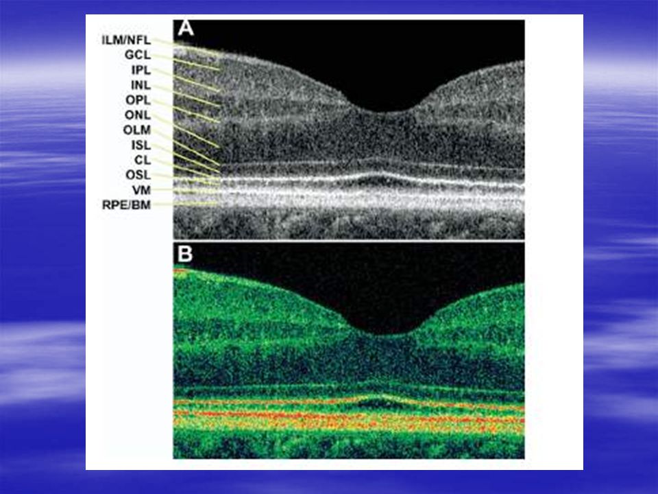
The internal structure of the retina consists of heterogeneous reflections, corresponding to varying ultrastructural anatomy.

20
The horizontally aligned NFL demonstrates a high tissue signal (red) that facilitates its identification.
 that facilitates its identification.")
21
High-flow intraretinal structures such as retinal blood vessels seen in this layer can result in a focal hyper reflective spot with optical shadowing of the retinal microstructures posterior to the vessele.

22
The (RPE), Bruch's membrane, and choriocapillaris complex collectively comprises the highly reflective external band
, Bruch s membrane, and choriocapillaris complex collectively comprises the highly reflective external band")
23
This thick band in the outer retina/anterior choroid appears as a red, linear stripe in OCT images

24
Just anterior to this band is another highly reflective line representing the junction between the photoreceptors' inner and outer segments

25
The outer retinal structures are better discriminated via ultra-high-resolution OCT, which is not yet commercially available.

26
The axially aligned cellular layers of the retina (inner nuclear, outer nuclear, and ganglion cell layers) demonstrate less back-scattering and back-reflection of incident OCT light
 demonstrate less back-scattering and back-reflection of incident OCT light")
27
This manifests as relatively low tissue signaIs (blue, green, yellow) compared to the horizontally aligned structures( internal limiting membrane, Henle's layer, and NFL).
 compared to the horizontally aligned structures( internal limiting membrane, Henle s layer, and NFL).")
28
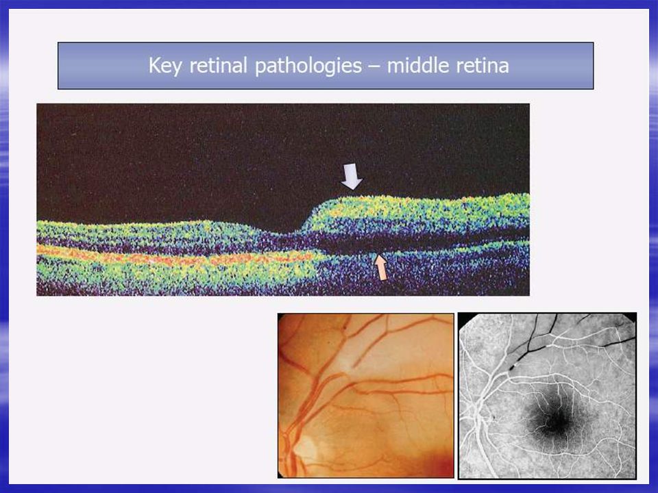
Reflectivity helps to identify anomalous structures. The sub-retinal fibrosis pictured below is identified by it's location, shape, and highly reflective structure.

29
Low reflective anomalous structures are areas of edema (fluid). These may be in the form of intraretinal cavities, cysts, diffuse intraretinal edema, or exudative detachments (image below).
..")
30
Black areas in the OCT scan may also be caused by shadowing. Shadowing occurs when a dense structure prevents light from penetrating below the structure, just like you or I cast a shadow on the ground in bright sunlight.

31
RETINAL THICKNESS ASSESSMENT The OCT unit determines the anterior and posterior surface of the retina in order to calculate retinal thickness

40
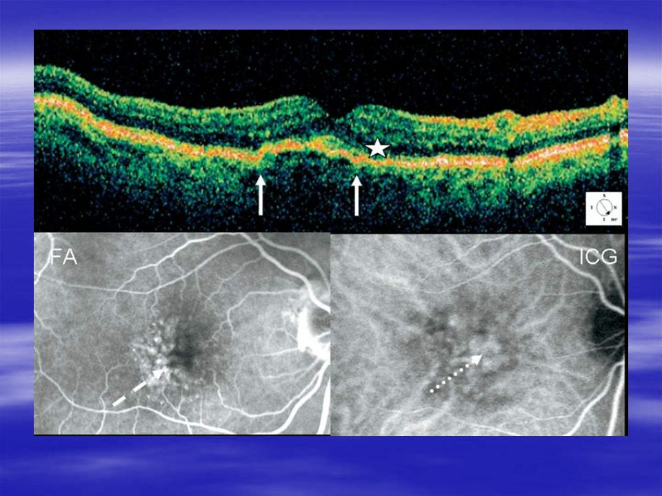
flat spreading PED (arrowhead) with irregularities of the retinal pigment epithelium (RPE) band, CME, subretinal fluid
 with irregularities of the retinal pigment epithelium (RPE) band, CME, subretinal fluid")
41
The PED is prominent, bulging, regular, and smooth. (Bottom left) Fluorescein angiogram (FA) showing smooth and regular PED with accentuated hyperfluorescence (dotted arrow) associated with an ill- delimited hyperfluorescent area of occult choroidal neovascularization
 Fluorescein angiogram (FA) showing smooth and regular PED with accentuated hyperfluorescence (dotted arrow) associated with an ill- delimited hyperfluorescent area of occult choroidal neovascularization.")
42
(PED) with chorioretinal anastomosis
 with chorioretinal anastomosis")
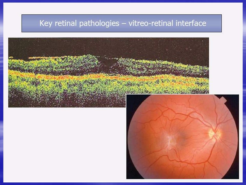
59
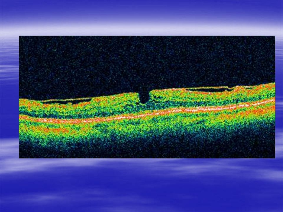
Two types of epiretinal macular membrane seen on OCT—one adherent to the retinal surface and one separated with a focal point of adhesion and traction

60
Vitreomacular traction syndrome on OCT. The vitreous, strongly adherent to the macular surface only, is highly reflective (white color).
..")
62
Rop foveal hypoplasia

64
Optical coherence tomography: coloboma and glaucoma Shown is a comparison of optical coherence tomography image of disc with (a) coloboma and one with (b) glaucoma. Arrow shows the glial tissue in colobomatous disc. Reprinted with permission from Ophthalmology (A clinical and optical coherence tomography study of the margins of choroidal colobomas, 2007;114: page 579, Fig. 8).
..")
74
limitations of OCT Corneal opacity Cataract Vitreous hemorrhage Uncooperative patients

75
Artifacts Artifacts in the OCT scan are anomalies in the scan that are not accurate images of actual physical structures, but are rather the result of an external agent or action.

76
large gap in the middle of the scan below. This is an artifact caused by a blink during scan acquisition. The was a high resolution scan, which takes about a second for the scan pass, which is plenty of time to record a blink.

77
The scan below has waves in the retinal contour. These are not retinal folds, but rather movement of the eye during the scan pass.

78
This scan has reduced brightness and detail on the right side. This is not intra-retinal edema. This is caused by a blockage of the light as it passes into the eye. The OCT was not centered well in the pupil, with some of the light striking the edge of the pupil.

Similar presentations


 pigment.>")
 1)Noninvasive 2) non-contact imaging 3)Millimeter penetration Aproximately 2-3 mm in tissue with micrometer scale (axial.>")


>")

 separated by the palpebral fissue Eyelashes Tarsal glands Lacrimal apparatus Vision Accessory structures.>")


 occludes a branch of the central retinal vein Blockage causes bleeding from that branch Concerned about neovascularization.>")


