Download presentation
Presentation is loading. Please wait.

1
Normal fetal anatomy

2
Anatomy Scan Indications –Identify fetal abnormalities –Identify IUGR –Dating –Placental localisation Timing –16-20 weeks –22-24 weeks Machine –Good quality machine –TGC sliders –Record images –High quality curvelinear probe Problems –Incorrect dates –Poor views –Fetal position

3
Routine History and consent from patient –Previous child with abnormality –Family history of abnormality –Exposure to teratogens eg drugs, infections –Explain limitations Growth scan Placental localisation Anatomy

4
Fetal head and face BPD and HC Cerebellar view –TCD = gestation –Foramen magna < 10mm –Nuchal fold < 6mm Lateral ventricles –Posterior aspect ventricle, Perpendicular to falx – 15mm severe Orbits –Inner to inner border, 1/3 ratio of eyes and bridge of nose Lips and nose –Exclude cleft lip Profile –Micrognathia, proboscis

10
Fetal head and face BPD and HC Cerebellar view –TCD = gestation –Foramen magna < 10mm –Nuchal fold < 6mm Lateral ventricles –Posterior aspect ventricle, Perpendicular to falx – 15mm severe Orbits –Inner to inner border, 1/3 ratio of eyes and bridge of nose Lips and nose –Exclude cleft lip Profile –Micrognathia, proboscis

11
Fetal trunk Coronal section –Heart, stomach, diaphragm and bladder Sagittal section of anterior abdominal wall Kidneys –Coronal –Transverse Diaphragms sagittal view Umbilical cord insertion Two umbilical arteries

16
Fetal trunk Coronal section –Heart, stomach, diaphragm and bladder Sagittal section of anterior abdominal wall Kidneys –Coronal –Transverse Diaphragms sagittal view Umbilical cord insertion Two umbilical arteries

17
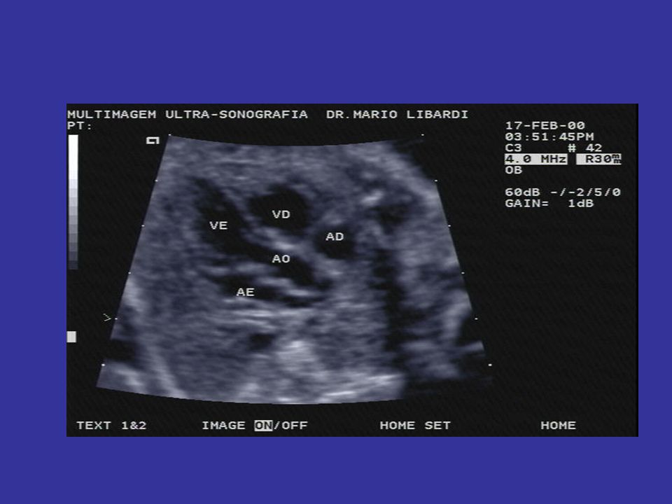
Fetal heart Reduce gain Situs 4 Chamber view –FHR and rhythm –Interventricular septum LVOT RVOT (Aortic arch) (Ductal arch)
 (Ductal arch)")
21
Fetal heart Situs 4 Chamber view –FHR and rhythm –Interventricular septum LVOT RVOT (Aortic arch) (Ductal arch)
 (Ductal arch)")
22
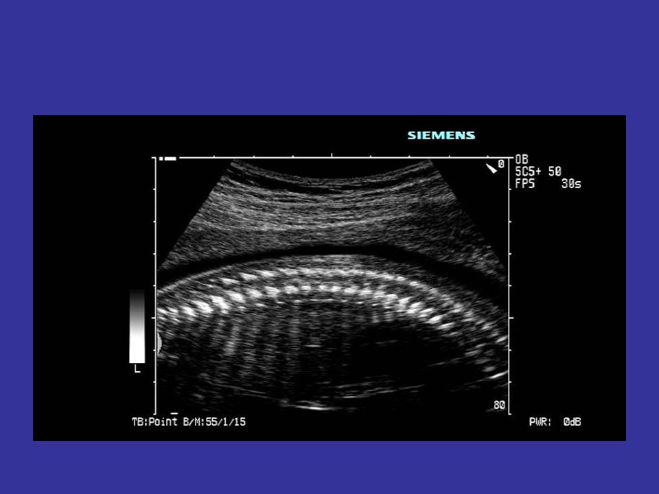
Fetal Spine and limbs Spine- reduce total gain Sagittal with skinline in view Coronal Transverse- 3 ossification centres ‘tight’, not splayed Follow limb from trunk noting side Full length all 12 bones Exclude club foot Feet- plantar view and count toes Hands- open if possible and count fingers and thumb

23
AND Coronal

26
OKAY!

Similar presentations


, UNSEPARATED VENTRICLE (18), LIVER (12), UMBILICAL VEIN (17), TRANSVERSE.>")


