Download presentation
Presentation is loading. Please wait.

1
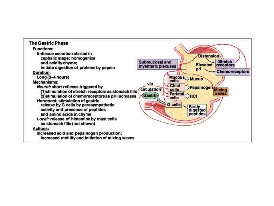
Phases of Gastric Secretion

2
Stomach Histology Rugae: Folds in stomach when empty
Gastric pits: Openings for gastric glands Contain cells Surface mucous: Mucus Mucous neck: Mucus Parietal: Hydrochloric acid and intrinsic factor Chief: Pepsinogen Endocrine: Regulatory hormones

5
Hydrochloric Acid Production

6
Movements in Stomach

8
Small Intestine Site of greatest amount of digestion and absorption
Divisions Duodenum Jejunum Ileum: Peyer’s patches or lymph nodules Modifications Circular folds or plicae circulares, villi, lacteal, microvilli Cells of mucosa Absorptive, goblet, granular, endocrine

9
Movement in small intestine:
Mixing: Segmental contraction that occurs in small intestine Secretion: Lubricate, liquefy, digest Digestion: Mechanical and chemical Absorption: Movement from tract into circulation or lymph Elimination: Waste products removed from body

10
Small Intestine Secretions
Mucus Protects against digestive enzymes and stomach acids Digestive enzymes Disaccharidases: Break down disaccharides to monosaccharides Peptidases: Hydrolyze peptide bonds Nucleases: Break down nucleic acids Duodenal glands Stimulated by vagus nerve, secretin, chemical or tactile irritation of duodenal mucosa

11
Duodenum and Pancreas

13
Duodenum Anatomy and Histology

14
Liver Lobes Ducts Major: Left and right Minor: Caudate and quadrate
Common hepatic Cystic From gallbladder Common bile Joins pancreatic duct at hepatopancreatic ampulla

17
Blood and Bile Flow

18
Duct System

19
Gallbladder Bile is stored and concentrated
Stimulated by cholecystokinin and vagal stimulation Dumps into small intestine Production of gallstones possible Drastic dieting with rapid weight loss

20
Pancreas Anatomy Secretions Endocrine Pancreatic juice (exocrine)
Pancreatic islets produce insulin and glucagon Exocrine Acini produce digestive enzymes Regions: Head, body, tail Secretions Pancreatic juice (exocrine) Trypsin Chymotrypsin Carboxypeptidase Pancreatic amylase Pancreatic lipases Enzymes that reduce DNA and ribonucleic acid
 Trypsin. Chymotrypsin. Carboxypeptidase. Pancreatic amylase. Pancreatic lipases. Enzymes that reduce DNA and ribonucleic acid.")
21
Bicarbonate Ion Production

22
Gastric hormones:

23
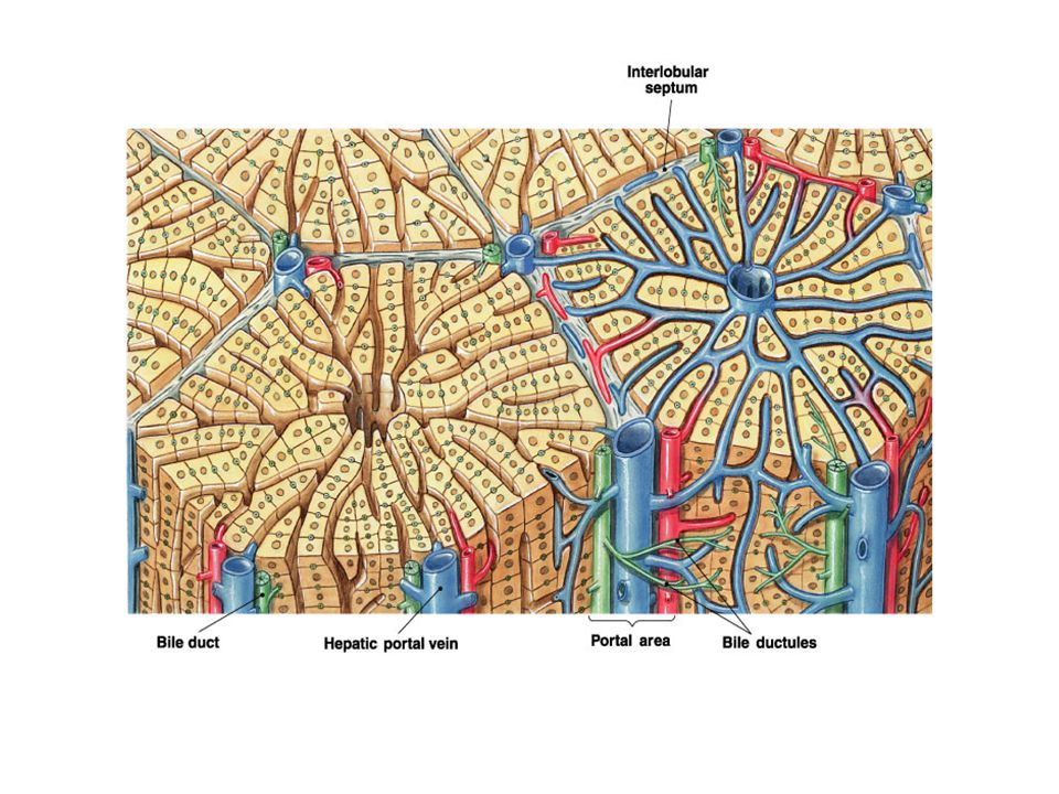
Liver Histology portal triad Figure 24.20a, b

24
3. Architecture of the Hepatic Parenchyma
The hepatic lobule is the structural unit of the liver. Portal vein Bile duct Sinusoids Central vein Liver cells (Hepatocytes) Portal area Hepatic artery
 Portal area. Hepatic artery.")
25
…each day around 600 ml of bile is produced…
Bile acid Phospholipids Cholesterol Bilirubin Waste products Electrolytes Mucin

26
Functions of the Liver Bile production Storage
Salts emulsify fats, contain pigments as bilirubin Storage Glycogen, fat, vitamins, copper and iron; huge blood reservoir of blood (storage); very high lymph flow Nutrient interconversion – Metabolic functions (see next slide) Detoxification Hepatocytes remove ammonia and convert to urea; metabolizes drugs, hormones; has the Cytochrome P-450 enzyme system. Phagocytosis – Cleans the blood Kupffer cells phagocytize worn-out and dying red and white blood cells, some bacteria Synthesis Albumins, fibrinogen, globulins, heparin, clotting factors The liver is the largest gland in the body and performs an astonishingly large number of tasks that impact all body systems. One consequence of this complexity is that hepatic disease has widespread effects on virtually all other organ systems. At the risk of losing sight of the forest by focusing on the trees, I will only briefly mention the vasculare and metabolic functions of the liver. It is a storage of cabohydrate, lipid copper and iron as well as it is able to interconvert nutrients. It detoxificate the bloocd by removing ammonia and converting it to ureal. The vascular functions include formation of lymph and it is part of phagocytic system. The Kupfeer cells phagocytize old blood cells and the liver also syntezise important components of the wound healing. But we have focused on the very first one, the secretory and excretory functions of the liver, because it is the only one that directly affects digestion - the bile production which plays a critical role in the digestion and absorption of dietary lipids. However, the understanding the vascular and metabolic functions of the liver is critical to appreciating the gland as a whole.
; very high lymph flow. Nutrient interconversion – Metabolic functions (see next slide) Detoxification. Hepatocytes remove ammonia and convert to urea; metabolizes drugs, hormones; has the Cytochrome P-450 enzyme system. Phagocytosis – Cleans the blood. Kupffer cells phagocytize worn-out and dying red and white blood cells, some bacteria. Synthesis. Albumins, fibrinogen, globulins, heparin, clotting factors. The liver is the largest gland in the body and performs an astonishingly large number of tasks that impact all body systems. One consequence of this complexity is that hepatic disease has widespread effects on virtually all other organ systems. At the risk of losing sight of the forest by focusing on the trees, I will only briefly mention the vasculare and metabolic functions of the liver. It is a storage of cabohydrate, lipid copper and iron as well as it is able to interconvert nutrients. It detoxificate the bloocd by removing ammonia and converting it to ureal. The vascular functions include formation of lymph and it is part of phagocytic system. The Kupfeer cells phagocytize old blood cells and the liver also syntezise important components of the wound healing. But we have focused on the very first one, the secretory and excretory functions of the liver, because it is the only one that directly affects digestion - the bile production which plays a critical role in the digestion and absorption of dietary lipids. However, the understanding the vascular and metabolic functions of the liver is critical to appreciating the gland as a whole.")
27
Liver’s Role in Metabolism
Carbohydrate Metabolism Storage of large amounts of glycogen Conversion of galactose and fructose to glucose Gluconeogenesis Formation of many chemical compounds from intermediate products of carbohydrate metabolism Fat Metabolism Beta-oxidation of fatty acids to supply energy to for other functions in the body Synthesis of cholesterol, phospholipids, and most lipoproteins (and their receptors) Synthesis of fats from proteins and carbohydrates
 Synthesis of fats from proteins and carbohydrates.")
28
Liver’s Role in Metabolism (cont’d)
Protein Metabolism Deamination of amino acids Formation of urea for removal of ammonia from the body fluids Formation of plasma proteins Interconversion of the various amino acids and synthesis of other compounds from amino acids

29
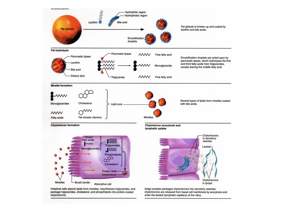
Exocrine Pancreas – Enzymes
Trypsinogen Chymotrysinogen Carboxypeptidases Pro-elastase Phospholipase pancreatic lipase Pancreatic amylase Pancreatic juice is composed of two secretory products critical to proper digestion: digestive enzymes and bicarbonate. The enzymes are synthesized and secreted from the exocrine ascinar cells, whereas bicarbonate is secreted from the epithelial cells lining small pancreatic ducts. Digestive Enzymes The pancreas secretes a magnificent battery of enzymes that collectively have the capacity to reduce virtually all digestible macromolecules into forms that are capable of, or nearly capable of being absorbed. Three major groups of enzymes are critical to efficient digestion: Proteases Digestion of proteins is initiated by pepsin in the stomach, but the bulk of protein digestion is due to the pancreatic proteases. Several proteases are synthesized in the pancreas and secreted into the lumen of the small intestine. The two major pancreatic proteases are trypsin and chymotrypsin, which are synthesized and packaged into secretory vesicles as an the inactive proenzymes trypsinogen and chymotrypsinogen. As you might anticipate, proteases are rather dangerous enzymes to have in cells, and packaging of an inactive precursor is a way for the cells to safely handle these enzymes. The secretory vesicles also contain a trypsin inhibitor which serves as an additional safeguard should some of the trypsinogen be activated to trypsin; following exocytosis this inhibitor is diluted out and becomes ineffective - the pin is out of the grenade. Once trypsinogen and chymotrypsinogen are released into the lumen of the small intestine, they must be converted into their active forms in order to digest proteins. Trypsinogen is activated by the enzyme enterokinase, which is embedded in the intestinal mucosa. Once trypsin is formed it activates chymotrypsinogen, as well as additional molecules of trypsinogen. The net result is a rather explosive appearance of active protease once the pancreatic secretions reach the small intestine. Trypsin and chymotrypsin digest proteins into peptides and peptides into smaller peptides, but they cannot digest proteins and peptides to single amino acids. Some of the other proteases from the pancreas, for instance carboxypeptidase, have that ability, but the final digestion of peptides into amino acids is largely the effect of peptidases in small intestinal epithelial cells. More on this later. Pancreatic Lipase The major form of dietary fat is triglyceride, or neutral lipid. A triglyceride molecule cannot be directly absorbed across the intestinal mucosa. Rather, it must first be digested into a 2-monoglyceride and two free fatty acids. The enzyme that performs this hydrolysis is pancreatic lipase, which is delivered into the lumen of the gut as a constituent of pancreatic juice. Sufficient quantities of bile salts must also be present in the lumen of the intestine in order for lipase to efficiently digest dietary triglyceride and for the resulting fatty acids and monoglyceride to be absorbed. This means that normal digestion and absorption of dietary fat is critically dependent on secretions from both the pancreas and liver. Pancreatic lipase has recently been in the limelight as a target for management of obesity. The drug orlistat (Xenical) is a pancreatic lipase inhibitor that interferes with digestion of triglyceride and thereby reduces absorption of dietary fat. Clinical trials support the contention that inhibiting lipase can lead to significant reductions in body weight in some patients. Amylase The major dietary carbohydrate for many species is starch, a storage form of glucose in plants. Amylase is the enzyme that hydrolyses starch to maltose (a glucose-glucose disaccharide), as well as the trisaccharide maltotriose and small branchpoints fragments called limit dextrins. The major source of amylase in all species is pancreatic secretions, although amylase is also present in saliva of some animals, including humans. Other Pancreatic Enzymes In addition to the proteases, lipase and amylase, the pancreas produces a host of other digestive enzymes, including ribonuclease, deoxyribonuclease, gelatinase and elastase. Bicarbonate and Water Epithelial cells in pancreatic ducts are the source of the bicarbonate and water secreted by the pancreas. The mechanism underlying bicarbonate secretion is essentially the same as for acid secretion parietal cells and is dependent on the enzyme carbonic anhydrase. In pancreatic duct cells, the bicarbonate is secreted into the lumen of the duct and hence into pancreatic juice.
 is a pancreatic lipase inhibitor that interferes with digestion of triglyceride and thereby reduces absorption of dietary fat. Clinical trials support the contention that inhibiting lipase can lead to significant reductions in body weight in some patients. Amylase. The major dietary carbohydrate for many species is starch, a storage form of glucose in plants. Amylase is the enzyme that hydrolyses starch to maltose (a glucose-glucose disaccharide), as well as the trisaccharide maltotriose and small branchpoints fragments called limit dextrins. The major source of amylase in all species is pancreatic secretions, although amylase is also present in saliva of some animals, including humans. Other Pancreatic Enzymes. In addition to the proteases, lipase and amylase, the pancreas produces a host of other digestive enzymes, including ribonuclease, deoxyribonuclease, gelatinase and elastase. Bicarbonate and Water. Epithelial cells in pancreatic ducts are the source of the bicarbonate and water secreted by the pancreas. The mechanism underlying bicarbonate secretion is essentially the same as for acid secretion parietal cells and is dependent on the enzyme carbonic anhydrase. In pancreatic duct cells, the bicarbonate is secreted into the lumen of the duct and hence into pancreatic juice.")
30
Bicarbonate Ion Production

37
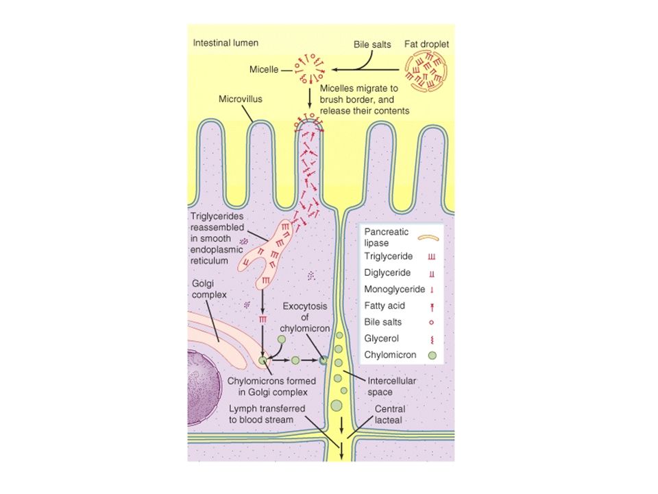
Lipoproteins Types Chylomicrons VLDL LDL HDL Enter lymph
Transports cholesterol to cells HDL Transports cholesterol from cells to liver

39
Water and Ions: Water Ions
Can move in either direction across wall of small intestine depending on osmotic gradients Ions Sodium, potassium, calcium, magnesium, phosphate are actively transported

40
Large Intestine: Extends from ileocecal junction to anus
Consists of cecum, colon, rectum, anal canal Movements sluggish (18-24 hours)
")
41
Large Intestine Cecum Colon Rectum Anal canal
Blind sac, vermiform appendix attached Colon Ascending, transverse, descending, sigmoid Rectum Straight muscular tube Anal canal Internal anal sphincter (smooth muscle) External anal sphincter (skeletal muscle) Hemorrhoids: Vein enlargement or inflammation
 External anal sphincter (skeletal muscle) Hemorrhoids: Vein enlargement or inflammation.")
42
Secretions of Large Intestine
Mucus provides protection Parasympathetic stimulation increases rate of goblet cell secretion Pumps Exchange of bicarbonate ions for chloride ions Exchange of sodium ions for hydrogen ions Bacterial actions produce gases called flatus

43
Histology of Large Intestine

44
Movement in Large Intestine
Mass movements Common after meals Local reflexes in enteric plexus Gastrocolic: Initiated by stomach Duodenocolic: Initiated by duodenum Defecation reflex Distension of the rectal wall by feces Defecation Usually accompanied by voluntary movements to expel feces through abdominal cavity pressure caused by inspiration

45
Reflexes in Colon and Rectum:

46
Digestion, Absorption, Transport
Breakdown of food molecules for absorption into circulation Mechanical: Breaks large food particles to small Chemical: Breaking of covalent bonds by digestive enzymes Absorption and transport Molecules are moved out of digestive tract and into circulation for distribution throughout body

47
Effects of Aging Death of myenteric plexus neurons
Atrophy of sphincter muscles Incontinence Decrease in mucus layer, connective tissue, muscles and secretions Increased susceptibility to infections and toxic agents Ulcerations and cancers

Similar presentations


 Chemical Digestion (mouth, stomach, intestines) Absorption (intestines) Assimilation (at each cell in the.>")


