Download presentation
Presentation is loading. Please wait.

1
CLEAR CELL ACANTHOMA CASE REPORT Floarea Sărac, Alin Meseşan, Constanţa Turda University of Oradea, Faculty of Medicine and Pharmacy, Dermatology Department, Romania

2
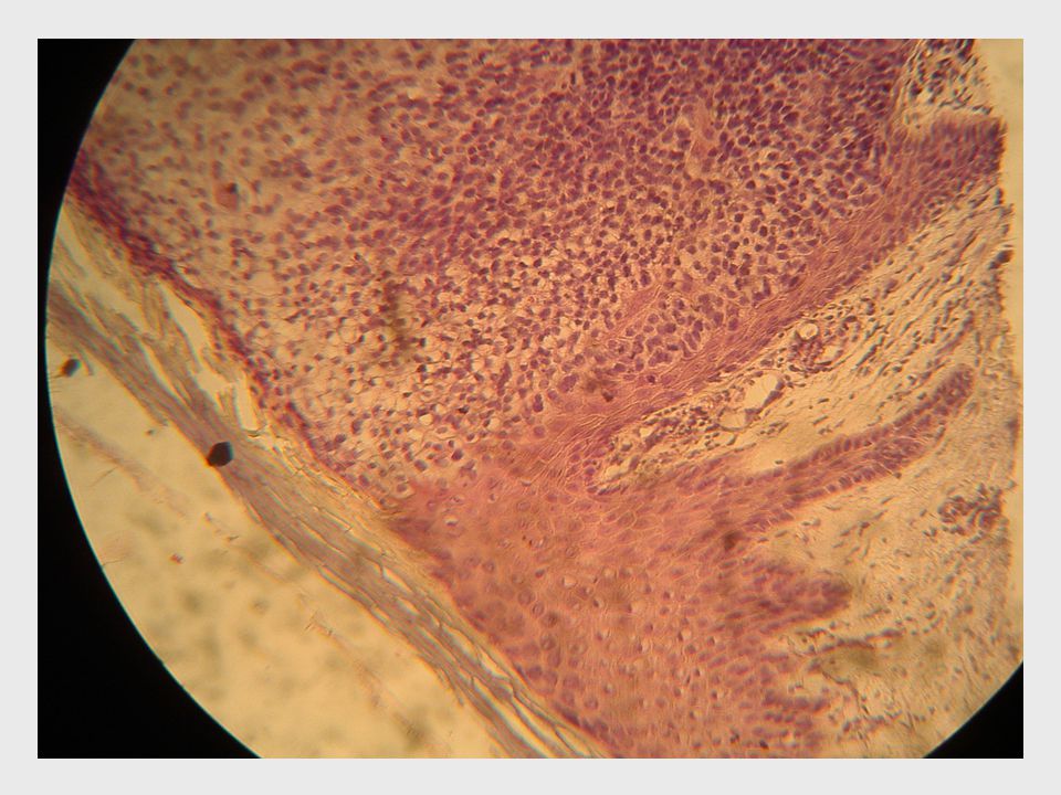
CASE REPORT We describe the case of a 70 years old man, who has multiple actinic keratoses of the face and upper limbs. For about six months the right arm shows a nodular formation, 1.5 cm diameter well delimited, crusted surface, easy to remove crust bleeding (Fig 1). It was suspected a squamous cell epithelioma. Lesion was excised and histopathological examination found : - a sharply demarcated psoriasiform epidermal hyperplasia composed of a proliferation of slightly enlarged keratinocytes with pale-staining cytoplasm. - although the basal cells at the base of these lesions are typically less clear,hey lack melanin much like the proliferating clear keratinocytes. - the intraepidermal adnexal structures are spared from the clear cell changes. - mild spongiosis is usually present along with scattered exocytosis of neutrophils and neutrophil debris (the neutrophils may form small ntraepidermal microabscesses ) - characteristic histologic changes are also found in the dermis with edematousdermal papillae, containing increased vascularity and a mixed inflammatory infiltrate including lymphocytes, plasma cells and neutrophils (Fig 2,3).
. It was suspected a squamous cell epithelioma. Lesion was excised and histopathological examination found : - a sharply demarcated psoriasiform epidermal hyperplasia composed of a proliferation of slightly enlarged keratinocytes with pale-staining cytoplasm. - although the basal cells at the base of these lesions are typically less clear,hey lack melanin much like the proliferating clear keratinocytes. - the intraepidermal adnexal structures are spared from the clear cell changes. - mild spongiosis is usually present along with scattered exocytosis of neutrophils and neutrophil debris (the neutrophils may form small ntraepidermal microabscesses ) - characteristic histologic changes are also found in the dermis with edematousdermal papillae, containing increased vascularity and a mixed inflammatory infiltrate including lymphocytes, plasma cells and neutrophils (Fig 2,3)..")
6
DISCUSSION, CONCLUSION Clear cell acanthoma is a distinct clinical and histological entity that was first described in 1962 by Degos. Although originally considered to be a benign neoplasm of the epidermis, some authors now believe that this lesion may actually represent a peculiar reactive dermatosis. Clear cell acanthoma is a rare benign (non-cancerous) epithelial skin tumourand and is a slow growing nodular or plaque-like hyperplasia of the epidermis characterized clinically by a predilection for the lower extremities of the middle aged and elderly, and histologically by a sharply demarcated intra-epidermal proliferation of glycogen-rich keratinocytes. Clinical features of the lesion include: slightly elevated to dome- shaped plaque or nodule; colour varies from pink to brown, but is most commonly blood red and shiny;can be from 3 to 20mm in diameter; wafer-like crusty scale may be stuck round the edges of the lesion. A moist or bleeding surface may result if scale is removed Although rare, they occur mostly in adults of middle-age or older. Both male and females can be affected. The diagnosis is rarely made before skin biopsy. They may persist for years and years without changing or causing any complications. They are easily excised.
 epithelial skin tumourand and is a slow growing nodular or plaque-like hyperplasia of the epidermis characterized clinically by a predilection for the lower extremities of the middle aged and elderly, and histologically by a sharply demarcated intra-epidermal proliferation of glycogen-rich keratinocytes. Clinical features of the lesion include: slightly elevated to dome- shaped plaque or nodule; colour varies from pink to brown, but is most commonly blood red and shiny;can be from 3 to 20mm in diameter; wafer-like crusty scale may be stuck round the edges of the lesion. A moist or bleeding surface may result if scale is removed Although rare, they occur mostly in adults of middle-age or older. Both male and females can be affected. The diagnosis is rarely made before skin biopsy. They may persist for years and years without changing or causing any complications. They are easily excised..")
Similar presentations


: Three cases Deba P Sarma, MD Omaha.>")


>")


