Download presentation
Presentation is loading. Please wait.

1
MULTIPLE KERATOACANTHOMAS ASSOCIATED WITH DISCOID LUPUS ERYTHEMATOSUS MA Benea, V Benea, SR Georgescu, A Rusu, A Ilie, A Udriste - “Prof. Dr. Scarlat Longhin” Clinical Hospital of Dermato-Venerology Bucharest

2
Keratoacanthoma is a benign tumor with origin in the pilo- sebaceous complex which must be differentiated from squamous cell carcinoma. The solitary form with spontaneous regression is the most common, but there are some forms with multiple lesions reported. Discoid lupus erythematosus is a chronic dermatosis that is characterised by atrophic or scarring lesions localised or widespread.

3
This is the case of a 36 year-old woman who was referred to our clinic for multiple well demarcated, exophytic, verrucous nodules, plaques and placards situated on the dorsum of the hands, forearms and face, which occurred 18 months ago simultaneously with the development of some hipopigmented-erythematous plaques and placards, on the presternal area and the upper limbs.

4
She had no biopsy and no treatment in this period of time. She denied any other skin conditions or internal diseases and the contact with any chemicals. Her family history was negative for skin diseases.

5
The physical examination revealed the presence of multiple lesions on the face, thorax and arms bilaterally. There were two distinct types of lesions: - one type appeared as hipopigmented atrophic plaques and placards, with erythematous patches, irregularly shaped, situated on the presternal area, arms, forearms and the dorsum of the hands; - the other type were tumor-like lesions ranging in size from 7 mm to 6 cm, with a mildly erythematous border and a yellow-brown verrucous exophytic surface located on the forearms, dorsum of the hands and face.

7
There were no other skin, mucous or nail changes. Laboratory tests failed to reveal any pathological changes (including the HIV test).
..")
8
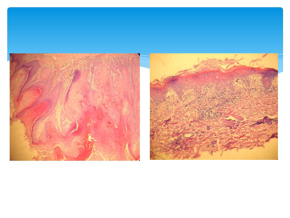
We performed two biopsies, one from each type of lesion. The one from the verrucous lesion revealed keratoacanthoma: central crater with keratin and overhanging epidermal“lips”, compact ortho- and parakeratosis, large keratinocytes with glassy, eosinophilic cytoplasms, inflammatory infiltrate at base, mostly with eosinophils, nuclear atypia in keratinocytes, especially at base.

9
The one from the scarring lesion showed alteration like in discoid lupus erythematosus: liquefaction degeneration of the basal cell layer, a patchy dermal infiltrate of lymphocytes with a few plasma cells and histiocytes, mostly near the appendages, which may be atrophic, degenerative alterations in the connective tissue (oedema, hyalinization, fibrinoid change) especially immediately under the epidermis.
 especially immediately under the epidermis.")
11
The clinical and pathologic data were consistent with the diagnosis of multiple keratoacanthomas and discoid lupus erythematosus. We treated the discoid lupus lesions with potent corticosteroid cream and the kerathoacanthomas with keratolitic creams and liquid nitrogen applications. After two months, the clinical appearance of the lesions improved.

Similar presentations


: Three cases Deba P Sarma, MD Omaha.>")


>")


.>")
. Erythema multiforme is a serious of acute, self-limited, recrudescent and inflammatory dermatopathy characterized by erythema,>")

