Download presentation
Presentation is loading. Please wait.

1
Prokaryotes and Viruses

2
West Nile Virus

3
West Nile Virus One of several mosquito-borne viruses in the United States that can infect people The virus exists in nature primarily through a transmission cycle involving mosquitoes and birds. Mosquitoes become infected with West Nile virus (WNV) when they feed on infected birds
 when they feed on infected birds.")
4
West Nile Virus Symptoms
Majority of people that become infected with the West Nile virus have no illness or experience only a mild flu-like illness May include fever, headache and body aches lasting only a few days Some persons may also have a mild rash or swollen lymph glands Less than one percent of those infected may develop meningitis or encephalitis, the most severe forms of the disease

5
How Long has the West Nile Virus Been Around?
Alexander the Great, 336 B.C., conquered a vast empire It’s speculated that his demise was due to West Nile encephalitis There is still no human vaccine for West Nile virus

6
West Nile Virus Takes Off
West Nile Virus is pathogenic, it invades its host and multiplies, causing disease It’s a flavivirus, traveling inside mosquitoes which act as the transferring agent from host to host

7
West Nile Virus Takes Off

8
West Nile Virus Takes Off

9
West Nile Virus Takes Off
Impacts, Issues Video West Nile Virus Takes Off

10
Microorganisms Single-celled organisms that are too small to be seen without a microscope Bacteria are the smallest living organisms Viruses are smaller but are not alive

11
The Prokaryotes Only two groups Archaebacteria and Eubacteria
Arose before the eukaryotes

12
Prokaryotic Characteristics
No membrane-bound nucleus Single chromosome Cell wall (in most species) Prokaryotic fission Metabolic diversity
 Prokaryotic fission. Metabolic diversity.")
13
Prokaryotic Body Plan DNA capsule plasma membrane
ribosomes in cytoplasm bacterial flagellum pilus cell wall cytoplasm

14
cytoplasm, with ribosomes
Prokaryotic Body Plan cytoplasm, with ribosomes DNA, in nucleoid pilus bacterial flagellum outer capsule cell wall plasma membrane

15
Prokaryotic Body Plan Prokaryotic body plan

16
Bacterial Shapes coccus bacillus spirillum

17
Bacterial Shapes

18
Bacterial Shapes

19
Bacterial Shapes

20
Bacterial Shapes sex pilus

21
Metabolic Diversity Photoautotrophs Chemoautotrophs Chemoheterotrophs

22
Gram Stain

23
Gram Stain Gram stain

24
Bacterial Genes Bacteria have a single chromosome
Circular molecule of DNA Many bacteria also have plasmids Self-replicating circle of DNA that has a few genes Can be passed from one cell to another

25
Prokaryotic Fission

27
Prokaryotic Fission

28
Prokaryotic Fission

29
Prokaryotic Fission

30
Prokaryotic Fission

31
Prokaryotic Fission

32
Prokaryotic Fission

33
Prokaryotic Fission

34
DNA replication begins DNA replication completed
Bacterium before DNA replication bacterial chromosome DNA replication begins parent DNA molecule DNA copy DNA replication completed

35
Membrane growth moves DNA molecules apart
New membrane and cell-wall material deposited Cytoplasm divided in two

36
Prokaryotic Fission - 3 Prokaryotic fission

37
Conjugation Transfer of plasmid

38
nicked plasmid conjugation tube

39
Prokaryotic conjugation

40
Prokaryotic Classification
EUBACTERIA (Bacteria) ARCHAEBACTERIA (Archaea) EUKARYOTES (Eukarya) Traditionally classified by numerical taxonomy Now increased use of comparative biochemistry
 ARCHAEBACTERIA. (Archaea) EUKARYOTES. (Eukarya) Traditionally classified by numerical taxonomy. Now increased use of comparative biochemistry.")
41
Prokaryotic Classification
to ancestors of eukaryotic cells DOMAIN BACTERIA DOMAIN BACTERIA biochemical and molecular origin of life

42
Eubacteria Includes most familiar bacteria
Have fatty acids in plasma membrane Most have cell wall; always includes peptidoglycan Classification based largely on metabolism

43
Eubacterial Diversity
Photoautotrophic Aerobic (Cyanobacteria) Anaerobic (Green bacteria) Chemoautotrophic Important in nitrogen cycle Chemoheterotrophic Largest group
 Anaerobic (Green bacteria) Chemoautotrophic. Important in nitrogen cycle. Chemoheterotrophic. Largest group.")
44
Examples of eubacteria

45
Eubacterial Diversity

46
Eubacterial Diversity

47
Eubacterial Diversity

48
Some Pathogenic Eubacteria
Most are chemoheterotrophs E. coli strains Clostridium botulinum Clostridium tetanus Borrelia burgdorferi Rickettsia rickettsii

49
Some Pathogenic Eubacteria
resting spore photo-synthetic cell heterocyst

50
Some Pathogenic Eubacteria

51
Some Pathogenic Eubacteria

52
Some Pathogenic Eubacteria
DNA spore coat capsule around cell wall

53
Bacterial Behavior Bacteria move toward nutrient-rich regions
Aerobes move toward oxygen; anaerobes avoid it Photosynthetic types move toward light Magnetotactic bacteria swim downward Myobacteria show collective behavior

54
Bacterial Behavior

55
Archaebacteria

56
Archaebacteria Methanogens Extreme halophiles Extreme thermophiles

57
Methanogens

58
Methanogens

59
Extreme Halophiles

60
Extreme Thermophiles

61
Extreme Thermophiles

62
Virus Noncellular infectious agent
Protein wrapped around a nucleic acid core Cannot reproduce itself; can only be reproduced using a host cell

63
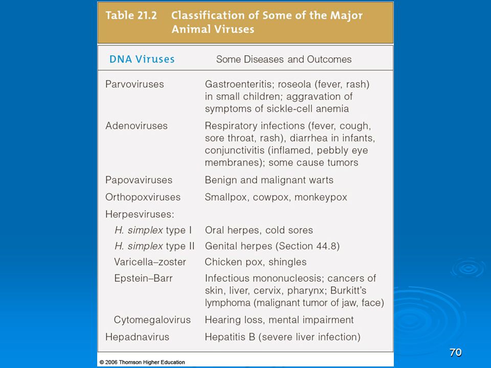
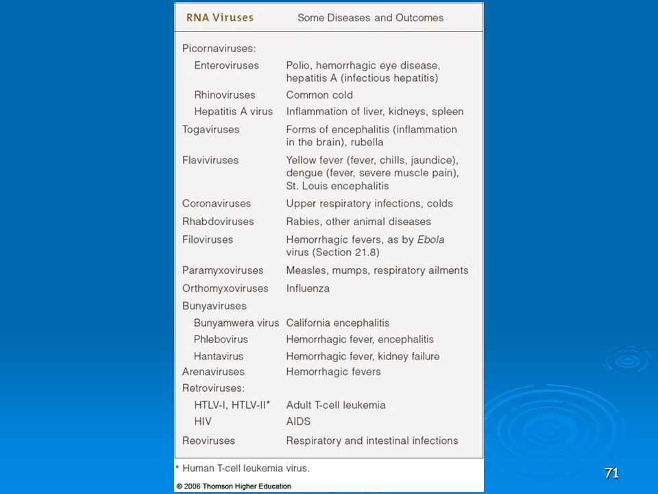
Viral Body Plans Genetic material is DNA or RNA Coat is protein
Complex virus (bacteriophage) Genetic material is DNA or RNA Coat is protein Helical virus Polyhedral virus
 Genetic material is DNA or RNA. Coat is protein. Helical virus. Polyhedral virus.")
64
Enveloped Virus (HIV) viral protein lipid envelope (derived from host)
viral RNA reverse transcriptase viral coat (proteins)
")
65
Enveloped Virus (HIV) viral RNA protein subunits of coat
18 nm diameter, 250 nm length

66
Enveloped Virus (HIV) 80 nm diameter
 80 nm diameter")
67
Enveloped Virus (HIV) 65–nm diameter head, 225 nm total length DNA
protein coat sheath base plate tail fiber

68
Enveloped Virus (HIV) viral coat (proteins) reverse transcriptase
nm diameter viral RNA lipid envelope: proteins span the envelope, line its inner surface, spike out above it

69
Viral Body Plans Body plans of viruses

72
Viruses

73
Viruses

74
Viruses

75
Viruses

76
Viral Multiplication - Basic Steps
Attach to host cell Enter host (virus or just genetic material) Direct host to make viral genetic material and protein Assemble viral nucleic acids and proteins Release new viral particles
 Direct host to make viral genetic material and protein. Assemble viral nucleic acids and proteins. Release new viral particles.")
77
Bacteriophage multiplication cycles
Viral Replication Bacteriophage multiplication cycles

78
e Lysis of host cell is induced; infectious particles escape.
Lytic Pathway d The coats get tail fibers, other parts. Lytic Pathway a Virus particle injects genetic material into a suitable host cell after binding to its wall. c Viral proteins are assembled into coats around viral DNA. b Viral DNA directs host cell to make viral proteins and replicate viral DNA. a-1 Viral DNA is integrated into the host’s chromosome. a-4 Viral DNA is excised from the chromosome. Lysogenic Pathway a-2 Before prokaryotic fission, the bacterial chromosome with the integrated viral DNA is replicated. a-3 After cell division, each daughter cell will have recombinant DNA. Fig , p.344

79
Lytic Pathway

80
Lysis of host cell is induced; infectious particles escape.
Lytic Pathway Tail fibers and other parts are added to coats. Virus particles bind to wall of suitable host. Viral genetic material enters cell cytoplasm. Viral protein molecules are assembled into coats; DNA is packaged inside. Viral DNA directs host machinery to produce viral proteins and viral DNA.

81
Lytic Pathway Lytic pathway

82
Viral DNA usually becomes integrated into the bacterial chromosome.
Lysogenic Pathway Viral DNA is excised from chromosome and cell enters lytic pathway. Prior to prokaryotic fission, the chromosome and integrated viral DNA are replicated. After binary fission, each daughter cell will have recombinant DNA.

83
Lysogenic Pathway Lysogenic pathway

84
Replication of an Enveloped Virus
DNA virus particle plasma membrane of host cell Transcription of viral DNA Replication of viral DNA Translation nuclear envelope some proteins for viral coat viral DNA other proteins for viral envelope

85
Replication of an Enveloped Virus

86
Replication of an Enveloped Virus
Enveloped DNA virus replication

87
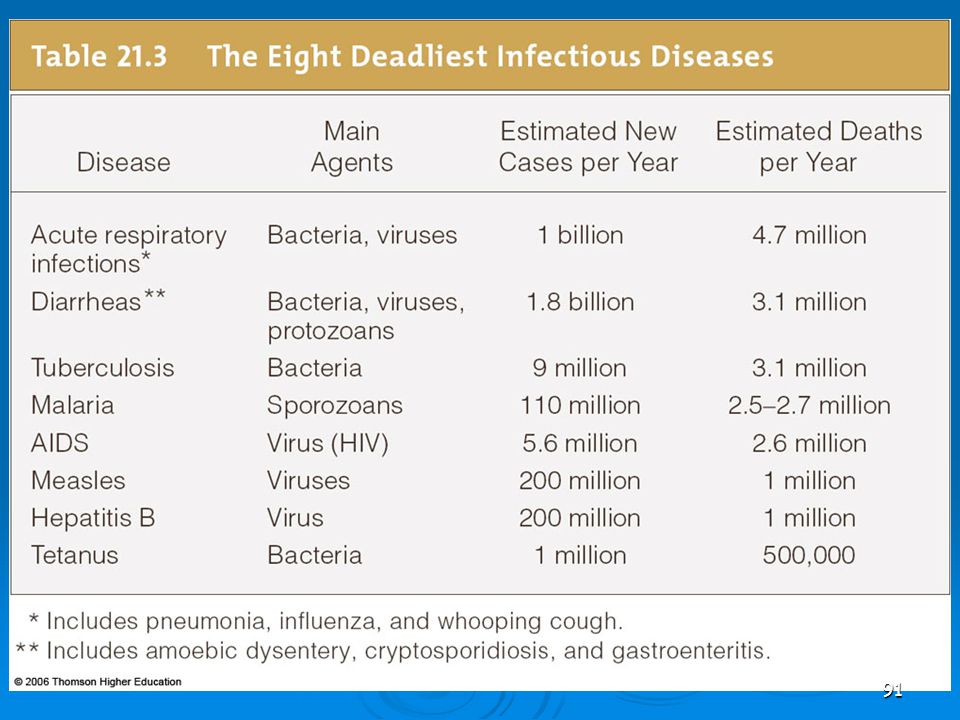
Nature of Disease Contagious disease pathogens must directly contact a new host Epidemic Pandemic (AIDS) Sporadic Endemic

88
Evolution and Disease Host and pathogen are coevolving
If a pathogen kills too quickly, it might disappear along with the individual host Most dangerous if pathogen Is overwhelming in numbers Is in a novel host Is a mutant strain

89
Mycobacterium tuberculosis

90
SARS virus

92
New Threats Emerging Pathogens Drug-resistant strains Food poisoning
Ebola virus Monkeypox virus SARS virus Drug-resistant strains Food poisoning E. coli Salmonella

93
Viroids Smaller than viruses Strands or circles of RNA
No protein-coding genes No protein coat Cause many plant diseases

94
Prions Small proteins Linked to human diseases Animal diseases Kuru
Creutzfeldt-Jakob disease (CJD) Animal diseases Scrapie in sheep Bovine spongiform encephalopathy (mad cow disease)
 Animal diseases. Scrapie in sheep. Bovine spongiform encephalopathy (mad cow disease)")
Similar presentations


.>")


 Prokaryotic cells (0.2–10 µm) cyanobacterium Viruses (0.05–0.2 µm) Escherichia.>")

 Found everywhere. Infect organisms in every kingdom Edward Jenner-first vaccine for smallpox.>")


