Download presentation
Presentation is loading. Please wait.

1
Skeletal System Axial Skeleton

3
Introduction Skull, vertebral column, thoracic cage (ribs & sternum)
")
4
Function 1. framework that supports organs 2. surface area for muscle attachment

5
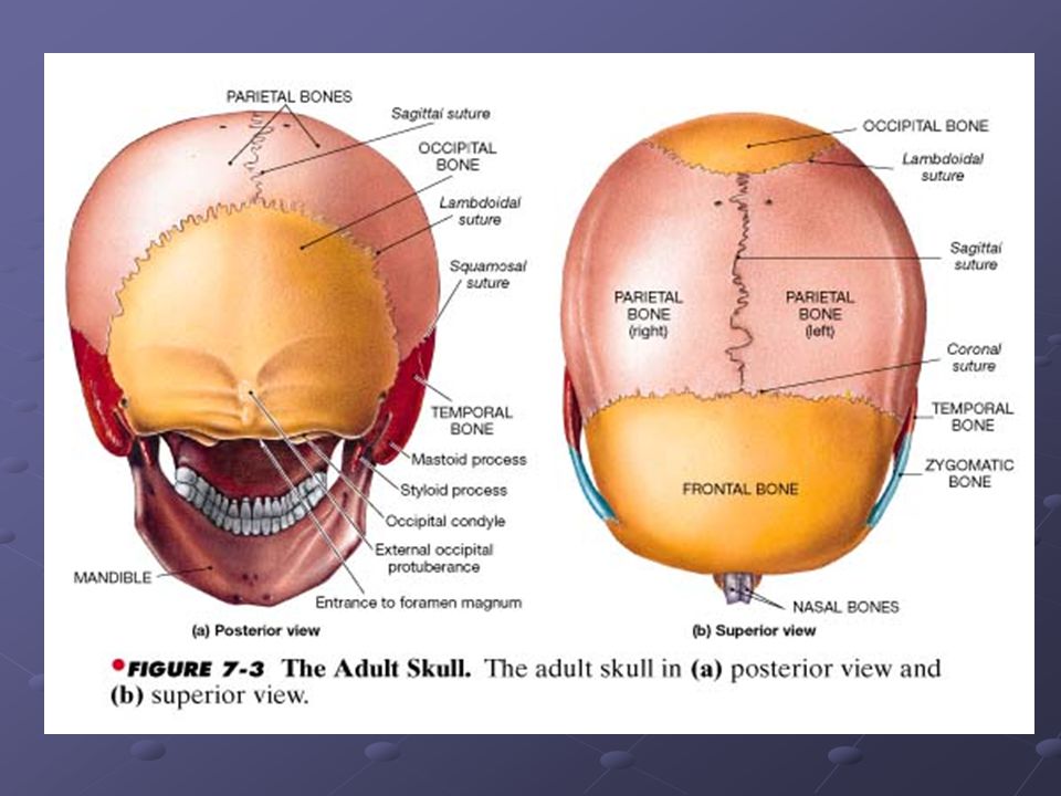
Skull Protects the brain and guards the entrance to the digestive and respiratory tract Cranial Bones – occipital, parietal (2), frontal, temporal (2), sphenoid & ethmoid bones Cranial bones protect the brain
, frontal, temporal (2), sphenoid & ethmoid bones. Cranial bones protect the brain.")
6
Skull Facial bones Function – protect and support entrances to digestive & respiratory tracts Superficial bones – maxillary, nasal, zygomatic, Mandible. Allow for attachment of muscles that control facial expressions and help in manipulation of food

14
Several bones of skull have sinuses
Deeper facial bones – palatine & vomer (separates oral and nasal cavities) Several bones of skull have sinuses Air filled chambers Function 1. helps to make bones lighter 2. has a mucus membrane that helps moisten and clean air
 Several bones of skull have sinuses. Air filled chambers. Function. 1. helps to make bones lighter. 2. has a mucus membrane that helps moisten and clean air.")
15
Sutures – immoveable joints that are connections b/n skull bones
4 major sutures 1. lamboidal suture – across posterior surface of skull. Separates occipital bone from the 2 parietal bones. 2. coronal suture – attaches frontal bone to parietal bones on either side 3. saggital suture – from lamboidal suture to coronal suture b/n the 2 parietals 4. squamosal suture – on each side of the skull; boundary b/n temporal and parietal bones Cranial and Facial Bones – see handout

16
Orbital and nasal complexes
1. Orbital complex – formed form cranial and facial bones which surround each eye 2. Nasal complex – surrounds nasal cavity

19
Skulls of Infants and Children
Right before birth, brain enlarges and bones cannot keep up. So at birth some bones are connected by fibrous connective tissue. Flexible so brain is not damaged. Fontanels – fibrous areas b/n cranial bones 4 types: 1. frontal fontanel – largest intersection of saggital, & coronal sutures 2. occipital fontanel – b/n lamboidal and saggital sutures

20
Skulls of Infants and Children
3. sphenoidal fontanel – junctions b/n squamousal and coronal sutures 4. mastoid fontanel – b/n squamousal and lamboidal sutures Occipital, sphenoid, and mastoid – disappear within 1-2 months after birth Frontal – until 2 years of age

22
Vertebral Column Adult vertebral column 26 bones – 24 vertebrae, sacrum & coccyx Function 1. support 2. bears weight of head, neck and trunk 3. transfers weight to appendicular skeleton 4. protects spinal cord 5. maintains upright position

23
Divided into 7 cervical 12 thoracic (articulates with ribs) 5 lumbar 1 sacrum 1 coccyx With development, sacrum is made up of 5 vertebrae until 25 than 1

26
Spinal curvature – 4 curves
Cervical Thoracic Lumbar Sacral Primary curves (accomodation curves) – thoracic and sacral b/c appear late in fetal development Secondary curves (compensation curves) – lumbar and cervical appear several months after birth Cervical – infant learns to balance head Lumbar – infants learns to stand
 – thoracic and sacral b/c appear late in fetal development. Secondary curves (compensation curves) – lumbar and cervical appear several months after birth. Cervical – infant learns to balance head. Lumbar – infants learns to stand.")
27
Cervical vertebrae 7 of them C1 – atlas; nod head “yes”
C2 – axis; rotation to say “no” C7 – vertebral prominens

29
Throracic Lumbar Sacrum Coccyx 12 of them
Contain facets for rib articulation Lumbar 5 largest Bear most weight Sacrum Fused components of 5 sacral vertebrae Coccyx “tailbone”

30
Thoracic Cage Ribs Sternum (breast bone) – flat 12 pairs
First 7 = true. Connected to sternum Ribs 8-12 = false b/c do not attach directly to sternum Ribs 11 & 12 = floating. Attach to vertebrae Sternum (breast bone) – flat Manubrium – articulates with clavicles; first pair of ribs Body – ribs 2-7 Xiphoid process – smallest part
 – flat. Manubrium – articulates with clavicles; first pair of ribs. Body – ribs 2-7. Xiphoid process – smallest part.")
Similar presentations