Download presentation
Presentation is loading. Please wait.

1
Chapter 5 Integumentary System
2
I. The Skin A. General Information Skin, Hair, Nails, Glands
Skin is the largest organ of the body (1.2 to 2.2 m2 or 22 ft2) Ranges in thickness from 1.5 to 4.0 mm with an average thickness of 1-2 mm It consists of the epidermis and the dermis Attached by fibers to the underlying layer or subcutaneous layer known as the hypodermis, which is mainly areolar and adipose connective tissues
 Ranges in thickness from 1.5 to 4.0 mm with an average thickness of 1-2 mm. It consists of the epidermis and the dermis. Attached by fibers to the underlying layer or subcutaneous layer known as the hypodermis, which is mainly areolar and adipose connective tissues.")
3
Hair shaft Dermal papillae Epidermis Subpapillary vascular plexus Papillary layer Pore Appendages of skin Dermis Reticular layer • Eccrine sweat gland • Arrector pili muscle Hypodermis (superficial fascia) • Sebaceous (oil) gland • Hair follicle Nervous structures • Hair root • Sensory nerve fiber Cutaneous vascular plexus • Pacinian corpuscle • Hair follicle receptor (root hair plexus) Adipose tissue
 • Sebaceous. (oil) gland. • Hair follicle. Nervous structures. • Hair root. • Sensory nerve fiber. Cutaneous vascular. plexus. • Pacinian corpuscle. • Hair follicle receptor. (root hair plexus) Adipose tissue.")
4
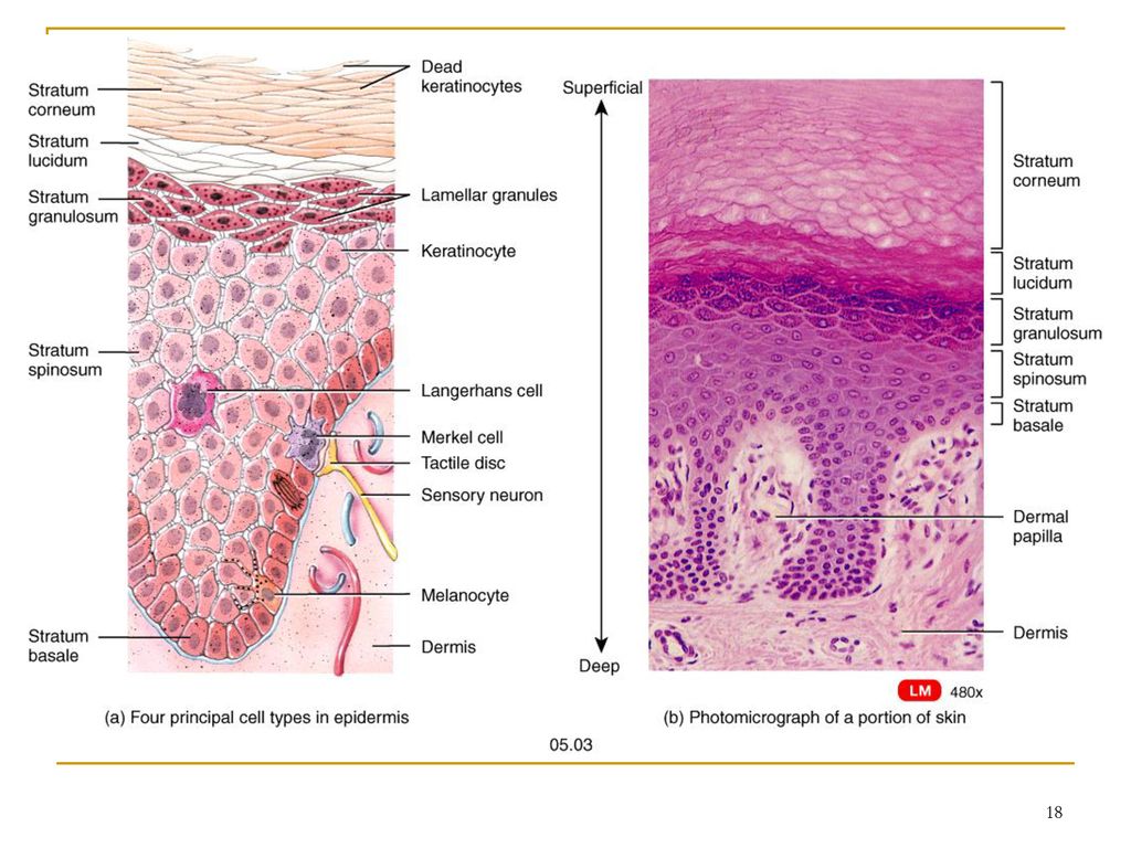
II. Epidermis It’s a keratinized stratified squamous epithelial tissue that includes four different cell types, and four to five distinct layers Avascular; nourished by diffusion from capillaries of the papillary layer of the dermis Separated from dermis by basement membrane

5
. Melanocyte Melanin granule Tactile (Merkel) cell Sensory nerve
ending Epidermal dendritic Dermis Keratinocytes Desmosomes (b) (a) Stratum corneum Most superficial layer; 20–30 layers of dead cells represented only by flat membranous sacs filled with keratin. Glycolipids in extracellular space. Stratum granulosum Three to five layers of flattened cells, organelles deteriorating; cytoplasm full of lamellated gran- ules (release lipids) and keratohyaline granules. Stratum spinosum Several layers of keratinocytes unified by desmosomes. Cells contain thick bundles of intermediate filaments made of pre-keratin. Stratum basale Deepest epidermal layer; one row of actively mitotic stem cells; some newly formed cells become part of the more superficial layers. See occasional melanocytes and epidermal dendritic cells.
 (a) Stratum corneum. Most superficial layer; 20–30 layers of dead. cells represented only by flat membranous. sacs filled with keratin. Glycolipids in. extracellular space. Stratum granulosum. Three to five layers of flattened cells, organelles. deteriorating; cytoplasm full of lamellated gran- ules (release lipids) and keratohyaline granules. Stratum spinosum. Several layers of keratinocytes unified by. desmosomes. Cells contain thick bundles of. intermediate filaments made of pre-keratin. Stratum basale. Deepest epidermal layer; one row of actively. mitotic stem cells; some newly formed cells. become part of the more superficial layers. See occasional melanocytes and epidermal. dendritic cells.")
6
A. Cells of the Epidermis
The epidermis contains the following four types of cells: 1. Keratinocytes They are arranged in 4 layers and represent the majority of the cells in the epidermis They start as cuboidal cells and become squamous as they reach the apical surface They produce keratin for strength as well as a water repellent sealant

7
Melanocyte Melanin granule Tactile (Merkel) cell Sensory nerve ending Epidermal dendritic cell Dermis Keratinocytes Stratum corneum Most superficial layer; 20–30 layers of dead cells represented only by flat membranous sacs filled with keratin. Glycolipids in extracellular space. Stratum granulosum Three to five layers of flattened cells, organelles deteriorating; cytoplasm full of lamellated granules (release lipids) and keratohyaline granules. Stratum spinosum Several layers of keratinocytes unified by desmosomes. Cells contain thick bundles of intermediate filaments made of pre-keratin. Stratum basale Deepest epidermal layer; one row of actively mitotic stem cells; some newly formed cells become part of the more superficial layers. See occasional melanocytes and epidermal dendritic cells. Desmosomes (b)
 and. keratohyaline granules. Stratum spinosum. Several layers of keratinocytes unified by. desmosomes. Cells contain thick bundles of. intermediate filaments made of pre-keratin. Stratum basale. Deepest epidermal layer; one row of actively. mitotic stem cells; some newly formed cells. become part of the more superficial layers. See. occasional melanocytes and epidermal. dendritic cells. Desmosomes. (b)")
9
2. Melanocytes Produce the pigment melanin which contributes to skin color Melanin granules cluster at the apical surface of the nucleus of keratinocytes and protect them from UV light Other cells present in the epidermis are: Langerhans cells and Merkel cells

11
B. Layers of the Epidermis
1. The Stratum basale (germinativum) It’s the deepest layer of the epidermis It consists of a single row of stem cells alternating with melanocytes (10-25%) and merkel cells The stem cells in this row are actively dividing (highly mitotic) and producing new keratinocytes that replace the cells that are lost at the apical surface of the skin
 It’s the deepest layer of the epidermis. It consists of a single row of stem cells alternating with melanocytes (10-25%) and merkel cells. The stem cells in this row are actively dividing (highly mitotic) and producing new keratinocytes that replace the cells that are lost at the apical surface of the skin.")
12
. Keratinocytes Stratum corneum
Melanocyte Melanin granule Tactile (Merkel) cell Sensory nerve ending Epidermal dendritic cell Dermis Keratinocytes Stratum corneum Most superficial layer; 20–30 layers of dead cells represented only by flat membranous sacs filled with keratin. Glycolipids in extracellular space. Stratum granulosum Three to five layers of flattened cells, organelles deteriorating; cytoplasm full of lamellated granules (release lipids) and keratohyaline granules. Stratum spinosum Several layers of keratinocytes unified by desmosomes. Cells contain thick bundles of intermediate filaments made of pre-keratin. Stratum basale Deepest epidermal layer; one row of actively mitotic stem cells; some newly formed cells become part of the more superficial layers. See occasional melanocytes and epidermal dendritic cells. Desmosomes (b)
 cell. Sensory. nerve ending. Epidermal. dendritic cell. Dermis. Keratinocytes. Stratum corneum. Most superficial layer; 20–30 layers of dead. cells represented only by flat membranous. sacs filled with keratin. Glycolipids in. extracellular space. Stratum granulosum. Three to five layers of flattened cells, organelles deteriorating; cytoplasm full of. lamellated granules (release lipids) and. keratohyaline granules. Stratum spinosum. Several layers of keratinocytes unified by. desmosomes. Cells contain thick bundles of. intermediate filaments made of pre-keratin. Stratum basale. Deepest epidermal layer; one row of actively. mitotic stem cells; some newly formed cells. become part of the more superficial layers. See. occasional melanocytes and epidermal. dendritic cells. Desmosomes. (b)")
15
2. The stratum spinosum It consists of several (8-10) rows of keratinocytes that may still be able to divide Projections or extensions of the melanocytes are located in this layer 3. The stratum granulosum Consists of 3-5 layers of flattened keratinocytes that begin to die (apoptosis) at the upper layers Keratin (provide strength) and Lamellar granules (produce water repellent) are produced in this layer
 at the upper layers. Keratin (provide strength) and Lamellar granules (produce water repellent) are produced in this layer.")
16
. Keratinocytes Stratum corneum
Melanocyte Melanin granule Tactile (Merkel) cell Sensory nerve ending Epidermal dendritic cell Dermis Keratinocytes Stratum corneum Most superficial layer; 20–30 layers of dead cells represented only by flat membranous sacs filled with keratin. Glycolipids in extracellular space. Stratum granulosum Three to five layers of flattened cells, organelles deteriorating; cytoplasm full of lamellated granules (release lipids) and keratohyaline granules. Stratum spinosum Several layers of keratinocytes unified by desmosomes. Cells contain thick bundles of intermediate filaments made of pre-keratin. Stratum basale Deepest epidermal layer; one row of actively mitotic stem cells; some newly formed cells become part of the more superficial layers. See occasional melanocytes and epidermal dendritic cells. Desmosomes (b)
 cell. Sensory. nerve ending. Epidermal. dendritic cell. Dermis. Keratinocytes. Stratum corneum. Most superficial layer; 20–30 layers of dead. cells represented only by flat membranous. sacs filled with keratin. Glycolipids in. extracellular space. Stratum granulosum. Three to five layers of flattened cells, organelles deteriorating; cytoplasm full of. lamellated granules (release lipids) and. keratohyaline granules. Stratum spinosum. Several layers of keratinocytes unified by. desmosomes. Cells contain thick bundles of. intermediate filaments made of pre-keratin. Stratum basale. Deepest epidermal layer; one row of actively. mitotic stem cells; some newly formed cells. become part of the more superficial layers. See. occasional melanocytes and epidermal. dendritic cells. Desmosomes. (b)")
17
4. The stratum lucidum It consists of 2-3 rows of dead, flattened, keratin rich keratinocytes It is present only in thick skin 5. The Stratum corneum This layer consists of rows of keratinocytes that are made up mainly of keratin These cells are continuously being shed and protect the inner layers

19
C. Thick and Thin Skin 1. Thick skin Has all 5 epithelial strata
Found in areas subject to pressure or friction Palms of hands, fingertips, soles of feet 2. Thin skin More flexible than thick skin Covers rest of body

20
III. The Dermis It’s the deeper portion of the skin
It consists mainly of connective tissue containing collagen and elastic fibers The dermis gives skin its structural strength. It contains the skin funtional accessories and organelles: nerves, blood vessels, hair follicles, smooth muscles, glands, temperature sensors, touch receptors, free nerve endings (pain receptors), and lymphatic vessels.
, and lymphatic vessels.")
21
Epidermis Hair shaft Dermis Reticular layer Papillary Hypodermis (superficial fascia) Dermal papillae Pore Subpapillary vascular plexus Appendages of skin • Eccrine sweat gland • Arrector pili muscle • Sebaceous (oil) gland • Hair follicle • Hair root Nervous structures • Sensory nerve fiber • Pacinian corpuscle • Hair follicle receptor (root hair plexus) Cutaneous vascular plexus Adipose tissue
 gland. • Hair follicle. • Hair root. Nervous structures. • Sensory nerve fiber. • Pacinian corpuscle. • Hair follicle receptor. (root hair plexus) Cutaneous vascular. plexus. Adipose tissue.")
22
A. The Layers of the Dermis
1. Papillary: Superficial (outer) 20% of the dermis. Consists of areolar c.t. with lots of elastic fibers. Arranged in dermal papillae 2. Reticular: Deep (inner) 80% of the dermis Consists of dense irregular C.T. with collagen and elastic fibers. May have pockets of adipose cells
 20% of the dermis. Consists of areolar c.t. with lots of elastic fibers. Arranged in dermal papillae. 2. Reticular: Deep (inner) 80% of the dermis. Consists of dense irregular C.T. with collagen and elastic fibers. May have pockets of adipose cells.")
23
B. Skin Color Skin color comes from the combination of three pigments present in the skin: 1) Melanin: produced by melanocytes with help of the enzyme tyrosinase 2) Carotene: from diet 3) Hemoglobin: the pigment in red blood cells (RBC’s) What is cyanosis? What is jaundice? What are decubitus ulcers or bedsores?
 Melanin: produced by melanocytes with help of the enzyme tyrosinase 2) Carotene: from diet 3) Hemoglobin: the pigment in red blood cells (RBC’s) What is cyanosis. What is jaundice. What are decubitus ulcers or bedsores .")
31
IV. Appendages of the Skin
1. Sweat (sudoriferous) glands They are very numerous (3 million) and distributed throughout the body There are two types of sweat glands: eccrine an apocrine sweat glands a) Eccrine sweat glands: They are the most widely distributed sweat glands; become active shortly after birth produce about 600 ml of sweat per day, which is a filtrate from blood Eccrine glands release sweat through pores in the skin
 glands. They are very numerous (3 million) and distributed throughout the body. There are two types of sweat glands: eccrine an apocrine sweat glands. a) Eccrine sweat glands: They are the most widely distributed sweat glands; become active shortly after birth. produce about 600 ml of sweat per day, which is a filtrate from blood. Eccrine glands release sweat through pores in the skin.")
32
Epidermis Hair shaft Dermis Reticular layer Papillary Hypodermis (superficial fascia) Dermal papillae Pore Subpapillary vascular plexus Appendages of skin • Eccrine sweat gland • Arrector pili muscle • Sebaceous (oil) gland • Hair follicle • Hair root Nervous structures • Sensory nerve fiber • Pacinian corpuscle • Hair follicle receptor (root hair plexus) Cutaneous vascular plexus Adipose tissue
 gland. • Hair follicle. • Hair root. Nervous structures. • Sensory nerve fiber. • Pacinian corpuscle. • Hair follicle receptor. (root hair plexus) Cutaneous vascular. plexus. Adipose tissue.")
33
The main function of eccrine sweat glands is thermoregulation: to assist in the regulation of body temperature and to a minor extent, the removal of waste (urea, uric acid, ammonia and lactic acid) b) Appocrine sweat glands: Found mainly in the axillary region, the groin and the bearded regions of adult males The sweat of apocrine glands contains the same components as sweat from eccrine glands but it also has lipids and proteins Apocrine glands start functioning at puberty and play no significant role in thermoregulation
 Appocrine sweat glands: Found mainly in the axillary region, the groin and the bearded regions of adult males. The sweat of apocrine glands contains the same components as sweat from eccrine glands but it also has lipids and proteins. Apocrine glands start functioning at puberty and play no significant role in thermoregulation.")
35
1. Sebaceous (Oil) glands
Distributed throughout the body, most of them associated with hair follicles Produce an oily secretion (sebum), which lubricates the skin and the hair, prevents water evaporation, and inhibits the growth of certain bacteria (bactericidal) 2. Hairs and Hair Follicles The main functions of the hair are: protection, sensation and filtering out of particles The root hair plexus: generates nerve impulses when the hair shaft is moved
, which lubricates the skin and the hair, prevents water evaporation, and inhibits the growth of certain bacteria (bactericidal) 2. Hairs and Hair Follicles. The main functions of the hair are: protection, sensation and filtering out of particles. The root hair plexus: generates nerve impulses when the hair shaft is moved.")
36
. Sweat pore Sebaceous gland Dermal connective tissue Eccrine
gland duct Eccrine gland Hair in hair follicle Secretory cells (a) Photomicrograph of a sectioned sebaceous gland (220x)
 Photomicrograph of a sectioned. sebaceous gland (220x)")
37
Epidermis Hair shaft Dermis Reticular layer Papillary Hypodermis (superficial fascia) Dermal papillae Pore Subpapillary vascular plexus Appendages of skin • Eccrine sweat gland • Arrector pili muscle • Sebaceous (oil) gland • Hair follicle • Hair root Nervous structures • Sensory nerve fiber • Pacinian corpuscle • Hair follicle receptor (root hair plexus) Cutaneous vascular plexus Adipose tissue
 gland. • Hair follicle. • Hair root. Nervous structures. • Sensory nerve fiber. • Pacinian corpuscle. • Hair follicle receptor. (root hair plexus) Cutaneous vascular. plexus. Adipose tissue.")
38
V. Functions of the Integumentary System
1. Protection: a) Chemical barriers: protection against UV light by melanin and against microorganisms by sebum b) Physical / mechanical barriers: protects against dehydration, abrasion or penetration of microorganisms; nails protect ends of digits c) Biological barriers: WBC’s
 Chemical barriers: protection against UV light by melanin and against microorganisms by sebum. b) Physical / mechanical barriers: protects against dehydration, abrasion or penetration of microorganisms; nails protect ends of digits. c) Biological barriers: WBC’s.")
39
2. Temperature Regulation
1) Sweat produced by sudoriferous glands causes evaporative cooling. 2) Arterioles in dermis change diameter as temperature changes. More or less blood flows through the dermis. 3. Cutaneous Sensation Pressure, temperature, pain, heat, cold, touch, movement of hairs.
 Sweat produced by sudoriferous glands causes evaporative cooling. 2) Arterioles in dermis change diameter as temperature changes. More or less blood flows through the dermis. 3. Cutaneous Sensation. Pressure, temperature, pain, heat, cold, touch, movement of hairs.")
42
4. Metabolic Functions The precursor molecule of vitamin D is produced in the skin with the help of sunlight It is then turned into calcitriol by the liver and kidneys Calcitriol is a hormone that aids in the absorption of calcium from the GI tract 5. Blood Reservoir 6. Excretion: Removal of waste products from the body. Sweat. Water, salt, urea, ammonia, uric acid

43
VI. Homeostatic imbalances of the Skin
A. Skin Cancer Occurs from mutations in the in the epidermal cells caused mainly by sun radiation (UV light) The three most common types of skin cancer are: 1) Basal cell carcinoma: which originates in the keratinocytes of the stratum basale It’s the most common but the least malignant accounting for 80% of skin cancers and developing relatively slowly
 The three most common types of skin cancer are: 1) Basal cell carcinoma: which originates in the keratinocytes of the stratum basale. It’s the most common but the least malignant accounting for 80% of skin cancers and developing relatively slowly.")
45
2) Squamous cell carcinoma: it’s the second most common skin cancer occurring in the keratinocytes at the stratum spinosum 3) Melanoma: which involves the melanocytes is the least common skin cancer It’s the most dangerous skin cancer because is highly metastatic and resistant to chemotherapy 1/3 of them develop from peexisting moles Assessment through the ABCD rule
 Melanoma: which involves the melanocytes is the least common skin cancer. It’s the most dangerous skin cancer because is highly metastatic and resistant to chemotherapy. 1/3 of them develop from peexisting moles. Assessment through the ABCD rule.")
47
B. Burns Classifications First-degree Second-degree Third-degree
Skin Grafts Split skin Artificial skin Cadavers or pigs
51
Figure 5.10 Partial thickness and full thickness burns.
Skin bearing partial thickness burn (1st and 2nd degree burns) (b) Skin bearing full thickness burn (3rd degree burn) 1st degree burn 2nd degree 3rd degree
55
Assessing burns - the "rule of nines"
Burns are assessed in terms of total body surface area (TBSA), which is the percentage affected by partial thickness or full thickness burns (superficial thickness burns are not counted). Head - 9% Anterior Torso - 18% Posterior Torso - 18% Each Leg - 18% Each Arm - 9% Genitalia/perineum - 1%
, which is the percentage affected by partial thickness or full thickness burns (superficial thickness burns are not counted). Head - 9% Anterior Torso - 18% Posterior Torso - 18% Each Leg - 18% Each Arm - 9% Genitalia/perineum - 1%")
56
Anterior and posterior
head and neck, 9% 4 1 / 2 % upper limbs, 18% lower limbs, 36% 100% Totals trunk, 36% Anterior trunk, 18% 9% (Perineum, 1%)
")
Similar presentations


 Largest organ of the body (15% of body weight) Skin thickness variable, normally 1-2 mm Protection –chemical barrier (waterproof)>")
 Adipose tissue>")


 like an onion?>")

 Skin derivatives>")

 Integument is nothing but the skin. Integument(=to cover in latin Skin and its appendages are the largest organ of the body Functions:>")
