Download presentation
Presentation is loading. Please wait.

1
Neuroradiology interactive lecture
366 RAD (Radiology)
")
2
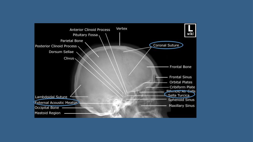
Name the structures L SKULL PA VIEW Skull X-RAY LAT. VIEW

4
Which is true on this brain CT regarding anatomy:
Internal capsule Caudate head Cerebral peduncle Putamen Thalamus 4th ventricle

5
Which is true on this brain CT regarding anatomy:
Anterior Horn of the Lateral Ventricle Caudate Nucleus Anterior Limb of the Internal Capsule Putamen and Globus Pallidus Posterior Limb of the Internal Capsule Third Ventricle Quadrigeminal Plate Cistern Cerebellar Vermis Occipital Lobe

6
Which is true in CT? Bone is black CSF is black
Gray matter is darker than white matter Gray and white matter can not be differentiated

7
Which is true in CT? Bone is black CSF is black
Gray matter is darker than white matter Gray and white matter can not be differentiated

9
Name the structures

10
Name the structures

11
Contraindication of MRI include all the following EXCEPT:
cardiac pacemaker cochlear implants metal close to the eye neurostimulators pregnancy (3rd trimester)
")
12
Contraindication of MRI include all the following EXCEPT:
cardiac pacemaker cochlear implants metal close to the eye neurostimulators pregnancy (3rd trimester)
")
13
MRI diffusion (DWI) is particularly helpful in assessment of all the following EXCEPT:
Brain infarction Brain abscess Brain tumors Hydrocephalus

14
MRI diffusion (DWI) is particularly helpful in assessment of all the following EXCEPT:
Brain infarction Brain abscess Brain tumors Hydrocephalus

15
MRI Diffusion.. DWI ADC map MR diffusion
Very helpful in assessment of: Early brain infarction. Brain abscess. Certain types of brain tumor. DWI ADC map

16
Which of the following is true?
This is CTA study This is MRA study This can only be done with contrast This is good to diagnose cerebral venous thrombosis

17
Which of the following is true?
This is CTA study This is MRA study This can only be done with contrast This is good to diagnose cerebral venous thrombosis

18
An MRI showed intra-axial lesion that is necrotic, irregular, strongly enhancing, and crossing midline. This lesion is most likely: Meningioma Infarction Multiple sclerosis Glioblastoma multiforme

19
An MRI showed intra-axial lesion that is necrotic, irregular, strongly enhancing, and crossing midline. This lesion is most likely: Meningioma Infarction Multiple sclerosis Glioblastoma multiforme

21
The lesion on this CT is:
Meningioma Abscess Multiple sclerosis Glioblastoma multiforme

22
The lesion on this CT is:
Meningioma Abscess Multiple sclerosis Glioblastoma multiforme

23
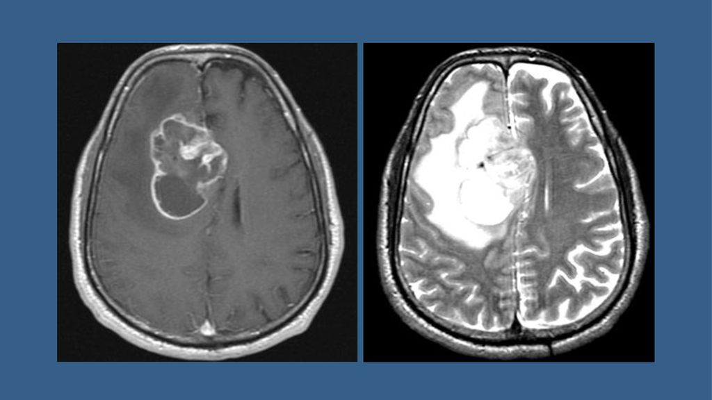
The lesion on this MRI is:
Meningioma Infarction Metastasis Abscess

24
The lesion on this MRI is:
Meningioma (extra-axial) Infarction Metastasis Abscess
 Infarction. Metastasis. Abscess.")
26
The lesion on this MRI is:
Pituitary adenoma Craniopharyngioma Meningioma Glioblastoma multiforme

27
The lesion on this MRI is:
Pituitary adenoma Craniopharyngioma (multi-cyctic) Meningioma Glioblastoma multiforme
 Meningioma. Glioblastoma multiforme.")
28
The abnormalities on this MRI are due to:
Multiple sclerosis Meningitis Brain tumor Encephalitis

29
The abnormalities on this MRI are due to:
Multiple sclerosis Meningitis Brain tumor Encephalitis

30
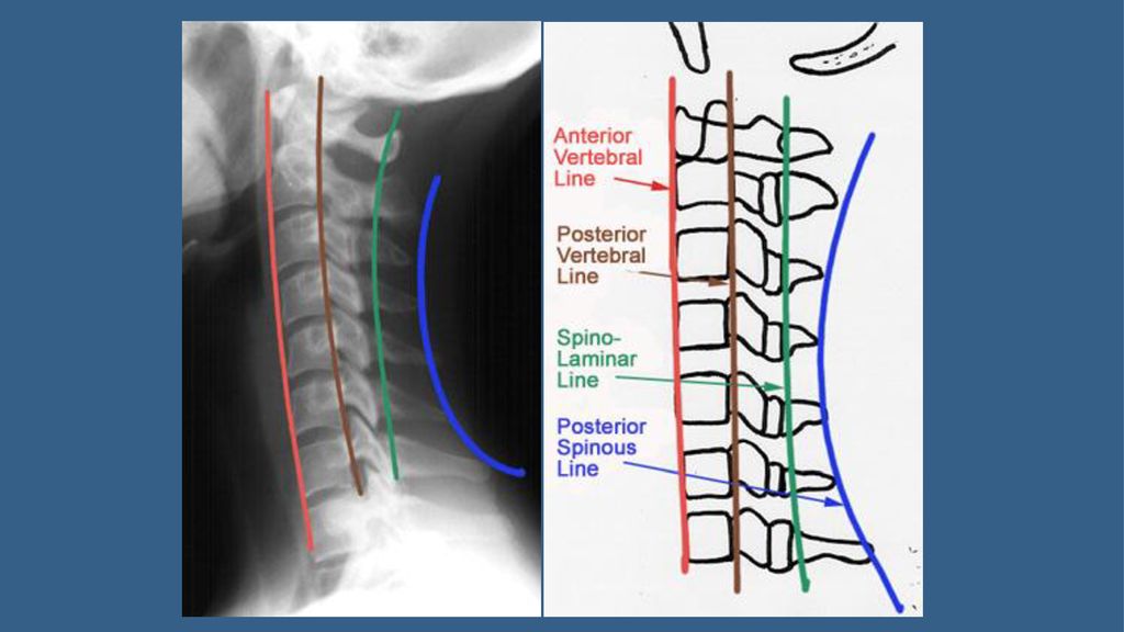
Which of the following is true about the lines of the cervical spine?
Red is intervertebral line Brown is posterior spinous line Green is spinolaminar line Blue is posterior vertebral line

31
Which of the following is true about the lines of the cervical spine?
Red is intervertebral line Brown is posterior spinous line Green is spinolaminar line Blue is posterior vertebral line

33
This MRI of the spine shows:
Meningocele Extradural tumor Discitis Vertebral fusion

34
This MRI of the spine shows:
Meningocele Extradural tumor Discitis Vertebral fusion

35
Patient C Patient A Patient B

36
EXTRA dural extra medullary (Epi dural ) Patient A
Patient C EXTRA dural extra medullary (Epi dural ) Patient A T 1 Intra dural intra medullary Patient B Intra dural extra medullary
 Patient A. T 1 Intra dural intra medullary. Patient B. Intra dural extra medullary.")
37
Intra dural intra medullary Intra dural extra medullary
EXTRA dural extra medullary (Epi dural )
")
38
What is the difference? Normal control Patient

39
What is the difference? Normal control Patient Cervical spondylosis

40
This MRI shows an infarction in the right basal ganglia.
T2WI FLAIR DWI This MRI shows an infarction in the right basal ganglia. The infarction is: Acute (recent) Chronic (old) Hemorrhagic In PCA territory
 Chronic (old) Hemorrhagic. In PCA territory.")
41
This MRI shows an infarction in the right basal ganglia.
T2WI FLAIR DWI This MRI shows an infarction in the right basal ganglia. The infarction is: Acute (recent) >> bright in all MRI sequence Chronic (old) Hemorrhagic In PCA territory
 >> bright in all MRI sequence. Chronic (old) Hemorrhagic. In PCA territory.")
42
This patient is most likely to have:
T2WI FLAIR DWI This patient is most likely to have: Left monoplegia Left hemiplegia Diplegia No symptoms

43
This patient is most likely to have:
T2WI FLAIR DWI This patient is most likely to have: Left monoplegia Left hemiplegia Diplegia No symptoms

44
This CT shows: Subdural hematoma Subarachnoid hemorrhage
Intraventricular hemorrhage All of the above

45
This CT shows: Subdural hematoma Subarachnoid hemorrhage
Intraventricular hemorrhage All of the above

46
The hematoma pointed by the arrow is:
Acute epidural Chronic epidural Acute subdural Chronic subdural None of the above

47
The hematoma pointed by the arrow is:
Acute epidural Chronic epidural Acute subdural Chronic subdural None of the above

48
This CT shows: Acute PCA infarct Chronic ACA infarct
Subarachnoid bleeding Meningioma Abscess

49
This CT shows: Acute PCA infarct Chronic ACA infarct
Subarachnoid bleeding Meningioma Abscess

50
Thank you

Similar presentations


 Prof. Ibrahim A. Alorainy.>")