Download presentation
Presentation is loading. Please wait.

1
Neuroradiology DR. Sharifa AL-Duraibi

2
Nuro-radiology anatomy

3
A. Orbit. B. Sphenoid Sinus. C. Temporal Lobe. D. External Auditory Canal. E. Mastoid Air Cells. F. Cerebellar Hemisphere.

4
A. Frontal Lobe. B. Frontal Bone (Superior Surface of Orbital Part). C. Dorsum Sellae. D. Basilar Artery. E. Temporal Lobe. F. Mastoid Air Cells. G. Cerebellar Hemisphere.

5
A. Frontal Lobe. B. Sylvian Fissure. C. Temporal Lobe. D. Suprasellar Cistern. E. Midbrain. F. Fourth Ventricle. G. Cerebellar Hemisphere.

6
A. Falx Cerebri. B. Frontal Lobe. C. Anterior Horn of Lateral Ventricle. D. Third Ventricle. E. Quadrigeminal Plate Cistern. F. Cerebellum.

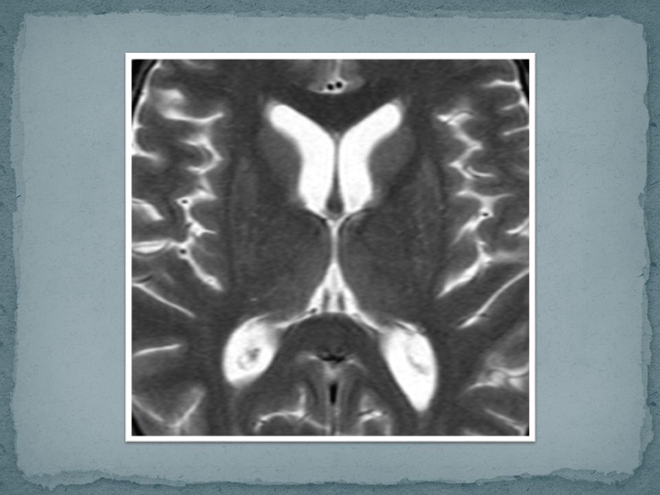
7
A. Anterior Horn of the Lateral Ventricle.
B. Caudate Nucleus. C. Anterior Limb of the Internal Capsule. D. Putamen and Globus Pallidus. E. Posterior Limb of the Internal Capsule. F. Third Ventricle. G. Quadrigeminal Plate Cistern. H. Cerebellar Vermis. I. Occipital Lobe.

8
A. Genu of the Corpus Callosum.
B. Anterior Horn of the Lateral Ventricle. C. Internal Capsule. D. Thalamus. E. Pineal Gland. F. Choroid Plexus. G. Straight Sinus.

9
A. Falx Cerebri. B. Sulcus. C. Gyrus. D. Superior Sagittal Sinus.

17
Head Trauma

18
Head Injury Head trauma is the leading cause of death in people under the age of 30. Males have 2-3 x frequency of brain injury than females

19
Not suitable in emergency situations.
Head injury Plain film Calvarial fractures. Penetrating injuries. Radiopaque foreign bodies. CT Disorientation. Neurological deficits. Unconsciousness. Sever headache. MR Not suitable in emergency situations.

20
Brain CT What is the initial radiological investigation for the evaluation of Comatose patient/sever headache?

21
CT has the advantage of being available 24 hours a day and is the gold standard for hemorrhage.
On CT 60% of infarcts are seen within 3-6 hrs and virtually all are seen in 24 hours. The overall sensitivity of CT to diagnose stroke is 64% and the specificity is 85%. The most important issue to determine when imaging a stroke patient is whether one is dealing with a hemorrhagic or ischemic event.

22
Subarachnoid hemorrhage (SAH)
Presents as sudden worse headache of life. Appears as linear high density in the sulci. Intracranial aneurysms cause of approximately 80% of non-traumatic SAH.

23
Subarachnoid hemorrhage (SAH)
SAH most commonly results from aneurysmal rupture. Appears as linear high density in the sulci, fissures, basal cisterns and ventricles.

25
Subdural haemorrhage Subdural haemorrhage (SDH) is a collection of blood accumulating in the potential space between the dura and arachnoid mater of the meninges around the brain.
 is a collection of blood accumulating in the potential space between the dura and arachnoid mater of the meninges around the brain.")
27
Extradural hemorrhage
An extradural haematoma (EDH) (also known as an epidural haematoma) is a collection of blood which forms between the inner surface of the skull and outer layer of dura.
 (also known as an epidural haematoma) is a collection of blood which forms between the inner surface of the skull and outer layer of dura.")
29
stroke Stroke is a clinical term for sudden, focal neurological deficit. Stroke accounts for one out of every 15 deaths in the United States.

30
stroke Strokes are classified into two major types: Hemorrhagic strokes: Are due to rupture of a cerebral blood vessel that causes bleeding into or around the brain. Hemorrhagic strokes account for 16% of all strokes. Ischemic stroke: Is caused by blockage of blood flow in a major cerebral blood vessel, usually due to a blood clot. Ischemic strokes account for about 84% of all strokes.

31
Ischemic stroke Caused by: Thrombosis. Hypoperfusion infarctions.

32
Ischemic stroke Changes of acute ischemia due to edema which include the following: Obscuration of the lentiform nuclei. Loss of insular ribbon. Loss of gray/white distinction. Sulcal effacement.

33
Ischemic stroke Loss of insular ribbon: Is the loss of the gray- white interface in the lateral margins of the insula.

34
Ischemic stroke A lacunar infarction occurs when the walls of small arteries thicken and cause the occlusion of the artery. These typically involve the small perforating vessels of the brain and result in lesions that are less than 1.5 cm in size.

35
Hemorrhagic strokes Intra-cerebral hemorrhage is the most common, accounting for 10% of all strokes. Subarachnoid hemorrhage, due to rupture of a cerebral aneurysm, accounts for 6% of strokes overall.

36
Hemorrhagic strokes The most common cause of non-traumatic intra- cerebral hematoma is hypertensive hemorrhage.

37
Hemorrhagic strokes It often appears as a high- density hemorrhage in the region of the basal ganglia. Blood may extend into the ventricular system. Intra-ventricular extension of the hematoma is associated with a poor prognosis.

39
From Mike Mosely (Stanford Radiology)
")
40
CT Head / brain Dark (black): Bright (white): CSF, old infarction,
old hematoma, cyst. Bright (white): Acute hemorrhage, calcification, contrast in vessels.
: Acute hemorrhage, calcification, contrast in vessels.")
41
Non contrast CT head Is the investigation of choice for Head trauma and acute stroke.

Similar presentations


>")


>")


