Download presentation
Presentation is loading. Please wait.

1
Neuroradiology interactive lecture 366 RAD (Radiology) Prof. Ibrahim A. Alorainy
 Prof. Ibrahim A. Alorainy")
2
Skull X-RAY LAT. VIEW L SKULL PA VIEW Name the structures

3
Which is true on this brain CT regarding anatomy : A.Internal capsule B.Caudate head C.Cerebral peduncle D.Putamen E.Thalamus F.4 th ventricle

4
Which is true in CT? A.Bone is black B.CSF is black C.Gray matter is darker than white matter D.Gray and white matter can not be differentiated

6
Name the structures

7
Contraindication of MRI include all the following EXCEPT: A.cardiac pacemaker B.cochlear implants C.metal close to the eye D.neurostimulators E.pregnancy (3 rd trimester)
")
8
MRI diffusion (DWI) is particularly helpful in assessment of: A.Brain infarction B.Brain abscess C.Brain tumors D.Hydrocephalus
 is particularly helpful in assessment of: A.Brain infarction B.Brain abscess C.Brain tumors D.Hydrocephalus")
9
MRI Diffusion.. DWI ADC map MR diffusion Very helpful in assessment of: Early brain infarction. Brain abscess. Certain types of brain tumor.

10
Which of the following is true ? A.This is CTA study B.This is MRA study C.This can only be done with contrast D.This is good to diagnose cerebral venous thrombosis

11
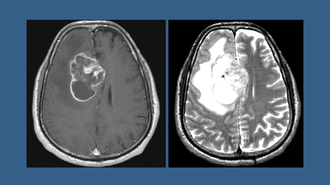
An MRI showed intra-axial lesion that is necrotic, irregular, strongly enhancing, and crossing midline. This lesion is most likely: A.Meningioma B.Infarction C.Multiple sclerosis D.Glioblastoma multiforme

13
The lesion on this CT is: A.Meningioma B.Abscess C.Multiple sclerosis D.Glioblastoma multiforme

14
The lesion on this MRI is: A.Meningioma B.Infarction C.Metastasis D.Abscess

16
Which of the following is true about the lines of the cervical spine? A.Red is intervertebral line B.Brown is posterior spinous line C.Green is spinolaminar line D.Blue is posterior vertebral line

18
This MRI of the spine shows: A.Meningocele B.Extradural tumor C.Discitis D.Vertebral fusion

19
Patient A Patient B Patient C

21
Normal control Patient What is the difference?

22
DWI FLAIRT2WI This MRI shows an infarction in the right basal ganglia. The infarction is: A.Acute (recent) B.Chronic (old) C.Hemorrhagic D.In PCA territory
 B.Chronic (old) C.Hemorrhagic D.In PCA territory.")
23
DWI FLAIRT2WI This patient is most likely to have: A.Left monoplegia B.Left hemiplegia C.Diplegia D.No symptoms

24
This CT shows: A.Subdural hematoma B.Subarachnoid hemorrhage C.Intraventricular hemorrhage D.All of the above

25
The hematoma pointed by the arrow is: A.Acute epidural B.Chronic epidural C.Acute subdural D.Chronic subdural E.None of the above

26
This CT shows: A.Acute PCA infarct B.Chronic ACA infarct C.Subarachnoid bleeding D.Meningioma E.Abscess

Similar presentations


>")