Download presentation
Presentation is loading. Please wait.

1
Chapter 13 Nuclear Magnetic Resonance Spectroscopy Jo Blackburn Richland College, Dallas, TX Dallas County Community College District 2003, Prentice Hall Organic Chemistry, 5 th Edition L. G. Wade, Jr. The slides used in this presentation are borrowed heavily from the great downloadable:

2
1 H NMR 1 GHz machine in France!

3
4 Essential points Re: NMR 1.Number of peaks = No of H environments (a peak may be split into many smaller peaks) 2.Position of the peak depends of ‘chemical’ (actually magnetic) environment. 3.Area under the peak = relative number of H in that chemical environment 4.Splitting patters reveals info about adjacent H’s

4
The NMR Spectrometer =>

5
The NMR Graph =>

6
Tetramethylsilane TMS is added to the sample. Since silicon is less electronegative than carbon, TMS protons are highly shielded. Signal defined as zero. Organic protons absorb downfield (to the left) of the TMS signal. =>
 of the TMS signal. =>.")
7
Number of Signals => Equivalent hydrogens have the same chemical shift. Each H in the methyl group at ‘a’ are equivalent to each other. (etc)
.")
8
Location of Signals - deshielding More electronegative atoms deshield more and give larger shift values. Effect decreases with distance. Additional electronegative atoms cause increase in chemical shift. =>

9
Delta Scale => This slide basically says peak position is independent of electromagnetic frequency OLD MACHINE NEWER MACHINE

10
Typical Values =>

11
Ethanol

13
Aromatic Protons, 7- 8 =>

14
Vinyl Protons, 5- 6 =>

15
Acetylenic Protons, 2.5 =>

16
Aldehyde Proton, 9- 10 => Electronegative oxygen atom

17
Carboxylic Acid Proton, 10+ =>

18
O-H and N-H Signals Chemical shift depends on concentration. Hydrogen bonding in concentrated solutions deshield the protons, so signal is around 3.5 for N-H and 4.5 for O-H. Proton exchanges between the molecules broaden the peak. =>

19
Intensity of Signals The area under each peak is proportional to the number of protons. Shown by integral trace. =>

20
Flv Video: InstrumentationInstrumentation

21
How Many Hydrogens? => When the molecular formula is known, each integral rise can be assigned to a particular number of hydrogens.

22
Spin-Spin Splitting Nonequivalent protons on adjacent carbons have magnetic fields that may align with or oppose the external field. This magnetic coupling causes the proton to absorb slightly downfield when the external field is reinforced and slightly upfield when the external field is opposed. All possibilities exist, so signal is split. =>

23
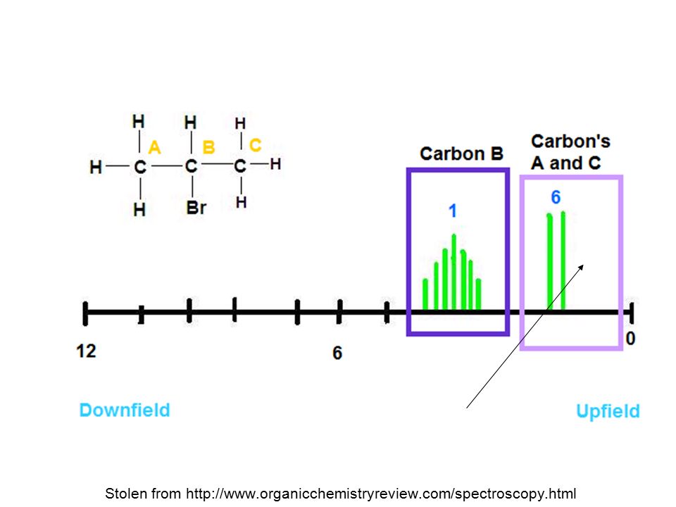
Range of Magnetic Coupling Protons on adjacent carbons normally will couple. Equivalent protons do not split each other. Protons separated by four or more bonds will not couple. Protons bonded to the same carbon will split each other only if they are not equivalent. (e.g CH2 next to a C*-H) =>
 =>.")
24
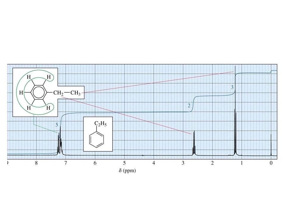
Simple spectra Stolen from http://www.organicchemistryreview.com/spectroscopy.html

26
Doublet: 1 Adjacent Proton =>

27
Triplet: 2 Adjacent Protons =>

28
No splitting – no adjacent H

29
1,1,2-Tribromoethane Nonequivalent protons on adjacent carbons. =>

30
ethanol

31
The N + 1 Rule If a signal is split by N equivalent protons, it is split into N + 1 peaks. =>

32
Pascal's triangle Helps you interpret the splitting Pattern. OR gives the splitting pattern (prediction) http://www.lincoln.k12.nv.us/alamo/high/Departments/Math/Pascal/Pascal's_Triangle_Webquest.html
 s_Triangle_Webquest.html.")
34
Splitting for Ethyl Groups =>

35
Splitting for Isopropyl Groups =>

36
http://www.absoluteastronomy.com/topics/Proton_NMR
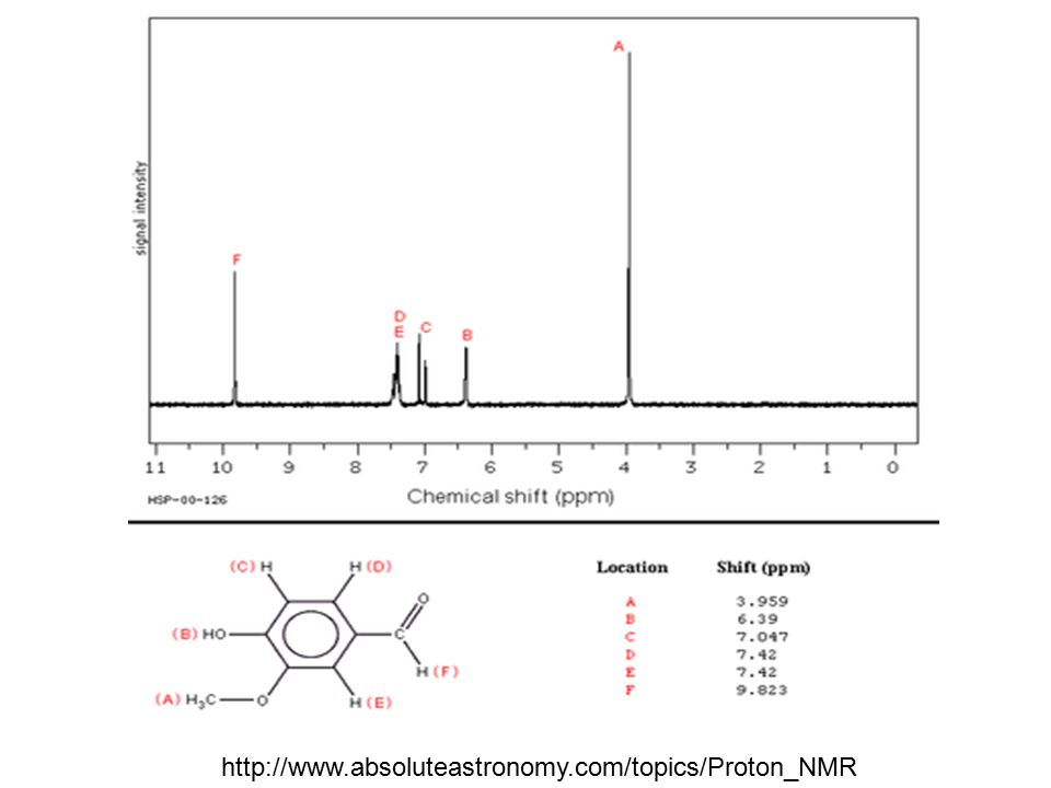
37
Double bond equivalents Sat d organic molecule has 2n+2 H’s for every C. Each double bond ‘takes 2 H away’ No H’s =2n e.g. ethene C 2 H 4 A ring also ‘takes 2 H’s away’ cyclehexane C 6 H 12 For every halogen, add 1 H For every N take away 1 H For every O, do nothing. Ethanol = C 2 H 5 OH =>C 2 H 6 Compare it to sat d formula for 2C’s No difference therefore molecule C 2 H 5 OH has no double bond equivalents. Ethanal = ? Methylbenzene = ? From molecular formula…

38
MRI Dangers of MRI

39
Functional group region >1400 cm -1 Stolen from http://www.organicchemistryreview.com/spectroscopy.html

40
Answer?Sure? 2-methylpropan-1-ol http://science.widener.edu/svb/nmr/seminar/isobutanol.html Quiz

41
High Performance (High Pressure) Liquid Chromatography (HPLC)
 Liquid Chromatography (HPLC)")
42
http://www.idex-hs.com/support/upchurch/i/hplcDiagram.gif HPLC schematic

43
The column http://www.goehler-hplc.de/images/parts.jpg

44
High Performance Liquid Chromatography (HPLC) (High Pressure) High pressure gives separation much faster than say gravity based chromatography (paper or column chromatography) Detector uses UV light to detect presence of chromophores
 (High Pressure) High pressure gives separation much faster than say gravity based chromatography (paper or column chromatography) Detector uses UV light to detect presence of chromophores")
45
High Performance Liquid Chromatography (HPLC) (High Pressure) Stationary phase in the form of a steel encased column (size of a straw) = silica gel, Al 2 O 3 or C 18 hydrocarbon – giving different polarities.Stationary phase in the form of a steel encased column (size of a straw) = silica gel, Al 2 O 3 or C 18 hydrocarbon – giving different polarities. Mobile phase = solventMobile phase = solvent Area under peak = measure of the relative abundance of the compoundArea under peak = measure of the relative abundance of the compound

46
Add a standard of known concentration to get it’s peak area.Add a standard of known concentration to get it’s peak area. The same species have the same retention time (if all other factors are kept constant e.g. pressure, solvent, column type)The same species have the same retention time (if all other factors are kept constant e.g. pressure, solvent, column type) Can separate chiral compoundsCan separate chiral compounds
The same species have the same retention time (if all other factors are kept constant e.g. pressure, solvent, column type) Can separate chiral compoundsCan separate chiral compounds.")
47
http://www.biocompare.com/Articles/ApplicationNote/1508/ANALYSIS-OF-AMINO-ACIDS-BY-REVERSED-PHASE- CHROMATOGRAPHY-WITH-PRECOLUMN-DERIVATISATION-AND-UV-VISIBLE-DETECTION.html UV detector set at λ=338nm Amino acids are poor at absorbing UV so they Have a UV chromophore bonded to them for the purpose of being HPLC’d

48
http://www.aldbot.com/HPLC-Hawaii.gif

49
Sample will pass quickly through the column IF: It has similar polarity to mobile phase Stationary phase in the column has a large particle size. Pressure of HPLC is increased Temp not really a factor as HPLC tends not to have a heater added Column is short. IN GENERAL LONGER COLUMN TIMES ARE BETTER (GIVE BETTER SEPARATION IN MISTURES SO REVERSE THE ABOVE CONDITIONS.

Similar presentations

 NMR Spectroscopy>")

>")
>")


 Spectroscopy>")


 Spectroscopy Structure Determination>")


