Download presentation
Presentation is loading. Please wait.

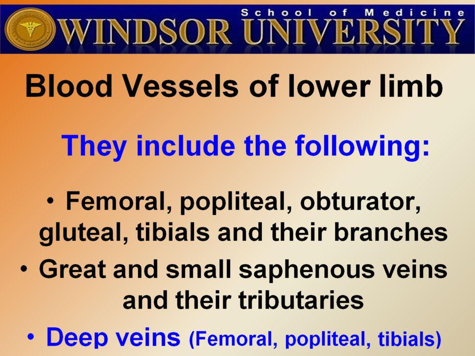
3
A A

5
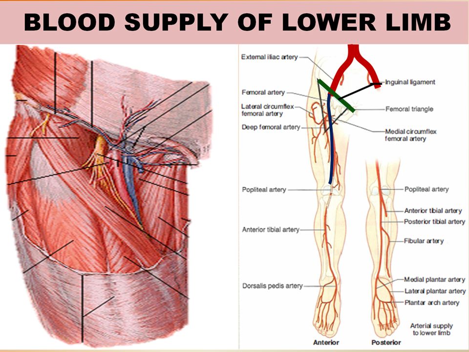
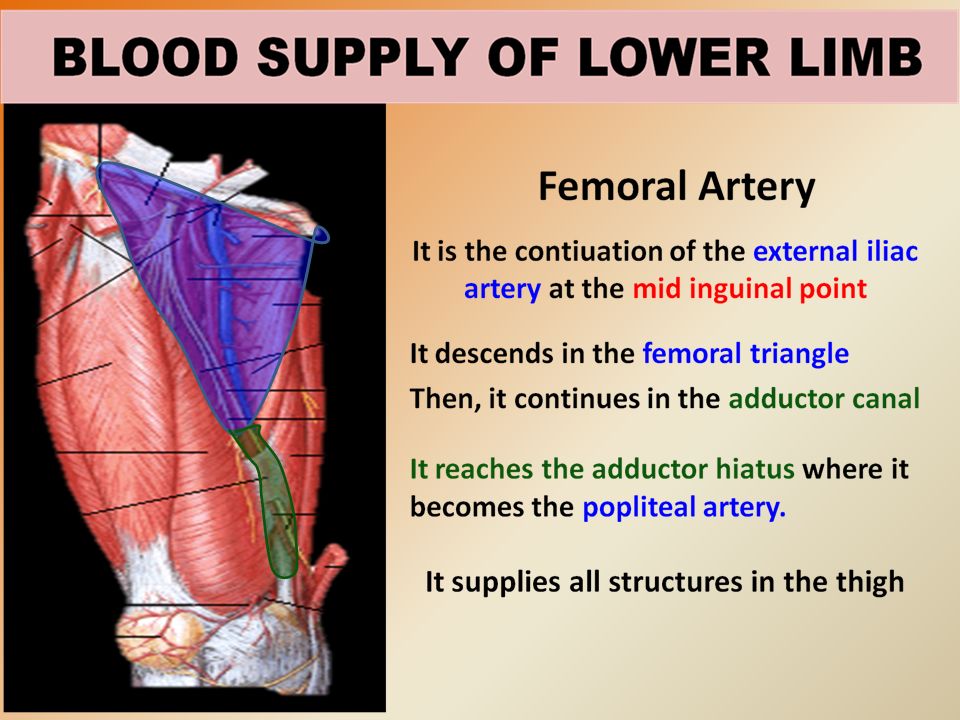
RELATIONS Anterior: Skin, fascia lata. Posterior: Hip joint, it is separated from it by Psoas muscle It lies on the muscles forming floor of the femoral triangle. Medial: Femoral vein. Lateral : Femoral nerve and its branches.

8
CRUCIATE ANASTOMOSIS It supplies blood to the lower limb in case of ligation of the femoral artery. It is formed by the union of Medial & Lateral circumflex femoral arteries + the Inferior gluteal artery superiorly + the First perforating artery inferiorly.

9
It is used for left cardiac angiography. A long catheter is inserted percutaneously into the artery and passed up the external iliac artery, common iliac artery and aorta to the left ventricle. CANNULATION OF FEMORAL ARTERY

10
The superficial position of the femoral artery in the femoral triangle makes it vulnerable to lacerations and gunshot wounds. Commonly, both the femoral artery and vein are lacerated in anterior thigh wounds because they lie so close together. As a result of communication between the injured vessels, an arteriovenous shunt occurs. LACERATION OF THE FEMORAL ARTERY

11
FEMORAL PULSE It can be palpated just inferior to the midinguinal point. How to Stop blood in the femoral artery? At this site, the femoral artery is by pressed directly posteriorly against the superior pubic ramus and the femoral head, this will stop the blood flow through the femoral artery and its branches.

13
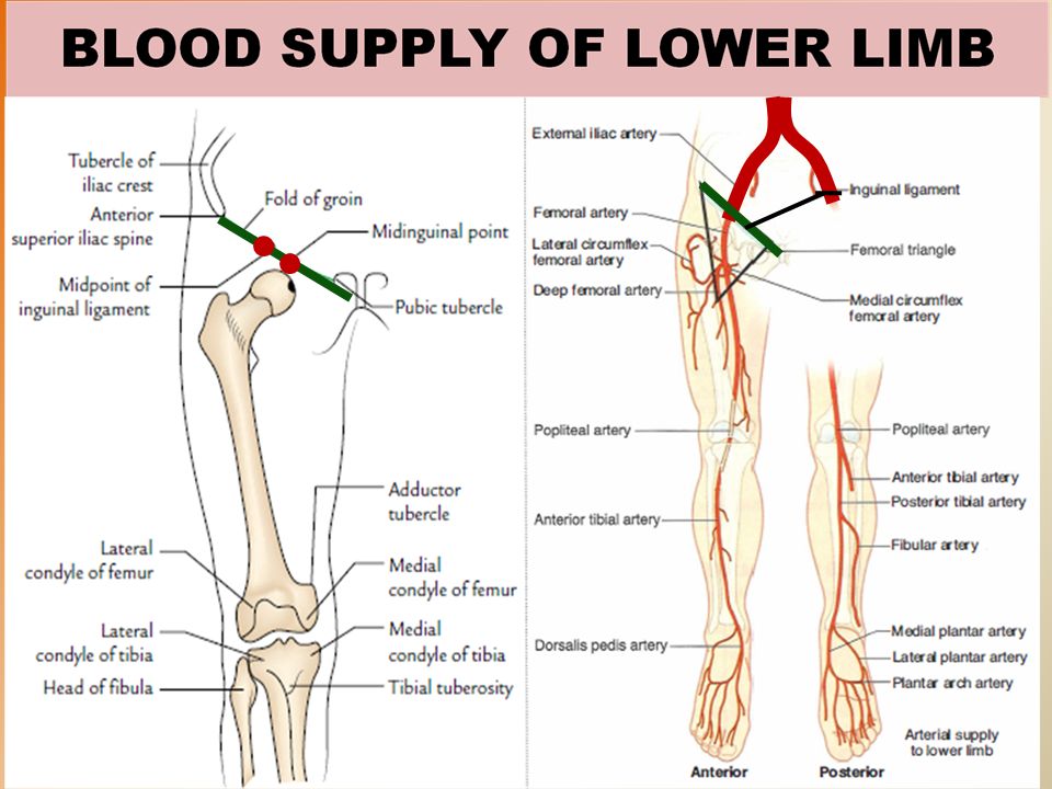
POPLITEAL ARTERY It is the continuation of femoral artery. It is the deepest structure in the Popliteal Fossa (posterior to the Popliteal vein & Tibial nerve), it runs close to the capsule of the knee joint.. Ends at the lower border of popliteus muscle by dividing into Anterior and Posterior Tibial Arteries
, it runs close to the capsule of the knee joint.. Ends at the lower border of popliteus muscle by dividing into Anterior and Posterior Tibial Arteries.")
16
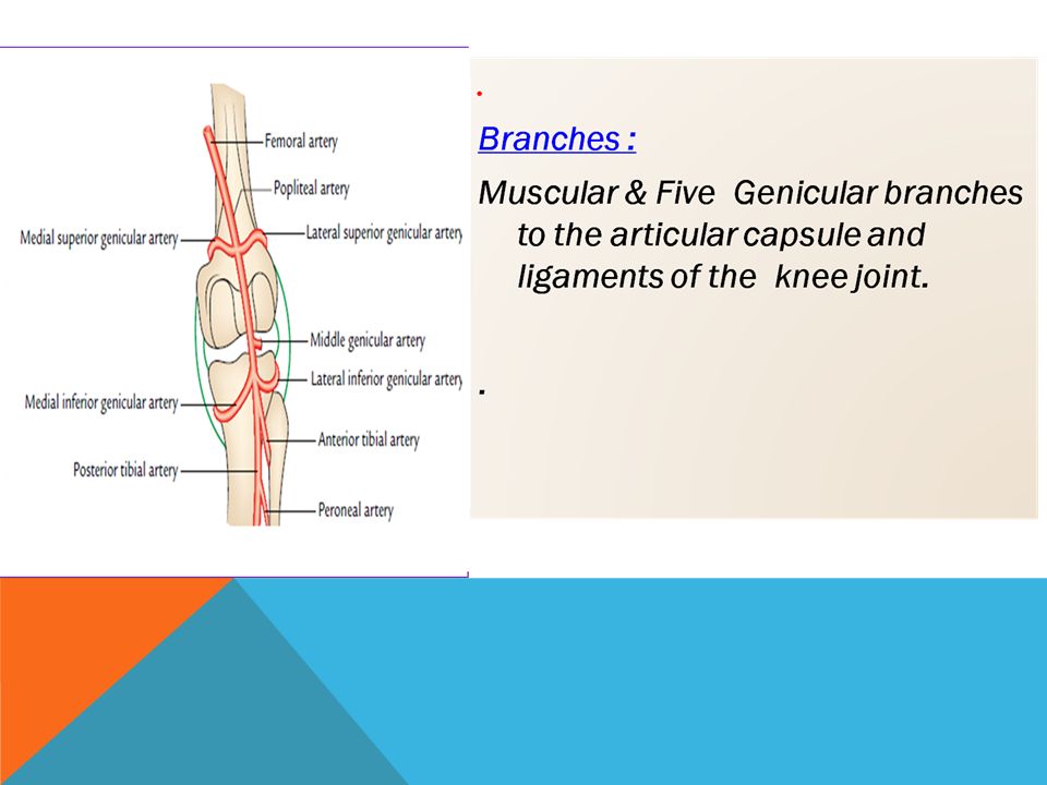
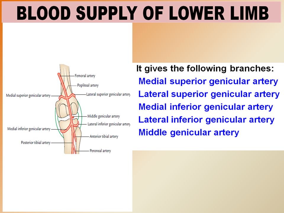
ANASTOMOSIS AROUND KNEE The Genicular arteries participate in the formation of the important Genicular Anastomosis around the knee, (it compensates for the narrowing of the Popliteal artery during flexion of the knee).
.")
17
ANASTOMOSIS AROUND THE KNEE JOINT Is made by the following branches: Descending branch of lateral circumflex femoral Descending genicular of femoral Anterior tibial recurrent Five branches of popliteal artery

18
Because of the deep position of the artery, its pulsations are best felt in the inferior part of the popliteal fossa where the artery is related to the tibia. Weakening or loss of the popliteal pulse is a sign of femoral artery obstruction. POPLITEAL PULSE

19
ANTERIOR TIBIAL ARTERY It is the smaller terminal branch of the popliteal artery. It enters the anterior compartment of the leg by passing through an opening in the upper part of ther interosseous membrane, in company with the Deep Peroneal nerve. It supplies structures in the Anterior Compartment of the leg. It ends at the ankle joint midway between the malleoli where it becomes the Dorsalis Pedis artery (dorsal artery of the foot.

20
POSTERIOR TIBIAL ARTERY It is the larger terminal branch of the popliteal artery and provides the main blood supply to the Foot. Descends Deep to muscles of the posterior compartment of the leg. Its lower part is covered by skin & fascia only. It Passes behind Medial Malleolus & Deep to Flexor Retinaculum.

21
POSTERIOR TIBIAL ARTERY It terminates by dividing into: Medial & Lateral plantar arteries. Branches: 1. Peroneal (fibular) artery: The largest and most important branch. It supplies a nutrient artery to the Fibula & Muscular branches to the muscles of the Lateral and Posterior Compartments of the Leg. 2. Nutrient artery to the Tibia ( it is the largest nutrient artery of the body). 3. Calcaneal arteries: supply the Heel.
 artery: The largest and most important branch. It supplies a nutrient artery to the Fibula & Muscular branches to the muscles of the Lateral and Posterior Compartments of the Leg. 2. Nutrient artery to the Tibia ( it is the largest nutrient artery of the body). 3. Calcaneal arteries: supply the Heel..")
22
Ii is taken Postero inferior to the medial malleolus (in the groove between the malleolus and the heel) The flexor retinaculum must be relaxed by inverting the foot. Palpation of PT pulse is essential for examining patients with occlusive peripheral arterial diseases. POSTERIOR TIBIAL PULSE

23
ARTERIES OF THE FOOT DORSALIS PEDIS ARTERY It is the main source of blood supply to the Toes. Begins in front of ankle joint as the direct continuation of the Anterior Tibial artery. It is superficial in position. Crossed by the inferior extensor retinaculum It passes to the 1 st interosseous space where it divides into a deep plantar artery to the sole and the first dorsal metatarsal artery.

24
It is easy to be felt being subcutaneous, over the tarsal bones between the tendons of extensor hallucis longus and extensor digitorum longus Some people have congenitally non palpable DP pulse, the anomaly is usually bilateral. A diminished or absent dorsalis pedis pulse usually suggests vascular insufficiency resulting from arterial diseas e. DORSALIS PEDIS PULSE

25
PLANTAR ARTERIES Medial P A The smaller of the two terminal branches of the posterior tibial artery. It supplies mainly the muscles of the great toe& the skin of the medial side of the sole. Lateral PA It is the larger terminal branch. At the base of the 5 th metatarsal bone, it curves medially to form the Plantar Arch (completed by the deep plantar artery b from Dorsalis Pedis artery). The arch supplies the skin, fascia and muscles in the sole and plantar digital arteries to the adjacent digits.
. The arch supplies the skin, fascia and muscles in the sole and plantar digital arteries to the adjacent digits..")
26
VEINS OF THE LOWER.LIMB The veins of the lower limb are two sets:. Superficial :in the subcutaneous tissue). Deep: deep to the deep fascia and accompany all major arteries The superficial & deep veins have valves which are numerous in the deep veins.
. Deep: deep to the deep fascia and accompany all major arteries The superficial & deep veins have valves which are numerous in the deep veins..")
27
GREAT SAPHENOUS VEIN The Longest Superficial vein of the body. Begins from the medial end of the dorsal venous arch (as the medial marginal vein). Ascends: In front of the Medial Malleolus accompanied by the (Saphenous nerve). Posterior the Medial Condyle of the femur. Passes through the Saphenous Opening (2.5-3.25) cm below and lateral to the pubic tubercle. Terminates in: Femoral Vein.
. Ascends: In front of the Medial Malleolus accompanied by the (Saphenous nerve). Posterior the Medial Condyle of the femur. Passes through the Saphenous Opening ( ) cm below and lateral to the pubic tubercle. Terminates in: Femoral Vein..")
28
SMALL SAPHENOUS VEIN Originates from the lateral end of the dorsal venous arch. Ascends: Behind the lateral Malleolus in company with the Sural nerve. Along the middle of the back leg. Termination : 1. It may join the Great Saphenous vein. 2. Or Bifurcates: One branch joins the Great saphenous and the other joins the. Popliteal vein.

29
Even when it is not visible in infants and obese persons or in patients in shock, the great saphenous vein can always be located Anterior to the Medial Malleolus. This procedure(saphenous cutdown) is used to insert a cannula for prolonged administration of blood, plasma or drugs. SAPHENOUS CUTDOWN
 is used to insert a cannula for prolonged administration of blood, plasma or drugs. SAPHENOUS CUTDOWN.")
30
DEEP VEINS (VENAE COMITANTES) Accompany all the major arteries and their branches. Usually they are paired. They are contained within the vascular sheath of the artery, whose pulsations help to compress and move during exercise, blood received from the veins.

31
PERFORATING VEINS They originate from the superficial veins & penetrate the deep fascia close to their origin. They contain valves which normally allow the blood to flow from the superficial to the deep veins. The perforating veins pass through the deep fascia at an oblique angle so during muscular contraction, they are compressed. This also prevents blood flowing from the superficial to the deep veins..

32
V ARICOSE VEINS It is Dilatation and Degeneration of the superficial veins that may be complicated by ulcers. It is more common in the postero medial part of the lower limb. It results because of incompetence of the valves in the perforating veins, Or valves within the great saphenous itself. This allows the passage of high pressure blood from the deep to the superficial veins.

Similar presentations

.>")

>")


