Download presentation
Presentation is loading. Please wait.

1
Anatomy of the female genital organs
Dr. Ali Abd El-Monsif Thabet

2
The female pelvis

3
A) Gynaecoid B) Android C) Anthropoid
 Gynaecoid B) Android C) Anthropoid")
4
Pelvis has "inlet" , "cavity" and "Outlet".
The female pelvis Pelvis has "inlet" , "cavity" and "Outlet". (A) Pelvic inlet (Pelvic brim)
 Pelvic inlet (Pelvic brim)")
5
Pelvic inlet 1. Anteroposterior diameters:
a) Anatomical anteroposterior diameter b) Obstetric anteroposterior diameter c) Oblique conjugate
 Anatomical anteroposterior diameter. b) Obstetric anteroposterior diameter. c) Oblique conjugate.")
6
The female pelvis

7
THE EXTERNAL GENITALIA
1. The Mons Veneris (Mons pubis) 2. The Labia Majora 3. The Labia Minora 4. The Clitoris 5. The vestibule 6. The external urethral meatus 7.The Hymen 8. Bartholin's Glands
 2. The Labia Majora. 3. The Labia Minora. 4. The Clitoris. 5. The vestibule. 6. The external urethral meatus. 7.The Hymen. 8. Bartholin s Glands.")
8
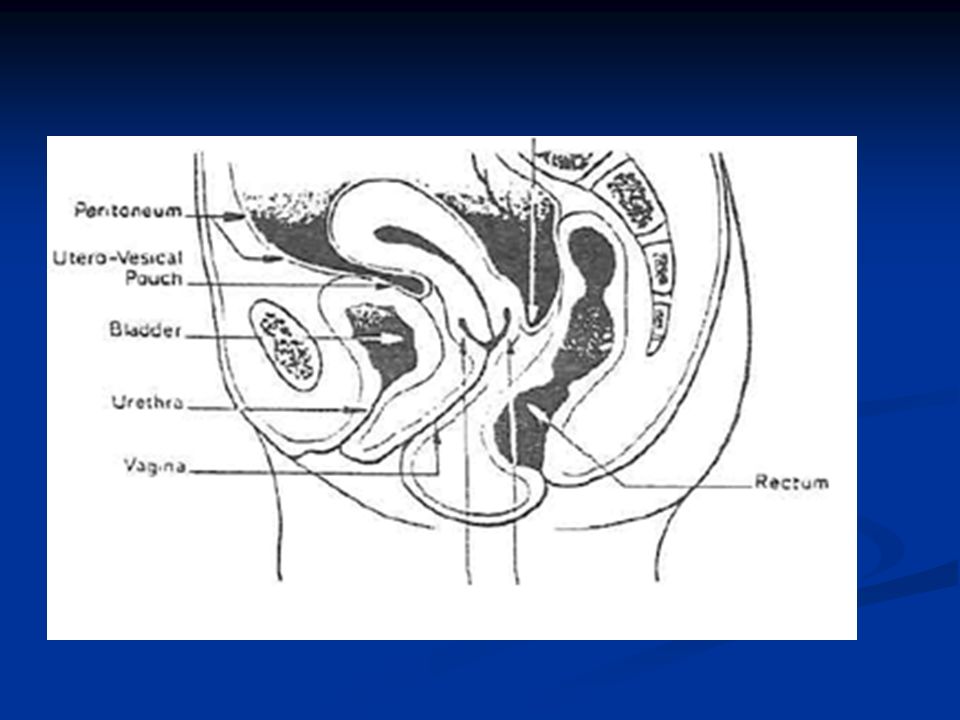
THE INTERNAL REPRODUCTIVE ORGANS
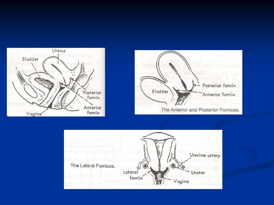
THE VAGINA The vagina is a fibromuscular canal composed of fibromuscular tissue, capable for great distention

10
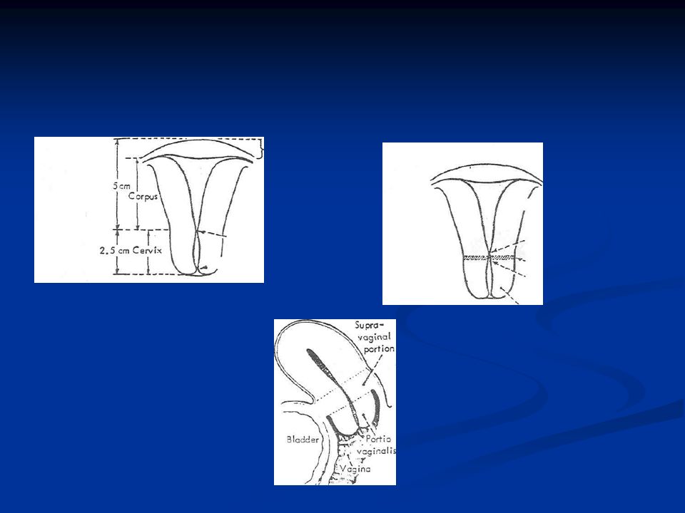
THE UTERUS The uterus is a hollow flattened pear-shaped muscular organ, which measures (1 x 2 x 3 inches) in a nullipara, and is slightly larger in a multipara (11/2 x 2 1/2 x 3 1/2 inches) ). Its cavity measures about 21/2-3 inches (6-71/2 cm) from the external os to the upper end
 in a nullipara, and is slightly larger in a multipara (11/2 x 2 1/2 x 3 1/2 inches) ). Its cavity measures about 21/2-3 inches (6-71/2 cm) from the external os to the upper end.")
13
THE FALLOPIAN TUBES Each tube is divided into four parts :
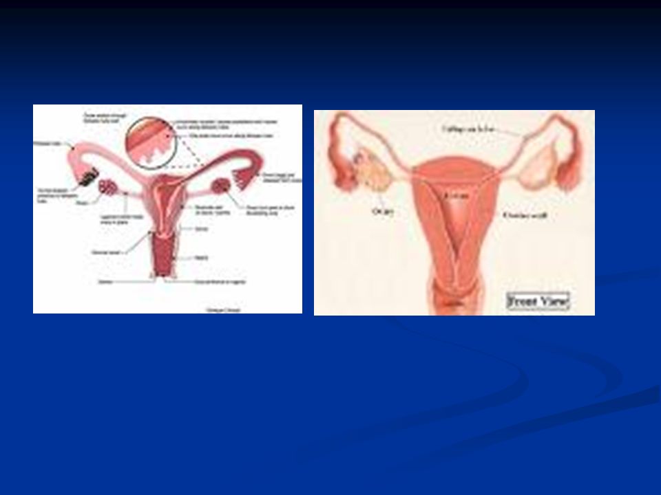
The tubes are two hollow organs 4 inches (10 cm) long, extending from the uterine cornu to open into the peritoneal cavity near to the ovaries. It's main function is that it is the site where fertilization occur, and it also transmit the fertilized ovum to be implanted into the endometrium Each tube is divided into four parts : The interstitial portion, which runs in the muscle wall of the uterus. It is the narrowest portion, being 1 mm in diameter, and it is about 1 cm long. The isthmus is the narrow portion immediately lateral to the uterus. It is characterized by thickness of its muscle layer. The ampulla is the widest part of the tube and has a much thinner musculature. The fimbriated end is the most distal portion.
 long, extending from the uterine cornu to open into the peritoneal cavity near to the ovaries. It s main function is that it is the site where fertilization occur, and it also transmit the fertilized ovum to be implanted into the endometrium. Each tube is divided into four parts : The interstitial portion, which runs in the muscle wall of the uterus. It is the narrowest portion, being 1 mm in diameter, and it is about 1 cm long. The isthmus is the narrow portion immediately lateral to the uterus. It is characterized by thickness of its muscle layer. The ampulla is the widest part of the tube and has a much thinner musculature. The fimbriated end is the most distal portion.")
15
THE FALLOPIAN TUBES

16
THE OVARIES The ovaries are two almond shaped bodies measuring 3 x 2 x 11/2 cm, attached to the uterine cornu behind the tubes by the ovarian ligament and to the posterior surface of the broad ligament by the mesovarium. The function of the ovaries is to produce ova and sex hormones (Oestrogen and Progesterone). These two functions are present only in reproductive period.
. These two functions are present only in reproductive period.")
17
the pelvic floor It is the soft structures which fill the pelvic outlet. It includes the following structures from above downwards. 1- Pelvic peritoneum; 2- Pelvic cellular tissue 3- Levator ani (pelvic diaphragm) muscle. 4- Perineum
 muscle. 4- Perineum.")
18
The cervical ligaments

19
4- Perineum

20
Thank you

Similar presentations